Abstract
Purpose:
The optimal systemic treatment for pulmonary large-cell neuroendocrine carcinoma (LCNEC) is still under debate. Previous studies showed that LCNEC with different genomic characteristics might respond differently to different chemotherapy regimens. In this study, we sought to investigate genomic subtyping using cell-free DNA (cfDNA) analysis in advanced LCNEC and assess its potential prognostic and predictive value.
Experimental design:
Tumor DNA and cfDNA from 63 patients with LCNEC were analyzed by target-captured sequencing. Survival and response analyses were applied to 54 patients with advanced-stage incurable disease who received first line chemotherapy.
Results:
The mutation landscape of frequently mutated cancer genes in LCNEC from cfDNA closely resembled that from tumor DNA, which led to a 90% concordance in genomic subtyping. The 63 LCNEC patients were classified into small cell lung cancer (SCLC)-like and non-small cell lung cancer (NSCLC)-like LCNEC based on corresponding genomic features derived from tumor DNA and/or cfDNA. Overall, patients with SCLC-like LCNEC had a shorter overall survival (OS) than those with NSCLC-like LCNEC despite higher response rate (RR) to chemotherapy. Furthermore, treatment with etoposide-platinum was associated with superior response and survival in SCLC-like LCNEC compared to pemetrexed-platinum and gemcitabine/taxane-platinum doublets, while treatment with gemcitabine/taxane-platinum led to a shorter survival compared to etoposide-platinum or pemetrexed-platinum in NSCLC-like LCNEC patients.
Conclusions:
Genomic subtyping has potentials in prognostication and therapeutic decision-making for patients with LCNEC and cfDNA analysis may be a reliable alternative for genomic profiling of LCNEC.
Keywords: cfDNA analysis, LCNEC, genomic subtyping, chemotherapy response, survival
Introduction
Pulmonary large-cell neuroendocrine carcinoma (LCNEC) is a high-grade neuroendocrine malignancy that accounts for about 1%~3% of all lung cancers (1,2). LCNEC tumors constitute a group of aggressive lung cancers characterized by high proliferation rate and poor prognosis (3,4). As a group of molecularly and biologically heterogeneous diseases, LCNEC tumors share many similar molecular and histological features with small cell lung cancer (SCLC) (5–7) as well as with non-small cell lung cancer (NSCLC) (8). Management of localized LCNEC tumors differs from that for SCLC in that surgical resection is considered standard for stage I and II diseases, similar to NSCLC, while surgery is only considered for node-negative stage I-IIA (T1–2, N0, M0) SCLC. For stages III and IV, treatment generally follows recommendations for SCLC, but the optimal chemotherapy regimen is still under debate. While some studies suggested that LCNEC should be treated as NSCLC, others have suggested LCNEC may respond better to etoposide-platinum, used in treating SCLC (9).
Previous pioneering studies indicate that LCNEC can be divided into different genomic subtypes, with similarities to either SCLC or NSCLC (8,10). More recently, Derks and colleagues reported that patients with LCNEC harbouring wild-type RB1 had improved progression-free survival (PFS) and overall survival (OS) when treated with gemcitabine or taxane partnered with platinum compared to etoposide with platinum (11). These interesting data suggested that genomic subtyping might have the potential to facilitate choosing chemotherapy regimens. However, although tissue sequencing remains the gold standard for genomic profiling, tumor tissues are often inadequate for next generation sequencing (NGS) after histologic diagnosis in advanced LCNEC. Recently, cell-free DNA (cfDNA) analysis has demonstrated great potential in genomic profiling and identifying targetable genomic aberrations (12–14), disease monitoring (15–17) and detection of minimal residual disease (18,19) across different cancer types. In the current study, we sought to investigate whether cfDNA analysis could serve as a reliable alternative for genomic subtyping of LCNEC.
Materials and Methods
Patients and samples collection
Patients who were diagnosed with LCNEC and treated with chemotherapy at Peking University Cancer Hospital (Beijing, China, referred as BJ-Cohort hereafter) or the University of Texas MD Anderson Cancer Center (Houston, TX, USA, referred as MDA-Cohort hereafter) from Jan 2007 to May 2018 were enrolled. The patients of MDA-Cohort were from GEMINI database, an MD Anderson Moon Shot Program-supported registry study. The study was performed in accordance with the ethical guidelines of the Declaration of Helsinki and approved by the Institutional Review Boards (IRB) of the Peking University Cancer Hospital or University of Texas MD Anderson Cancer Center, and all patients signed informed consent.
The pathological diagnosis was reviewed and confirmed by independent pulmonary pathologists according to the 4th edition of the World Health Organization Classification of Lung Tumors (20) and tumors with histological components other than LCNEC were excluded, and neuroendocrine features were confirmed by at least one of the following neuroendocrine markers: CD56, chromogranin A, or Synaptophysin in >10% of tumor cells. Tumor specimens and peripheral blood were collected for NGS analysis. 41 out of 45 tumor tissues and 37 out 41 plasma samples were collected prior to initiation of any therapy. Tissue samples from 4 patients (MDA20, MDA23, MDA27 and MDA30 in MDA-Cohort) and blood samples from 4 patients (A03, A09, A11 in BJ-Cohort and MDA21 in MDA-Cohort) were collected after chemotherapy. All samples were procured before targeted therapy when applicable. Tissue specimens containing a minimum of 50% tumor cells were used for NGS analysis.
Sample processing and NGS from BJ-Cohort
Tumor DNA was extracted from formalin-fixed, paraffin-embedded (FFPE) specimens. Matched peripheral blood was collected in EDTA Vacutainer tubes (BD Diagnostics, Franklin Lakes, NJ, USA) and processed within 3 hours of receiving. Plasma was separated by centrifugation at 2,500 g for 10 min, transferred to microcentrifuge tubes, and centrifuged at 16,000 g for 10 min to remove remaining cell debris. Peripheral blood lymphocytes (PBLs) from the first centrifugation were used for the extraction of germline genomic DNA as normal control. PBL DNA and tumor tissue DNA were extracted using the DNeasy Blood & Tissue Kit (Qiagen, Hilden, Germany). cfDNA was isolated from 0.5–2.0 mL plasma using QIAamp Circulating Nucleic Acid Kit (Qiagen, Hilden, Germany).
PBL or tissue DNA was sheared to 300-bp fragments with a Covaris S2 ultrasonicator followed by the Indexed Illumina NGS library preparation using Illumina TruSeq DNA Library Preparation Kit (Illumina, San Diego, CA) following the manufacture’s instruction. Sequencing libraries were prepared for cfDNA using the KAPA DNA Library Preparation Kit (KapaBiosystems, Wilmington, MA, USA). Libraries were hybridized to custom-designed biotinylated oligonucleotide probes (Roche NimbleGen, Madison, WI, USA) covering genes as indicated in Supplementary Table S1. DNA sequencing was performed using the HiSeq 3000 Sequencing System with 2×151-bp paired-end reads according to the manufacturer’s recommendations using Hiseq 3000/4000 PE Cluster Generation Kit and Hiseq 3000/4000 SBS Kit (Illumina, San Diego, CA, USA).
Terminal adaptor sequences and low-quality reads were removed before BWA (version 0.7.12-r1039) was employed to align the clean reads to the reference human genome (hg19). Picard (version 1.98) was used to mark PCR duplicates. Realignment and recalibration were performed using GATK (version 3.4–46-gbc02625). Single nucleotide variants (SNV) were called using MuTect (version 1.1.4) and NChot, a software developed in-house to review hotspot variants (21). Small insertions and deletions (Indels) were called by GATK. Somatic copy-number alterations were identified with CONTRA (v2.0.8). Significant copy number variation was expressed as the ratio of adjusted depth between cfDNA and control gDNA. The final candidate variants were all manually verified in the Integrative Genomics Viewer (IGV).
Sample processing and NGS from MDA-Cohort
Tumor DNA from MDA-Cohort was applied to a CLIA-certified institutional NGS test as previously described (22). Briefly, 10–20 ng DNA from each FFPE specimen was analyzed with the Oncomine Cancer Panel (Thermo Fisher Scientific,Waltham, MA, USA) at an average amplicon depth of at least 100×, providing critical exon coverage of 50~146 genes for the detection of SNVs, Indels and copy number amplifications (23) (Supplementary Table S2). One sample MDA24 was tested for FoundationOne® that covered 315 cancer genes (24).
cfDNA was analyzed using Guardant360 assay (Guardant health, CA, USA), a proprietary cfDNA NGS assay that detects SNVs of 70 genes as well as selected actionable or informative copy number aberrations, indels, and fusions (Supplementary Table S2). cfDNA was extracted from the entire plasma aliquot prepared from a single 10ml Streck Cell-Free DNA BCT (Streck, Inc.) (QIAmp Circulating Nucleic Acid Kit, Qiagen, Inc.). 5–30ng of extracted cfDNA was used to prepare sequencing libraries, which were then enriched by hybrid capture (Agilent Technologies, Inc.), pooled, and sequenced by paired-end sequencing of 160 – 170 base pair DNA strands with average coverage of 8,000×-15,000× (NextSeq 500 and/or HiSeq 2500, Illumina, Inc.). Germline variants were quantitatively excluded, as previously described (25).
Subclonal analysis
PyClone (26) was applied to mutations from 20 patients from Peking University Cancer Hospital with paired tumor DNA and cfDNA to infer the subclonal architecture of these specimens. Briefly, PyClone was run with 20,000 iterations and default parameters. The copy number information of each SNV was used as input for PyClone analysis (15,27), and variants were clustered as previously described (26). Clonal mutations were defined as variants in the cluster with greatest mean CCF (cancer cell fraction), otherwise subclonal (26).
Statistical analysis
Tumor responses (CR = complete remission, PR = partial response, SD = stable disease, and PD = progressive disease) were evaluated according to the Response Evaluation Criteria in Solid Tumors (RECIST version 1.1) (28). Overall response rate was defined as total number of responders, including complete and partial responders, divided by the response-evaluable patients. PFS was defined as the time from the starting day of first-line chemotherapy to the first day of documented disease progression or death from any cause. Patients without progressive disease at the time of analysis were censored at the time of the last follow-up. Survival curves were generated using the Kaplan-Meier method, and the log-rank test was used to compare the survival curves. The clinic pathologic characteristics and response rates of chemotherapy were compared between the two groups using chi-square tests. Differences in categorical variables between two groups for statistical significance were evaluated using the c2 test or Fisher exact test. All multivariate statistical analyses were performed with SPSS (v.23.0; IBM, College Station, TX), and survival analyses were performed with GraphPad Prism (v. 6.0; GraphPad Software, La Jolla, CA) software. Statistical significance was defined as a two-sided P-value < 0.05. The group including less than 5 samples was not used to do statistical analyses. Survival and response analyses were only applied to patients with advanced disease (stage IIIB and IV) who received first line chemotherapy.
Results
Patient characteristics
In this retrospective study, a total of 63 patients with LCNEC were enrolled including 44 patients from Peking University Cancer Hospital (BJ-Cohort) and 19 patients from University of Texas MD Anderson Cancer Center (MDA-Cohort). Among these patients, 54 of 63 patients with incurable stage IIIB or IV LCNEC received first-line chemotherapy (Table 1). The chemotherapy regimens include: etoposide-platinum commonly used for SCLC (hereafter referred as SCLC-PE); pemetrexed with platinum usually used for non-squamous cell NSCLC (hereafter referred as NSCLC-PEM); and gemcitabine, docetaxel, or paclitaxel with platinum commonly used for NSCLC and sometimes in SCLC (hereafter referred as NSCLC-GEM/TAX) (Supplementary Table S3).
Table 1.
Patient clinical characteristics
| Age (y) | Median | 58 |
| Range | 33~82 | |
| Gender | Male | 70% (43) |
| Female | 30% (20) | |
| Smoking Status | Smoker | 70% (44) |
| Non-smoker | 30% (19) | |
| Stage IIIB or IV diseases; received first line chemotherapy (n=54) | NSCLC-GEM/TAX | 14 |
| SCLC-PE | 27 | |
| NSCLC-PEM | 13 |
Mutational landscape of LCNEC
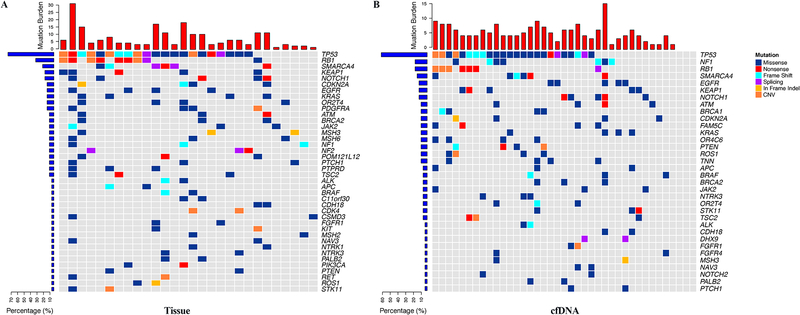
To depict the mutation landscape of LCNEC tumors, we first analyzed the mutations from tumor DNA of 28 patients from BJ-Cohort, which were subjected to NGS of a panel of 179 known cancer genes (Supplementary Table S1). The average sequencing depth was 919× (103× - 3,008×). Mutations were identified in all 28 tumors for a total of 205 somatic variants (7.4 variants per sample on average), including 146 missense, 20 nonsense, 7 splice sites, 13 frame-shift mutations, 3 deletions, 1 insertion, 1 gene fusion, 4 copy number gains and 10 copy number losses with a mean nonsynonymous mutation burden of 13.6 mutations/mega base (Supplementary Table S4), comparable to previous report (10). Commonly altered genes in these LCNEC tumors included TP53 (n=21, 75%), RB1 (n=9, 32.1%), SMARCA4 (n=6, 21.4%), NOTCH1 (n=5, 17.9%) and KEAP1 (n=5, 17.9%), etc. In addition, KRAS, EGFR and CDKN2A mutations were found in 4 patients and STK11 mutation or loss was detected in 2 patients (Fig. 1A and Supplementary Table S4).
Figure 1. Cancer gene mutation landscape of LCNEC tumors from BJ-Cohort derived from NGS of tumor DNA(n=28) (A) and cfDNA(n=39) (B).
Patients were arranged along the x-axis. Mutation burden (number of mutations) is shown in the upper panel. Genes with somatic mutations detected in more than one patient were shown. Mutation frequencies of each gene were shown on the left.
There were 32 cancer genes covered by both cancer gene panels used in sequencing tumor DNA specimens from MDA-Cohort and BJ-Cohort (Supplementary Table S5). Since smaller gene panels were utilized and fewer patients were tested, we did not attempt to depict the genomic landscape of LCNEC tumors from MDA-Cohort. Nevertheless, the two most important cancer genes for LCNEC TP53 (76% in MDA-Cohort vs 75% in BJ-Cohort) and RB1 (29% in MDA-Cohort vs 32% in BJ-Cohort) had similar mutation rates compared to BJ-Cohort. On the other hand, mutations of STK11 were more common in MDA-Cohort (35% in MDA-Cohort versus 7% in BJ-Cohort, p = 0.04) that may reflect the ethnic and etiological differences between these two patient populations.
cfDNA analysis may be a potentially promising alternative for genomic profiling of LCNEC
To investigate the feasibility of genomic profiling of LCNEC using cfDNA, cfDNA from 39 patients from BJ-Cohort was subjected to NGS of the same 179 cancer genes at an average sequencing depth of 1,325× (range 447× - 2,457×). A total of 182 somatic variants were identified (ranging from 5 to 38 per sample) in 36 of 39 (92.3%) cfDNA samples including 171 SNVs/Indels, 4 copy number gains and 7 copy number losses with an average of 9.6 mutations/mega base, which was slightly lower than that from tumor DNA. As shown in Fig. 1B and Supplementary Table S6, the overall mutational landscape derived from cfDNA was similar to that from tumor DNA. TP53 (n=24, 66.7%), RB1 (n=7, 19.4%), NF1 (N=7, 19.4%), SMARCA4 (n=6, 16.7%), EGFR (n=5, 13.9%), KEAP1 (n=5, 13.9%) and NOTCH1 (n=5, 13.9%) were the most frequently mutated genes in cfDNA in this patient cohort. In addition, somatic alterations in KRAS, ATM, BRCA1 or CDKN2A were detected in 4 of 39 patients and mutations in STK11 or BRCA2 were identified in 3 patients respectively. No mutations were detected in cfDNA samples of A03, A09 and A11.
To further assess the reliability of genomic profiling of LCNEC tumors using cfDNA, we next analyzed the 23 patients from BJ-Cohort, who had paired tumor and cfDNA subjected to NGS of the same panel of 179 cancer genes. Somatic variants were identified in all 23 tumor DNA and 20 of the 23 cfDNA specimens (No variants were detected from cfDNA samples A03, A09 and A11 as described above.) (Fig. 2). Interestingly, only 19% of tumor DNA-specific mutations and 31% of cfDNA-specific mutations were clonal while 56% of mutations shared by tumor DNA and cfDNA were clonal, indicating the substantial genomic heterogeneity of LCNEC tumors. Importantly, the concordence rate for the most frequently mutated genes such as RB1, TP53, PTEN, FGFR1 and FGFR4 was 90% (18/20), 85% (17/20), 95% (19/20), 90% (18/20) and 95% (19/20), respectively, suggesting cfDNA sequencing may be a promising alternative for genomic profiling of advanced LCNEC tumors, particularly for the frequently mutated cancer genes.
Figure 2. Cancer gene mutations detected in paired tumor DNA and cfDNA from 20 patients.
Genes with somatic mutations are listed on the x-axis, and samples are shown on the y-axis. Mutations detected in tumor tissue DNA only, in cfDNA only or shared were shown in blue, red and orange, respectively.
Genomic subtyping of LCNEC
Several pioneer studies (8,10,11) have demonstrated that mutations in RB1 and TP53 are critical biomarkers for subclassification of LCNEC. We next classified patients of BJ-Cohort into two groups based on the mutational status of RB1 and TP53 (Criterion 1). Considering the high concordance between tumor DNA and cfDNA profiling particularly in the most frequently mutated genes, NGS data from tumor DNA and cfDNA was merged for subsequent genomic subtyping analyses. Patients with mutations or copy number loss in both RB1 and TP53 were classified as SCLC-like LCNEC and patients without RB1+TP53 co-alterations were classified as NSCLC-like LCNEC. Based on this criterion, 20 of the 28 patients from BJ-Cohort were classfied as NSCLC-like LCNEC and 8 patients as SCLC-like LCNEC. From the 36 patients with mutations detected from cfDNA, 28 patients were classified as NSCLC-like LCNEC and 8 as SCLC-like LCNEC. Importantly, among the 20 patients who had somatic variants detected in paired tumor DNA and cfDNA, the genomic classification based on tumor DNA or cfDNA was concordant in 90% (18/20) of patients (Supplementary Table S7) once again highlighting the reliability of cfDNA analysis for genomic subtyping of LCNEC. Similarly, based on this criterion, 15 patients from MDA-Cohort were classified as NSCLC-like LCNEC and 4 patients as SCLC-like LCNEC.
Genomic subtyping is associated with survival and chemotherapy response
We next sought to investigate the impact of genomic characteristics on clinical outcome of LCNEC patients. Based on the presence of co-alterations in TP53 and RB1 (Criterion 1), 15 of 63 patients were classified as SCLC-like LCNEC and 48 as NSCLC-like LCNEC. Interestingly, the SCLC-like LCNEC tumors displayed significantly higher level of Ki67, a marker for cell proliferation, than NSCLC-like tumors (p<0.05) (Supplementary Fig. S1). Fifty-four of the 63 patients with incurable stage IIIB and IV LCNEC received platinum-based chemotherapy as first line treatment. The overall response rate (RR = CR + PR) was 46.7% (7/15) in patients with SCLC-like LCNEC and 25.6% (10/39) in patients with NSCLC-like LCNEC. However, despite higher RR, the OS of patients with SCLC-like LCNEC appeared to be shorter than those with NSCLC-like LCNEC although it did not reach statistical significance (median 9.8 months vs 14.4 months, p=0.18).
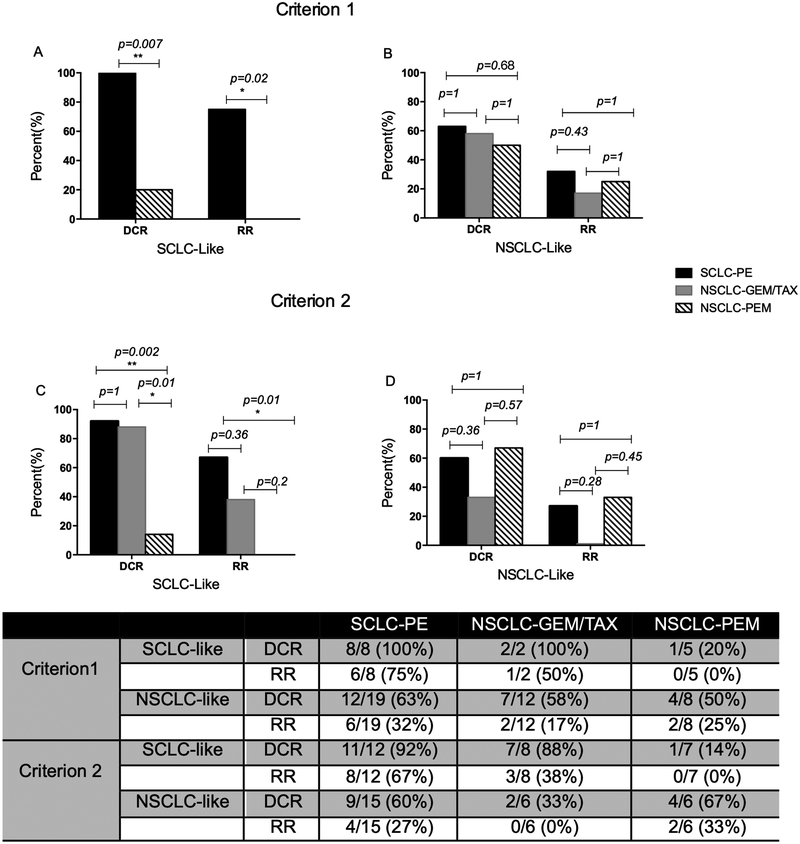
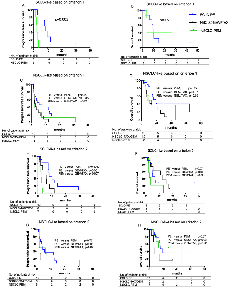
Next, we investigated whether genomic subtyping is associated with response to different chemotherapy regimens as previously reported (10,11). In SCLC-like LCNEC, the disease control rate (DCR = CR + PR + SD) and RR to SCLC-PE regimen were significantly higher than those to the NSCLC-PEM regimen (100% vs 20%, p=0.007 for DCR and 75% vs 0%, p=0.02 for RR, respectively) (Fig. 3A). In addition, the PFS of patients treated with SCLC-PE was significantly longer than patients treated with NSCLC-PEM (8.3 vs 2.4 months, p=0.002, Fig. 4A). The OS of patients treated with SCLC-PE regimen also appeared to be longer than those treated with NSCLC-PEM although the difference was not significant (9.7 vs 4.1 months, p=0.6, Fig. 4B).
Figure 3. Genomic subtyping was associated with response to different chemotherapy regimens.
DCR: disease control (complete response + partial response + stable disease) rate. RR: response (complete response + partial response) rate. Criterion 1: LCNEC tumors with RB1+TP53 co-alterations were classified as SCLC-like LCNEC (n=15), otherwise as NSCLC-like (n=) (39). Criterion 2: LCNEC tumors harboring any of the following alterations: RB1 mutation or loss, PTEN loss/mutation, FGFR1/FGFR4 mutation/amplification, TP53 loss were classified as SCLC-like LCNEC (n=27), otherwise as NSCLC-like (n=27). SCLC-PE: etoposide-platinum doublets. NSCLC-GEM/TAX: gemcitabine or taxane-platinum doublets. NSCLC-PEM: pemetrexed-platinum doublets. Note: Only 2 SCLC-like LCNEC patients based on criterion 1 were treated with NSCLC-GEX/TAX, who were not included in the statistical analysis.
Figure 4. Genomic subtyping was associated with survival of LCNEC patients treated with different chemotherapy regimens.
A-D: the genomic subtyping was based on the presence (SCLC-like) or absence (NSCLC-Like) of co-alterations in TP53 and RB1 (Criterion 1). E-H: the genomic subtyping was based on the presence (SCLC-like) or absence (NSCLC-Like) of any of the following alterations: RB1 mutation or loss, PTEN loss/mutation, FGFR1/FGFR4 mutation/amplification, TP53 loss (Criterion 2). SCLC-PE: etoposide-platinum doublets. NSCLC-GEM/TAX: gemcitabine or taxane-platinum doublets. NSCLC-PEM: pemetrexed-platinum doublets. 54 patients with first line chemotherapy were included in survival analysis.
For the NSCLC-like LCNEC, on the other hand, neither DCR nor RR was significantly different between patients treated with 3 different chemotherapy regimens (Fig. 3B). However, compared to NSCLC-GEM/TAX regimen, SCLC-PE was associated with significantly longer PFS (median 5.5 vs 2.5 months, p=0.045, Fig. 4C) and longer OS with borderline significance (median 19.6 vs 9.4 months, p=0.07, Fig. 4D).
Optimized genomic subtyping
Compared to previous studies (10,11), SCLC-like LCNEC seemed be underrepresented in our cohorts: 24% (15/63) compared to 42% (18/43) in MSKCC cohort (10). We next explored whether we could further classify NSCLC-like LCNEC by analyzing additional genomic aberrations that were more frequently associated with SCLC including PTEN loss/mutation, FGFR4/FGFR1 mutation/amplification and TP53 loss (29–31). We therefore classified LCNEC harboring any of these alterations: RB1 mutation or loss, PTEN loss/mutation, FGFR1/FGFR4 mutation/amplification, TP53 loss, as “SCLC-like” tumors, otherwise as NSCLC-like (Criterion 2). With these criteria, 27 patients were classified as SCLC-like and 36 as “NSCLC-like”. Once again, despite higher RR to chemotherapies in SCLC-like LCNEC compared with NSCLC-like LCNEC (41% vs 22%), patients with SCLC-like LCNEC had shorter OS than patients with NSCLC-like LCNEC although the difference was not significant (10.3 vs 14.4 months, p=0.32).
For SCLC-like LCNEC based on Criterion 2, SCLC-PE once again demonstrated superiority compared to NSCLC-PEM with regards to DCR (92% vs 14%, p=0.002), RR (67% vs 0%, p=0.01, Fig. 3C), PFS (median 8.3 vs 2.3 months, p=0.0002, Fig. 4E) and OS (median 10.3 vs 5 months, p=0.07, Fig. 4F). In addition, NSCLC-GEM/TAX also appeared to be superior to NSCLC-PEM with higher DCR (88% vs 14%, p=0.01) and RR (38% vs 0%, p=0.2, Fig. 3C) as well as longer PFS (median 5.9 vs 2.3 months, p=0.007, Fig. 4E) and OS (median 23.9 vs 5 months, p=0.06, Fig. 4F).
In the NSCLC-like LCNEC based on Criterion 2, no differences were observed in the DCR or RR for different regimens (Fig. 3D). However, compared to NSCLC-GEM/TAX, SCLC-PE was associated with significantly longer PFS (median 4.1 vs 1.9 months, p=0.03) and borderline significantly longer OS (median 16 vs 7.3 months, p=0.08). NSCLC-PEM also appeared to be superior to NSCLC-GEM/TAX in PFS (median 4 vs 1.9 months, p=0.07) and OS (median 29.8 vs 7.3 months, p=0.22) but the difference did not reach statistical significance (Fig. 4G–H).
Combining NSCLC-regimen treatment subgroups
Given the small sample size in this study, we combined NSCLC-GEM/TAX and NSCLC-PEM subgroups and redid the analyses. As shown in the Supplementary Fig. S2, SCLC-PE was associated with significantly longer PFS than NSCLC regimens (GEM/TAX or PEM) in SCLC-like LCNEC patients based on either criterion 1 (median 8.3 months versus 3.0 months, p=0.0013, Supplementary Fig. S2A) or criterion 2 (median 8.3 months versus 3.0 months, p=0.0032, Supplementary Fig. S2E), but OS did not seem to be different (Supplementary Fig. S2 C and G). For NSCLC-like LCNEC patients, SCLC-PE also appeared to be superior or at least equivalent to NSCLC regimens for both PFS and OS (Supplementary Fig. S2 B, D, F, H) suggesting that SCLC-PE might be the most effective regimen for the majority of LCNEC patients.
Sensitive gene mutations indicating target therapy
In this cohort of LCNEC, targetable genomic aberrations were detected in 5 patients including EGFR L858R mutation in patients A04, A21 and A35, an EGFR T790M mutation in patient A15 and EML4-ALK fusion in patient A11 from BJ-Cohort. All these 5 patients were wild type for RB1. Patients A04, A21 and A35 carrying EGFR L858R mutation received EGFR tyrosine kinase inhibitor gefitinib or erlotinib at the time of disease progression after chemotherapy and all achieved a PR, while patient A15 with EGFR T790M and patient A11 with EML4-ALK fusion have not received target therapy agents yet. Of particular interest, the classic targetable mutations in EGFR or ALK were only detected in patients from BJ-Cohort, but not from MDA-Cohort. Similarly, such mutations were rarely reported in Caucasian patients (32–34). Taken together, these results implied the potential enthinic and etiologic differences between Caucasian and Asian patients and suggested genomic profiling is warranted at least for Asian patients with advanced LCNEC to potentially identify underappreciated treatment options.
Discussion
Currently, the diagnosis of LCNEC is largely dependent on histology and immunohistochemistry (IHC) assessment, which may not always accurately reflect the true tumor biology. Genomic features may provide additional information for the clinical and biological behaviors of LCNEC. TP53 and RB1 were the most commonly altered cancer genes in LCNEC. The prevalence of genomic alterations was reported to be as high as 71% to 92% for TP53 and 26% to 47% for RB1 (8,10,11,35). In addition to point mutations, inactivation of RB1 can occur via large chromosomal losses, which are poorly detected by NGS. IHC may potentially capture those LCNEC with large chromosomal loss-mediated RB1 loss (8). We attempted to test RB1 by IHC, however, were unsuccessful due to depletion of specimens, once again highlighting this challenge in studying advanced LCNEC. Nevertheless, the frequency of genomic alterations in TP53 (81% in MDA-Cohort and 75% in BJ-Cohort) and RB1 (25% in MDA-Cohort and 32% in BJ-Cohort) in current study was comparable with previous studies (8,10,11,35).
A practical challenge for genomic profiling in advanced LCNEC is the insufficient tissue for NGS-based assays. In the current study, we demonstrated that the mutation landscape of the most frequently mutated cancer genes in LCNEC derived from cfDNA resembled that from tumor DNA, which led to a 90% concordance in genomic subtyping of LCNEC suggesting that cfDNA sequencing may be a reliable modality for detecting somatic mutations and defining genomic subtypes of LCNEC. Furthermore, cfDNA sequencing is non-invasive and “real-time”, which makes it an ideal tool for monitoring response and investigating genomic evolution of LCNEC over time, particularly during treatment. There were 3 patients in our cohort who had mutations detected in tumors, but not in cfDNA. Interestingly, all 3 patients received chemotherapy and achieved a PR before blood collection. Therefore, the “failure to detect mutations from cfDNA” may have reflected treatment response.
Pioneer studies have demonstrated that genomic features may be able to classify LCNEC into different molecular subtypes that may be associated with distinct response to different chemotherapy regimens (8,10,11,35). Using previously defined criteria of co-alterations of RB1 and TP53, or our extended criteria, the OS appeared to be shorter in patients with SCLC-like LCNEC tumors despite higher RR to chemotherapy, which is reminiscent of classic SCLC and NSCLC. In addition, chemotherapy with etoposide-platinum commonly used for treating SCLC demonstrated superiority compared to NSCLC regimens. These data suggest that a proportion of LCNEC may truly resemble classic SCLC, therefore, probably should be treated as SCLC.
On the other hand, a bigger proportion of LCNEC tumors were classified as NSCLC-like tumors by either co-alterations in TP53 plus RB1 or our extended criterion. While SCLC-PE and NSCLC-PEM demonstrated similar efficacy in these patients (Fig. 3 and Fig. 4), NSCLC-TAX/GEM appeared to be inferior to SCLC-PE or NSCLC-PEM. NSCLC-TAX/GEM is commonly used for NSCLC, particularly squamous cell carcinoma and sometimes in SCLC. In a previous study (11), Derks et al reported that patients with wild-type RB1 treated with NSCLC-TAX/GEM had a significantly longer OS than those treated with SCLC-PE, which is contrary to the findings in the current study. Reanalysis of our data based solely on the mutation status of RB1 as in Derks’s study, SCLC-PE still outperformed NSCLC-GEM/TAX (Supplementary Table S8). The underlying reasons for such discrepancy between these two studies are not clear. Difference in patient clinical characteristics, ethnicity, or concomitant therapy like radiation etc. could be potential factors that have contributed to the difference. However, since the sample size was small in both Derks study (15 and 13 RB1 wild-type LCNEC patients treated with NSCLC-GEM/TAX or SCLC-PE respectively) and the current study (11 and 17 RB1 wild-type LCNEC patients treated with NSCLC-GEM/TAX or SCLC-PE respectively), these data should be interpreted with extreme caution. Future studies with large sample size are needed to address this critical question.
Overall, SCLC-PE appeared to be a “good regimen” for the majority of LCNEC patients, both SCLC-like and NSCLC-like in our cohort by either three therapeutic subgroups (Fig. 3 and Fig. 4) or by two therapeutic subgroups (Supplementary Fig. S2), consistent with multiple previous studies (8–10,35). However, NSCLC regimens are still worth investigating, particularly in future studies with larger cohorts considering potentially different efficacy and toxicity profiles associated with different regimens. For example, in our study, although the difference did not reach statistical significance (p=0.07), likely due to small sample size, some NSCLC-like LCNEC based on Criterion 2, may have had better or comparable clinical benefit from pemetrexed-platinum (Fig. 3 and Fig. 4). As such, larger NGS panels when available should be considered to allow further classification of LCNEC to identify subgroup of patients who may benefit from different NSCLC regimens.
A disappointing fact, however, is how poor these patients with advanced LCNEC did no matter which chemotherapy regimen was used in our study and others (11). Therefore, a new paradigm in treating these patients is needed. Immune checkpoint inhibitors (ICIs) have changed the therapeutic landscape of many cancer types (36–38). Encouraging data has emerged on the efficacy of ICIs in patients with advanced LCNEC (39,40). However, our knowledge for ICIs in LCNEC is rudimentary as very few LCNEC patients were included in prospective immunotherapy trials (36,41) and the data on predictive biomarkers to ICIs in LCNEC patients is sparse. Among promising predictive biomarkers to ICIs established in other cancer types (38,42–45), PD-L1 and TMB were most scrutinized. There were case reports on durable response to ICIs in LCNEC with low PD-L1, but high TMB (40,46). Given high TMB was reported to be independent of PD-L1 and associated with benefit across multiple cancer types including both NSCLC and SCLC, it is not unreasonable to speculate that TMB may be predictive to ICIs in both SCLC-like and NSCLC-like LCNEC. To address these critical questions regarding the clinical benefit from ICIs in patients with LCNEC and optimal predictive biomarkers, prospective studies on larger cohort of patients are eventually warranted. Although an integrative approach should be applied to understand the mechanisms underlying response or resistance to ICIs in LCNEC patients, NGS using large gene panels that cover critical cancer gene mutations and are able to accurately estimate TMB should be considered for correlative studies not only because genomic features could have profound impact on response to ICIs, but also because genomic alterations are potential optimal biomarkers, thanks to the facts that DNA is stable and technologies for DNA analyses are relatively mature. With these wealthy data from these studies, the impact of genomic subtyping on response to ICIs in LCNEC patients can be further studied.
Supplementary Material
Translational Relevance.
LCNECs constitute a group of aggressive lung cancers with poor prognosis. The optimal chemotherapy regimen for advanced LCNEC is still under debate. Genomic subtyping may help to facilitate choosing chemotherapy regimens. However, tumor tissues are often inadequate for genomic profiling. Our results showed that mutation landscape from cfDNA closely resembled that from tumor DNA, which led to a 90% concordance in genomic subtyping. Furthermore, patients with SCLC-like LCNEC and patients with NSCLC-like LCNEC based on genomic subtyping had different survival and treatment response to different chemotherapy regimens. These results supported the potential of genomic subtyping in prognostication and therapeutic decision-making for patients with LCNEC and indicated that cfDNA analysis may be a reliable alternative for genomic profiling of LCNEC.
Acknowledgements
We are grateful to Dr. Ke Wang, Tao Liu and Yanyan Zhang from Geneplus-Beijing Institute for help data analysis and statistics. We also appreciate supports from the neuroendocrine carcinoma & neuroendocrine tumor multidiscipline team of Peking University Cancer Hospital & Institute.
Grant support:
This study was supported by National Key R&D Program of China (No. 2018YFC0910700); The generous philanthropic contributions to The University of Texas MD Anderson Lung Moon Shot Program and the MD Anderson Cancer Center Support Grant (P30 CA01667); Cancer Prevention and Research Institute of Texas Multi-Investigator Research Award grant (RP160668), the Cancer Prevention and Research Institute of Texas (R120501), the MD Anderson Physician Scientist Program, the Khalifa Scholar Award, the UT Lung Specialized Programs of Research Excellence Grant (P50CA70907), the T.J. Martell Foundation, the Beijing Natural Science Foundation (Commission No. 7162038) and the Beijing Municipal Administration of Hospitals’ Youth Program (Commission No. QML20161101).
Footnotes
Conflicts of interest:
Y.F.G., Y.Q.W., L.P.C., Y.H.G., L.Y., and X.Y are current employees of Geneplus-Beijing. J.V.H. is a consultant for AstraZeneca, Abbvie, Boehringer Ingelheim, Bristol-Myers Squibb, Medivation, ARIAD, Synta, Oncomed, Novartis, Genentech, and Calithera Biosciences, holds stock in Cardinal Spine LLC and Bio-Tree, and has received funding from AstraZeneca. I.I.W. receives honoraria from Roche/Genentech, Ventana, GlaxoSmithKline, Celgene, Bristol-Myers Squibb, Synta Pharmaceuticals, Boehringer Ingelheim, Medscape, Clovis, AstraZeneca, and Pfizer, and research support from Roche/Genentech, Oncoplex, and HGT. J.Z. serves as a consultant for AstraZeneca and Geneplus-Beijing and received honoraria and funding from Bristol-Myers Squibb, Merck, Roche, OrigiMed, Innovent, Bristol-Myers Squibb, Merck & Co. and Adaptive Biotechnologies Inc. outside the submitted work. The other authors declare no conflicts of interest.
References
- 1.Travis WD, Linnoila RI, Tsokos MG, Hitchcock CL, Cutler GB Jr., Nieman L, et al. Neuroendocrine tumors of the lung with proposed criteria for large-cell neuroendocrine carcinoma. An ultrastructural, immunohistochemical, and flow cytometric study of 35 cases. Am J Surg Pathol 1991;15(6):529–53. [DOI] [PubMed] [Google Scholar]
- 2.Derks JL, Hendriks LE, Buikhuisen WA, Groen HJ, Thunnissen E, van Suylen RJ, et al. Clinical features of large cell neuroendocrine carcinoma: a population-based overview. Eur Respir J 2016;47(2):615–24 doi 10.1183/13993003.00618-2015. [DOI] [PubMed] [Google Scholar]
- 3.Travis WD, Brambilla E, Burke AP, Marx A, Nicholson AG. Introduction to The 2015 World Health Organization Classification of Tumors of the Lung, Pleura, Thymus, and Heart. Journal of thoracic oncology : official publication of the International Association for the Study of Lung Cancer 2015;10(9):1240–2 doi 10.1097/JTO.0000000000000663. [DOI] [PubMed] [Google Scholar]
- 4.Asamura H, Kameya T, Matsuno Y, Noguchi M, Tada H, Ishikawa Y, et al. Neuroendocrine neoplasms of the lung: a prognostic spectrum. Journal of clinical oncology : official journal of the American Society of Clinical Oncology 2006;24(1):70–6 doi 10.1200/JCO.2005.04.1202. [DOI] [PubMed] [Google Scholar]
- 5.Jones MH, Virtanen C, Honjoh D, Miyoshi T, Satoh Y, Okumura S, et al. Two prognostically significant subtypes of high-grade lung neuroendocrine tumours independent of small-cell and large-cell neuroendocrine carcinomas identified by gene expression profiles. Lancet 2004;363(9411):775–81 doi 10.1016/S0140-6736(04)15693-6. [DOI] [PubMed] [Google Scholar]
- 6.Cerilli LA, Ritter JH, Mills SE, Wick MR. Neuroendocrine neoplasms of the lung. Am J Clin Pathol 2001;116 Suppl:S65–96. [DOI] [PubMed] [Google Scholar]
- 7.Matsumura Y, Umemura S, Ishii G, Tsuta K, Matsumoto S, Aokage K, et al. Expression profiling of receptor tyrosine kinases in high-grade neuroendocrine carcinoma of the lung: a comparative analysis with adenocarcinoma and squamous cell carcinoma. J Cancer Res Clin Oncol 2015;141(12):2159–70 doi 10.1007/s00432-015-1989-z. [DOI] [PMC free article] [PubMed] [Google Scholar]
- 8.George J, Walter V, Peifer M, Alexandrov LB, Seidel D, Leenders F, et al. Integrative genomic profiling of large-cell neuroendocrine carcinomas reveals distinct subtypes of high-grade neuroendocrine lung tumors. Nature communications 2018;9(1):1048 doi 10.1038/s41467-018-03099-x. [DOI] [PMC free article] [PubMed] [Google Scholar]
- 9.Fasano M, Della Corte CM, Papaccio F, Ciardiello F, Morgillo F. Pulmonary Large-Cell Neuroendocrine Carcinoma: From Epidemiology to Therapy. Journal of thoracic oncology : official publication of the International Association for the Study of Lung Cancer 2015;10(8):1133–41 doi 10.1097/JTO.0000000000000589. [DOI] [PMC free article] [PubMed] [Google Scholar]
- 10.Rekhtman N, Pietanza MC, Hellmann MD, Naidoo J, Arora A, Won H, et al. Next-Generation Sequencing of Pulmonary Large Cell Neuroendocrine Carcinoma Reveals Small Cell Carcinoma-like and Non-Small Cell Carcinoma-like Subsets. Clinical cancer research : an official journal of the American Association for Cancer Research 2016;22(14):3618–29 doi 10.1158/1078-0432.CCR-15-2946. [DOI] [PMC free article] [PubMed] [Google Scholar]
- 11.Derks JL, Leblay N, Thunnissen E, van Suylen RJ, den Bakker M, Groen HJM, et al. Molecular Subtypes of Pulmonary Large-cell Neuroendocrine Carcinoma Predict Chemotherapy Treatment Outcome. Clinical cancer research : an official journal of the American Association for Cancer Research 2018;24(1):33–42 doi 10.1158/1078-0432.CCR-17-1921. [DOI] [PubMed] [Google Scholar]
- 12.Sundaresan TK, Sequist LV, Heymach JV, Riely GJ, Janne PA, Koch WH, et al. Detection of T790M, the Acquired Resistance EGFR Mutation, by Tumor Biopsy versus Noninvasive Blood-Based Analyses. Clinical cancer research : an official journal of the American Association for Cancer Research 2016;22(5):1103–10 doi 10.1158/1078-0432.CCR-15-1031. [DOI] [PMC free article] [PubMed] [Google Scholar]
- 13.Murtaza M, Dawson SJ, Tsui DW, Gale D, Forshew T, Piskorz AM, et al. Non-invasive analysis of acquired resistance to cancer therapy by sequencing of plasma DNA. Nature 2013;497(7447):108–12 doi 10.1038/nature12065. [DOI] [PubMed] [Google Scholar]
- 14.Tabernero J, Lenz HJ, Siena S, Sobrero A, Falcone A, Ychou M, et al. Analysis of circulating DNA and protein biomarkers to predict the clinical activity of regorafenib and assess prognosis in patients with metastatic colorectal cancer: a retrospective, exploratory analysis of the CORRECT trial. The Lancet Oncology 2015;16(8):937–48 doi 10.1016/S1470-2045(15)00138-2. [DOI] [PMC free article] [PubMed] [Google Scholar]
- 15.Murtaza M, Dawson SJ, Pogrebniak K, Rueda OM, Provenzano E, Grant J, et al. Multifocal clonal evolution characterized using circulating tumour DNA in a case of metastatic breast cancer. Nature communications 2015;6:8760 doi 10.1038/ncomms9760. [DOI] [PMC free article] [PubMed] [Google Scholar]
- 16.Bettegowda C, Sausen M, Leary RJ, Kinde I, Wang Y, Agrawal N, et al. Detection of circulating tumor DNA in early- and late-stage human malignancies. Science translational medicine 2014;6(224):224ra24 doi 10.1126/scitranslmed.3007094. [DOI] [PMC free article] [PubMed] [Google Scholar]
- 17.Mok T, Wu YL, Lee JS, Yu CJ, Sriuranpong V, Sandoval-Tan J, et al. Detection and Dynamic Changes of EGFR Mutations from Circulating Tumor DNA as a Predictor of Survival Outcomes in NSCLC Patients Treated with First-line Intercalated Erlotinib and Chemotherapy. Clinical cancer research : an official journal of the American Association for Cancer Research 2015;21(14):3196–203 doi 10.1158/1078-0432.CCR-14-2594. [DOI] [PubMed] [Google Scholar]
- 18.Garcia-Murillas I, Schiavon G, Weigelt B, Ng C, Hrebien S, Cutts RJ, et al. Mutation tracking in circulating tumor DNA predicts relapse in early breast cancer. Sci Transl Med 2015;7(302):302ra133 doi 10.1126/scitranslmed.aab0021. [DOI] [PubMed] [Google Scholar]
- 19.Tie J, Wang Y, Tomasetti C, Li L, Springer S, Kinde I, et al. Circulating tumor DNA analysis detects minimal residual disease and predicts recurrence in patients with stage II colon cancer. Sci Transl Med 2016;8(346):346ra92 doi 10.1126/scitranslmed.aaf6219. [DOI] [PMC free article] [PubMed] [Google Scholar]
- 20.Travis WD, Brambilla E, Nicholson AG, Yatabe Y, Austin JHM, Beasley MB, et al. The 2015 World Health Organization Classification of Lung Tumors: Impact of Genetic, Clinical and Radiologic Advances Since the 2004 Classification. Journal of thoracic oncology : official publication of the International Association for the Study of Lung Cancer 2015;10(9):1243–60 doi 10.1097/JTO.0000000000000630. [DOI] [PubMed] [Google Scholar]
- 21.Yang X, Chu Y, Zhang R, Han Y, Zhang L, Fu Y, et al. Technical Validation of a Next-Generation Sequencing Assay for Detecting Clinically Relevant Levels of Breast Cancer-Related Single-Nucleotide Variants and Copy Number Variants Using Simulated Cell-Free DNA. The Journal of molecular diagnostics : JMD 2017;19(4):525–36 doi 10.1016/j.jmoldx.2017.04.007. [DOI] [PubMed] [Google Scholar]
- 22.Lam VK, Tran HT, Banks KC, Lanman RB, Rinsurongkawong W, Peled N, et al. Targeted Tissue and Cell-Free Tumor DNA Sequencing of Advanced Lung Squamous-Cell Carcinoma Reveals Clinically Significant Prevalence of Actionable Alterations. Clinical lung cancer 2018. doi 10.1016/j.cllc.2018.08.020. [DOI] [PubMed] [Google Scholar]
- 23.Lih CJ, Harrington RD, Sims DJ, Harper KN, Bouk CH, Datta V, et al. Analytical Validation of the Next-Generation Sequencing Assay for a Nationwide Signal-Finding Clinical Trial: Molecular Analysis for Therapy Choice Clinical Trial. J Mol Diagn 2017;19(2):313–27 doi 10.1016/j.jmoldx.2016.10.007. [DOI] [PMC free article] [PubMed] [Google Scholar]
- 24.Chalmers ZR, Connelly CF, Fabrizio D, Gay L, Ali SM, Ennis R, et al. Analysis of 100,000 human cancer genomes reveals the landscape of tumor mutational burden. Genome medicine 2017;9(1):34 doi 10.1186/s13073-017-0424-2. [DOI] [PMC free article] [PubMed] [Google Scholar]
- 25.Lanman RB, Mortimer SA, Zill OA, Sebisanovic D, Lopez R, Blau S, et al. Analytical and Clinical Validation of a Digital Sequencing Panel for Quantitative, Highly Accurate Evaluation of Cell-Free Circulating Tumor DNA. PLoS One 2015;10(10):e0140712 doi 10.1371/journal.pone.0140712. [DOI] [PMC free article] [PubMed] [Google Scholar]
- 26.Roth A, Khattra J, Yap D, Wan A, Laks E, Biele J, et al. PyClone: statistical inference of clonal population structure in cancer. Nature methods 2014;11(4):396–8 doi 10.1038/nmeth.2883. [DOI] [PMC free article] [PubMed] [Google Scholar]
- 27.Jamal-Hanjani M, Wilson GA, McGranahan N, Birkbak NJ, Watkins TBK, Veeriah S, et al. Tracking the Evolution of Non-Small-Cell Lung Cancer. The New England journal of medicine 2017;376(22):2109–21 doi 10.1056/NEJMoa1616288. [DOI] [PubMed] [Google Scholar]
- 28.Eisenhauer EA, Therasse P, Bogaerts J, Schwartz LH, Sargent D, Ford R, et al. New response evaluation criteria in solid tumours: revised RECIST guideline (version 1.1). European journal of cancer (Oxford, England : 1990) 2009;45(2):228–47 doi 10.1016/j.ejca.2008.10.026. [DOI] [PubMed] [Google Scholar]
- 29.McFadden DG, Papagiannakopoulos T, Taylor-Weiner A, Stewart C, Carter SL, Cibulskis K, et al. Genetic and clonal dissection of murine small cell lung carcinoma progression by genome sequencing. Cell 2014;156(6):1298–311 doi 10.1016/j.cell.2014.02.031. [DOI] [PMC free article] [PubMed] [Google Scholar]
- 30.Takeuchi T, Minami Y, Iijima T, Kameya T, Asamura H, Noguchi M. Characteristics of loss of heterozygosity in large cell neuroendocrine carcinomas of the lung and small cell lung carcinomas. Pathol Int 2006;56(8):434–9 doi 10.1111/j.1440-1827.2006.01985.x. [DOI] [PubMed] [Google Scholar]
- 31.Ninomiya H, Nomura K, Satoh Y, Okumura S, Nakagawa K, Fujiwara M, et al. Genetic instability in lung cancer: concurrent analysis of chromosomal, mini- and microsatellite instability and loss of heterozygosity. British journal of cancer 2006;94(10):1485–91 doi 10.1038/sj.bjc.6603121. [DOI] [PMC free article] [PubMed] [Google Scholar]
- 32.De Pas TM, Giovannini M, Manzotti M, Trifiro G, Toffalorio F, Catania C, et al. Large-cell neuroendocrine carcinoma of the lung harboring EGFR mutation and responding to gefitinib. Journal of clinical oncology : official journal of the American Society of Clinical Oncology 2011;29(34):e819–22 doi 10.1200/JCO.2011.36.2251. [DOI] [PubMed] [Google Scholar]
- 33.Hoton D, Humblet Y, Libbrecht L. Phenotypic variation of an ALK-positive large-cell neuroendocrine lung carcinoma with carcinoid morphology during treatment with ALK inhibitors. Histopathology 2018;72(4):707–10 doi 10.1111/his.13388. [DOI] [PubMed] [Google Scholar]
- 34.Aroldi F, Bertocchi P, Meriggi F, Abeni C, Ogliosi C, Rota L, et al. Tyrosine Kinase Inhibitors in EGFR-Mutated Large-Cell Neuroendocrine Carcinoma of the Lung? A Case Report. Case reports in oncology 2014;7(2):478–83 doi 10.1159/000365413. [DOI] [PMC free article] [PubMed] [Google Scholar]
- 35.Miyoshi T, Umemura S, Matsumura Y, Mimaki S, Tada S, Makinoshima H, et al. Genomic Profiling of Large-Cell Neuroendocrine Carcinoma of the Lung. Clinical cancer research : an official journal of the American Association for Cancer Research 2017;23(3):757–65 doi 10.1158/1078-0432.CCR-16-0355. [DOI] [PubMed] [Google Scholar]
- 36.Gandhi L, Rodriguez-Abreu D, Gadgeel S, Esteban E, Felip E, De Angelis F, et al. Pembrolizumab plus Chemotherapy in Metastatic Non-Small-Cell Lung Cancer. The New England journal of medicine 2018;378(22):2078–92 doi 10.1056/NEJMoa1801005. [DOI] [PubMed] [Google Scholar]
- 37.Wolchok JD, Kluger H, Callahan MK, Postow MA, Rizvi NA, Lesokhin AM, et al. Nivolumab plus ipilimumab in advanced melanoma. The New England journal of medicine 2013;369(2):122–33 doi 10.1056/NEJMoa1302369. [DOI] [PMC free article] [PubMed] [Google Scholar]
- 38.Rosenberg JE, Hoffman-Censits J, Powles T, van der Heijden MS, Balar AV, Necchi A, et al. Atezolizumab in patients with locally advanced and metastatic urothelial carcinoma who have progressed following treatment with platinum-based chemotherapy: a single-arm, multicentre, phase 2 trial. Lancet 2016;387(10031):1909–20 doi 10.1016/S0140-6736(16)00561-4. [DOI] [PMC free article] [PubMed] [Google Scholar]
- 39.Levra MG M J, Valette CA, et al. P1.07–012 Efficacy of Immune Checkpoint Inhibitors in Large Cell Neuroendocrine Lung Cancer: Results from a French Retrospective Cohort[J]. Journal of Thoracic Oncology 2017;12(1):S702–S3. [Google Scholar]
- 40.Wang VE, Urisman A, Albacker L, Ali S, Miller V, Aggarwal R, et al. Checkpoint inhibitor is active against large cell neuroendocrine carcinoma with high tumor mutation burden. Journal for immunotherapy of cancer 2017;5(1):75 doi 10.1186/s40425-017-0281-y. [DOI] [PMC free article] [PubMed] [Google Scholar]
- 41.Reck M, Mok TSK, Nishio M, Jotte RM, Cappuzzo F, Orlandi F, et al. Atezolizumab plus bevacizumab and chemotherapy in non-small-cell lung cancer (IMpower150): key subgroup analyses of patients with EGFR mutations or baseline liver metastases in a randomised, open-label phase 3 trial. Lancet Respir Med 2019;7(5):387–401 doi 10.1016/S2213-2600(19)30084-0. [DOI] [PubMed] [Google Scholar]
- 42.Riaz N, Havel JJ, Makarov V, Desrichard A, Urba WJ, Sims JS, et al. Tumor and Microenvironment Evolution during Immunotherapy with Nivolumab. Cell 2017;171(4):934–49 e16 doi 10.1016/j.cell.2017.09.028. [DOI] [PMC free article] [PubMed] [Google Scholar]
- 43.Yarchoan M, Hopkins A, Jaffee EM. Tumor Mutational Burden and Response Rate to PD-1 Inhibition. The New England journal of medicine 2017;377(25):2500–1 doi 10.1056/NEJMc1713444. [DOI] [PMC free article] [PubMed] [Google Scholar]
- 44.Le DT, Durham JN, Smith KN, Wang H, Bartlett BR, Aulakh LK, et al. Mismatch repair deficiency predicts response of solid tumors to PD-1 blockade. Science (New York, NY) 2017;357(6349):409–13 doi 10.1126/science.aan6733. [DOI] [PMC free article] [PubMed] [Google Scholar]
- 45.Shin DS, Zaretsky JM, Escuin-Ordinas H, Garcia-Diaz A, Hu-Lieskovan S, Kalbasi A, et al. Primary Resistance to PD-1 Blockade Mediated by JAK1/2 Mutations. Cancer discovery 2017;7(2):188–201 doi 10.1158/2159-8290.CD-16-1223. [DOI] [PMC free article] [PubMed] [Google Scholar]
- 46.Mauclet C, Duplaquet F, Pirard L, Rondelet B, Dupont M, Pop-Stanciu C, et al. Complete tumor response of a locally advanced lung large-cell neuroendocrine carcinoma after palliative thoracic radiotherapy and immunotherapy with nivolumab. Lung cancer 2019;128:53–6 doi 10.1016/j.lungcan.2018.12.006. [DOI] [PubMed] [Google Scholar]
Associated Data
This section collects any data citations, data availability statements, or supplementary materials included in this article.


