

Abstract
Objective:
The aim of this study was to evaluate the influence of humidity on the intra-tubular penetration, bond strength and failure mode associated with AH Plus (AH) and MTA Fillapex (MTAF) sealers.
Methods:
For this analysis, an apparatus was created to maintain the humidity of the specimens. Sixty bovine single-rooted teeth with similar anatomy were used. The teeth were randomly divided into 4 groups (n=15): G1 - AH/moist, G2 - AH/dry, G3 - MTAF/moist and G4 - MTAF/dry. A laser scanning confocal microscope was used to analyse the penetrability of the sealers into the dentinal tubules. A push-out test was performed to examine the diameter and height of the root canal fillings. The failure mode was analysed under a stereomicroscope at 40× magnification. The data were submitted to non-parametric Kruskal-Wallis and Dunn’s tests. The significance level was P<0.05.
Results:
The MTAF sealer exhibited higher intra-tubular penetration values compared to AH Plus (P<0.05) sealer. AH Plus showed the highest bond strength values. Regarding the type of failure mode, a majority of cohesive failures was identified, irrespective of the moisture conditions, which were not statistically significant among the sealers (P<0.05).
Conclusion:
Humidity conditions did not influence the intra-tubular penetration, bond strength and failure mode of AH Plus and MTAF sealers.
Keywords: Dentin, endodontics, humidity, material testing, root canal obturation
HIGHLIGHTS.
MTA Fillapex was introduced by Ângelus (Ângelus Ind e Comércio, Londrina, Paraná, Brazil) as an endodontic sealer, with calcium silicate and salicylate resin as its main active components.
The aim of this study was to evaluate the influence of humidity on intra-tubular penetration, bond strength and type of failure associated with endodontic sealers.
The results of this study showed statistically significant differences concerning the degree of intra-tubular penetration and bond strength of the endodontics sealers compared in this study, irrespective of the moisture conditions.
INTRODUCTION
Some authors have reported that endodontic sealers play an important role in filling areas of difficult access, such as the dentinal tubules, and anatomically complex areas (1). Studies have observed that penetration of these materials could reduce the bacteria within the dentinal tubules due to their anti-bacterial properties (2). One of the main purposes of endodontic filling is to promote sealing of the root canal system to hinder tissue fluid, bacteria and/or their products’ penetration into canal, thereby preventing it from being re-infected after debridement (3). Epoxy resin-based sealers are extensively used in endodontics because of their excellent physical-chemical and biological properties (4). The AH Plus sealer is considered the gold standard because of its low-solubility, adequate dimensional stability and micro-retention to dentin (5).
Although resin-based sealers have good properties, some researchers have shown increasing interest in materials capable of enhancing periapical tissue repair. Hence, the MTA Fillapex sealer, with calcium silicate and salicylate resin as its main active components, was introduced by Ângelus (Ângelus Ind e Comércio, Londrina, Paraná, Brazil). When MTA Fillapex, considered an endodontic sealer with acceptable physical-chemical properties, was compared with AH Plus, it showed lower flow rates, setting time and solubility (6). Thus, it is considered a suitable sealer for endodontic treatment (7).
Many factors could be associated with endodontic treatment success or failure. The presence or absence of humidity in the radicular dentin during the filling process is a decisive factor for achieving an adequate filling (8, 9). The bond strength between the sealer and the dentinal walls could be affected by the presence of humidity, possibly leading to bond failures and leakage (10). In literature, there have been discussions regarding the different levels of residual humidity and how they could interfere with the sealing ability of endodontic sealers, particularly relative to resin-based sealers, known for their hydrophobic properties (11, 12).
The aim of this study was to evaluate the influence of humidity on the intra-tubular penetration, bond strength and type of failure mode associated with endodontic sealers. The null hypothesis tested was that the penetrability, bond strength and failure mode associated with AH Plus and MTA Fillapex sealers would not be influenced by the presence or absence of humidity.
MATERIALS AND METHODS
Approval from the ethics committee of the Bauru School of Dentistry was obtained for this study (CEP 191-2011). Sixty bovine incisor teeth were stored in 1% thymol solution. Extremely flattened or curved teeth were excluded from this study. Teeth with similar root canal diameters were selected, the crowns were sectioned, and the roots were reduced to a standardised length of 16 mm. The root canals were manually prepared with K-files using a step-back technique, until a #80 K-file reached the working length. The root canals were irrigated with a 30-gauge endodontic irrigation needle (Endo-Eze Tips, Ultradent, Utah, USA) with 2 mL of 2.5% sodium hypochlorite (NaOCl) solution at every change of file. In addition, the root canals were flushed with 5 mL of 17% ethylenediaminetetraacetic acid (EDTA; for 3 minutes) followed by saline solution.
The specimens were randomly divided into four groups (N=15) according to the endodontic sealers: AH Plus (Dentsply-Detrey, Konstanz, Germany) and MTA Fillapex (Ângelus Odontológica, Londrina, PR, Brazil) and the moisture conditions to be analysed: G1 - AH/moist, G2 - AH/dry, G3- MTAF/moist and G4 - MTAF/dry. In the teeth filled under moist conditions (G1 and G3), the root apex was covered with utility wax and two layers of nail varnish. To promote humidity of the specimens, the teeth were stored in an aqueous solution of sodium chloride, and to maintain it, an apparatus of polyvinyl chloride (PVC) with acrylic resin assembled with a damp sponge was set up. For this purpose, 10 mL of deionised water was added to each specimen (Fig. 1). The root canals were aspirated and dried with paper points, according to the condition to be studied in each group. In addition, the teeth were stored in an incubator at 37°C and 100% humidity. Thus, the dental tubules could be maintained hydrated before the filling process. In contrast, in Groups G2 and G4, all the procedures were performed under dry conditions. To standardise and control the amount of sealer to be applied during root canal filling, the endodontic sealers were weighed (AY220-Shimadzu, Alpax Comércio de Protudos para Laboratório Ltda, São Paulo, Brazil). Furthermore, a fluorescent agent (0.1% rhodamine B) was applied to both sealers for laser scanning confocal microscopy (LSCM) analysis, as proposed by D Alpino et al. (13). The teeth were filled using a cold lateral compaction technique, with the aid of a #30 K-file and gutta-percha cones (#80 as the master cone and R7 as secondary cones). Finally, the teeth were incubated at 37°C for 72 hours to allow the sealers to set. After the incubation time, dentin fragments measuring 2 mm in diameter were obtained. They were cut with a water-cooled diamond wheel at 200 rpm from different sections of the specimens at distances of 2, 4 and 6 mm from the root apex. Subsequently, water abrasive papers (600-, 900- and 1,200-grit) were used for the finishing and polishing of the dentin fragments for further analysis.
Figure 1.

Apparatus made of polyvinyl chloride and acrylic resin with a damp sponge
Laser scanning confocal microscopy analysis
A laser scanning confocal microscope was used (Leica Microsystems GmbH, Mannheim, Germany) with emission and absorption wavelengths between 540 nm and 590 nm. The specimens were observed at a distance of 10 μm under the surface at 10× magnification. The images were saved in a TIFF format with a resolution of 1024×1024 using the computer program Leica Application Suite (Leica Microsystems GmbH). Subsequently, the images were transferred to Image J (National Institute of Health, USA) and analysed twice, relative to sealer penetrability into the dentinal tubules.
Push-out test
The diameter and height of the root canal fillings of specimens were also studied. The dentin fragments were placed in a universal testing machine (Instron 3342; Instron Corporation, Canton, MA, USA) with the larger diameter of the fragment facing downwards. Load was applied with a load cell of 5 kN that was in contact with only the filling during loading. A push-out test was performed at a speed of 1.0 mm/min until the filling was observed to be displaced, indicating a bond failure. Each specimen was separately observed to calculate the area filled with gutta-percha and sealers. To calculate the area and height of the specimens, a digital pachymeter (Profield 150/6×0.01 mm/0.0005, Ciudad del Este, Paraguay) was used. As suggested by Nagas et al. (8), the values were calculated in mega Pascals (mPa) by dividing the load in Newtons (N) by the area of the bonded interface. The bond area of each section was calculated by using the following formula: area=2π r×h, where π=3.14, r=radius of the intra-radicular space, and h=height of the section in mm. However, the bond strength (SL) was calculated using the formula: SL=π (R+r) √ h2+(R-r)2 (19). SL - Lateral surface of the area occupied by the gutta-percha R - Radius of the filling material measured at the coronal level r - Radius of the filling material measured at the apical level h - Diameter of the specimen
Failure mode analysis
The failure mode was analysed under a stereomicroscope (Carl Zeiss Microscopy GmbH, Jena, Germany) at 40× magnification. Each specimen was identified according to the failure mode and was classified as adhesive (failure at the sealer-dentin or the sealer-core material interface), cohesive (failure within sealer or dentin) or mixed (failure in both the sealer and dentin) failures (8).
Statistical analysis
The sample calculation was performed using the G*Power v3.1 for Mac (Heinrich Heine, Universität Düsseldorf) by selecting the Wilcoxon-Mann Whitney test of the T test family. The data of a previous study were used, and the effect size (1.42) in the present study was established (14). The alpha type error of 0.05, a beta power of 0.95 and a ratio N2/N1 of 1 were also stipulated. A total of 12 samples per group were indicated as the ideal size required for noting significant differences. An additional 20% of the sample was used to compensate for possible loss of samples during the experiment. For data analysis the Graph Pad Prism 6 (GraphPad Software, Inc., California, USA) software and non-parametric Kruskal-Wallis and Dunn’s tests were used. The level of significance adopted was P<0.05.
RESULTS
The intra-tubular penetration values of the sealers are shown in Table 1. The penetrability of AH Plus and MTA Fillapex was analysed by means of LSCM images (Figs. 2-5). Concerning this variable, MTA Fillapex showed higher penetrability values irrespective of the moisture conditions, being greater in the cervical third (6 mm) and showing statistically significant difference when compared with AH Plus (P<0.05). However, the penetrability of AH Plus was not affected by the moisture conditions. The push-out bond strength values (mPa) are presented in Table 2. AH Plus presented higher bond strength values than did MTA Fillapex, not being influenced by the moisture conditions (P<0.05). The types of failure of the experimental groups are listed in Figure 6. The failure mode analysis identified a majority of cohesive failures, irrespective of the moisture conditions. However, in the AH Plus groups, many cohesive dentin failures were observed, while in the MTA Fillapex groups, only cohesive failures of the filling material could be identified (P<0.05).
TABLE 1.
Medium±min/max of AH Plus and MTA Fillapex penetration depths
| Penetration depth (μm) (Medium±min/max) | ||||||
|---|---|---|---|---|---|---|
| 2 mm | 4 mm | 6 mm | ||||
| Dry | Moist | Dry | Moist | Dry | Moist | |
| AH Plus | 487.3 (0.0-1258)aA | 0.0 (0.0-1091)aA | 963.9 (474.7-1313)aA | 805.4 (0.0-1216)aA | 986.6 (761.6-1389)aA | 915.7 (0.0-1371)aA |
| MTA Fillapex | 927(0.0-1463)aB | 887.8 (0.0-1164)aB | 970.1 (0.0-1193)aB | 1011 (694.5-1177)aB | 1050 (0.0-1215)aB | 1009 (651.7-1168)aB |
Lower case identifiers show statistical difference in the intra-group comparison of the same section level. Upper case identifiers show statistical difference between the groups (sealers) (P<0.05)
Figure 2.

(a-c) MTA Fillapex under dry conditions: 6 mm (a), 4 mm (b), and 2 mm (c)
Figure 4.

(a-c) AH Plus under dry conditions: 6 mm (a), 4 mm (b), and 2 mm (c)
Figure 5.

(a-c) AH PLUS under moist conditions: 6 mm (a), 4 mm (b), and 2 mm (c)
TABLE 2.
Medium±min/max of the push-out test
| Push-out test (mPa) (Medium±min/max) | ||||||
|---|---|---|---|---|---|---|
| 2 mm | 4 mm | 6 mm | ||||
| Dry | Moist | Dry | Moist | Dry | Moist | |
| AH Plus | 9.3 (0.02-46.29)aA | 12.32 (5.21-28.31)aA | 7.21 (2.12-30.69)aA | 6.9 (2.57-18.30)aA | 5.65(2.95-17.02)aA | 9.26 (2.76-14.92)aA |
| MTA Fillapex | 0.48 (0.0-17.14)aB | 0.65 (0.0-8.04)aB | 0.43 (0.11-25.97)aB | 0.77 (0.13-2)aB | 0.42 (0.13-7.31)aB | 0.46 (0.09-1.2)aB |
Lower case identifiers show statistical difference in the intra-group comparison of the same section level. Upper case identifiers show statistical difference between groups (sealers) (P<0.05)
Figure 6.
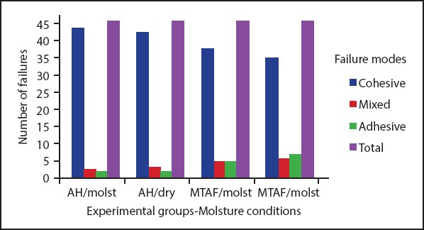
Types of failure mode related to the moisture conditions
AH: AH Plus, MTAF: MTA Fillapex
Figure 3.

(a-c) MTA Fillapex under moist conditions: 6 mm (a), 4 mm (b), and 2 mm (c)
DISCUSSION
Results of this study showed statistically significant differences in the intra-tubular penetration and bond strength values of the endodontic sealers compared in this study. When considering the penetrability and bond strength of AH Plus and MTA Fillapex, the null hypothesis was accepted, because there was no statistically significant difference relat-ed to the humidity conditions on the properties of these seal-ers. In addition the moisture conditions did not affect the failure mode, confirming the null hypothesis of our study.
Camargo et al. (15) explained the ethical aspects related to the use of extracted human teeth for scientific research and suggested the use of bovine teeth as an alternative to resolve these issues. Human teeth are morphologically and histologically similar to other mammalian teeth, but size and availability make bovine incisors preferable for research (16, 17). Therefore, the use of bovine teeth is an approved method in literature (15). According to Camargo et al. (15), bovine teeth show a significantly higher number of dentin tubules compared to human teeth; however, no statistically significant difference associated with the diameter of dentin tubules, has been found between them. Nevertheless, some of the morphological differences between human and bovine teeth have been correlated to the faster development of these structures (18).
Adhesive materials are frequently studied by means of bond strength tests (8). Push-out testing has been used to evaluate the bond strength of different endodontic materials to the radicular dentin and is considered a reliable technique (8). The adhesive properties and penetrability of some sealers into the dentinal tubules have also been analysed. LSCM has been used for detailed image acquisition, rendering the detection of the distribution of these materials inside the dentinal tubules of the root canal walls (18). As a result, LSCM is considered the ideal method for analysing the adaptation and penetrability of endodontic sealers (18). In addition, LSCM provides detailed information about the presence and distribution of sealers or dental adhesives inside dentinal tubules throughout the total circumference of the root canal walls by using fluorescent rhodamine-marked sealers (13, 18). Consequently, this information could be efficiently obtained from LSCM images (13, 18).
In the present study, MTA Fillapex showed higher penetrability values when compared with AH Plus, as confirmed by other studies (6, 19). Also, the penetrability of MTA Fillapex and AH Plus was not affected by moisture conditions, showing statistically significant difference (P<0.05). Therefore, the penetrability of these sealers into the dentin tubules was greater at the 6 mm level and lower at the 2 mm level. Based on the results, it has been observed that the penetrability of these sealers was higher in the absence of humidity. However, it was not sufficient to show a statistically significant difference. These results could be justified by the different composition and smaller particle size of MTA Fillapex, which has been correlated to greater flowability compared to AH Plus (19). In addition, this could be related to higher lateral condensation forces or due to the dentin structure in these parts of the roots (20). Furthermore, MTA Fillapex has been correlated to a higher ratio of salicylate resin compared to MTA, which affects the chemical reaction among these components and explains the extended setting time of this material (21). This leads to dimensional changes and formation of gaps between root canal areas and filling materials (21). In our study, when considering the AH Plus sealer independently, it exhibited higher penetrability in the absence of humidity. These results could be explained by the hydrophobic nature of this sealer (11, 12). As both are resin-based sealers, excessive moisture contamination could decrease the monomer conversion, leading to incomplete resin polymerisation and affecting their sealing ability as a consequence (8). The resultant entrapment of water droplets between the sealer-dentin interfaces would also lead to disruption of the bond, possibly explaining their behaviour in the presence of humidity (8). Amoroso-Silva et al. (22) found that both sealers were statistically similar relative to their penetration into the dentinal tubules, showing a lower percentage of penetrability at the 2 mm level and the highest at the 6 mm level. Therefore, these authors suggested that AH Plus and MTA Fillapex had high flowability, facilitating their penetration into the dentinal tubules (22). These results are in contrast with other studies, where the moist conditions did not affect the sealing ability of AH Plus and MTA Fillapex at the apical level of the root canal (23, 24).
When compared with MTA Fillapex, the push-out test revealed superior bond strength values for AH Plus irrespective of the moisture conditions. It was observed that the bond strength of these sealers was higher in the presence of humidity. However, it was not enough to show statistically significant difference. Studies have observed that these results are related to its capacity to interact chemically with the collagen network and form covalent bonds between the epoxy rings and the amine groups of the exposed collagen (25). Similarly, AH Plus has been associated with long-term stability and efficient cohesion between molecules, increasing its micromechanical retention to the root dentin. Accordingly, it has been suggested that it may be advantageous to leave root canals slightly moist before filling procedures to enhance the sealing properties of endodontic sealers (8). The findings of this study are in agreement with previous research, in which MTA Fillapex showed the lowest pushout values (3, 20-21, 23, 25-27). Another study showed no difference between the bond strength values of AH Plus and MTA Fillapex (28). Thus, considering MTA Fillapex, a paste-topaste sealer, two chemical reactions have been identified as being responsible for the setting and physical-mechanical properties of this material: the progressive hydration of orthosilicate ions occurs followed by a reaction between MTA and salicylate resin (21, 27). This allows the formation of an ionic polymer containing calcium silicate particles, which reacts with water (7, 29). The final reaction leads to the formation of calcium hydroxide and a nanoporous amorphous calcium silicate hydrate gel that polymerises and creates a solid network (29). Water diffusion into sealers may result in the deterioration of their physical-mechanical properties, reducing the durability of the interfaces by hydrolysis and microcrack formation (30). However, water absorption could be beneficial because it promotes an expansion of the material, which may promote proper sealing (27). Therefore, the presence of moist conditions may have enhanced the chemical reactions between the sealer components. This may explain the results for the push-out test in the presence of humidity for MTA Fillapex.
The analysis of the failure mode showed a majority of cohesive failures, irrespective of the moisture conditions. These results are in line with a study by de Paula et al. (23), wherein mainly cohesive failures of the filling materials were observed. Therefore, in the AH Plus groups, many cohesive dentin failures were observed, while in the MTA Fillapex groups, only cohesive failures of the filling material could be identified (Fig. 6). Moreover, as found by Nagas et al. (8), under dry conditions, the majority of specimens showed both adhesive failures at the sealer/dentin interface and cohesive failures within the sealer. Under moist conditions, the most common failure mode was adhesive failure along the sealer/core material interface. Regarding the types of failure identified in the push-out test, some studies have suggested that these results could be associated with a higher resistance to the displacement of the filling materials, reducing the occurrence of interference at the dentin-sealer interface. Therefore, the observation of failures of the filling materials rather than of the dentin surface was expected (28).
CONCLUSION
Based on the experimental methods and limitations of this study, the authors suggest that humidity conditions did not influence the intra-tubular penetration, bond strength and failure mode of AH Plus and MTA Fillapex sealers. Consequently, MTA Fillapex exhibited higher intra-tubular penetration values when compared to AH Plus, irrespective of the moisture conditions. Moreover, AH Plus showed bond strength values higher than MTA Fillapex, not being influenced by the moisture conditions. The failure mode analysis identified a majority of cohesive failures. Further studies are needed to understand the influence of moisture conditions on intra-tubular penetration, bond strength and failure mode associated with AH Plus (AH) and MTA Fillapex (MTAF) to support these findings.
Footnotes
Ethics Committee Approval: Ethics committee approval was received for this study from the ethics committee of Bauru School of Dentistry (CEP 191-2011).
Conflict of interest: No conflict of interest was declared by the authors.
Peer-review: Externally peer-reviewed.
Financial Disclosure: The authors declared that this study has received no financial support.
Authorship contributions: Concept – R.R.V., M.A.H.D.; Design – M.P.A., B.P.; Supervision – R.R.V., M.A.H.; Materials – M.A.H.D., B.C.V.; Data Collection and/or Processing – B.P., M.E.R.P.; Analysis and/or interpretation – I.G.M., M.P.A.; Literature search – B.C.V., M.E.R.P.; Writing – M.E.R.P., B.P.; Critical Review – I.G.M., R.R.V.
REFERENCES
- 1.Silva RV, Silveira FF, Horta MCR, Duarte MAH, Cavenago BC, de Morais IG, et al. Filling effectiveness and dentinal penetration of endodontic sealers:a stereo and confocal laser scanning microscopy study. Braz Dent J. 2015;26(5):541–46. doi: 10.1590/0103-6440201300138. [DOI] [PubMed] [Google Scholar]
- 2.Wang Z, Shen Y, Haapasalo M. Effectiveness of endodontic disinfecting solutions against young and old Enterococcus faecalis biofilms in dentin canals. J Endod. 2012;38:1376–79. doi: 10.1016/j.joen.2012.06.035. [DOI] [PubMed] [Google Scholar]
- 3.Taşdemir T, Er K, Çelik D, Tahan E, Serper A, Ceyhanli KT, et al. Bond strength of calcium silicate-based sealers to dentine dried with different techniques. Med Princ Pract. 2014;23:373–76. doi: 10.1159/000362619. [DOI] [PMC free article] [PubMed] [Google Scholar]
- 4.Duarte MA, de O Demarchi AC, de Moraes IG. Determination of pH and calcium ion release provided by pure and calcium hydroxide-containing AHPlus. Int Endod J. 2004;37(1):42–45. doi: 10.1111/j.1365-2591.2004.00756.x. [DOI] [PubMed] [Google Scholar]
- 5.Dias KC, Soares CJ, Steier L, Versiani MA, Rached-Júnior FJA, Pécora JD, et al. Influence of drying protocol with isopropyl alcohol on the bond strength of resin-based sealers to the root dentin. J Endod. 2014;40(9):1454–58. doi: 10.1016/j.joen.2014.02.021. [DOI] [PubMed] [Google Scholar]
- 6.Silva EJNL, Rosa TP, Herrera DR, Jacinto RC, Gomes BPFA, Zaia AA. Evaluation of citotoxicity and physicochemical properties of calcium silicate-based endodontic sealer mta fillapex. J Endod. 2013;39(2):274–77. doi: 10.1016/j.joen.2012.06.030. [DOI] [PubMed] [Google Scholar]
- 7.Vitti RP, Prati C, Sinhoreti MA, Zanchi CH, e Silva MGS, Ogliari FA, et al. Chemical-physical properties of experimental root canal sealers based on butyl ethylene glycol disalicylate and MTA. Dent Mater. 2013;29(12):1287–94. doi: 10.1016/j.dental.2013.10.002. [DOI] [PubMed] [Google Scholar]
- 8.Nagas E, Uyanik MO, Eymirli A, Cehreli ZC, Vallittu PK, Lassila LV, et al. Dentin moisture conditions affect the adhesion of root canal sealers. J Endod. 2012;38(2):240–44. doi: 10.1016/j.joen.2011.09.027. [DOI] [PubMed] [Google Scholar]
- 9.Zmener O, Pameijer CH, Serrano SA, Vidueira M, Macchi RL. Significance of moist root canal dentin with the use of methacrylate-based endodontic sealers:an in vitro coronal dye leakage study. J Endod. 2008;34:76–9. doi: 10.1016/j.joen.2007.10.012. [DOI] [PubMed] [Google Scholar]
- 10.Horning TG, Kessler JR. A comparison of three different root canal sealers when used to obturate a moisture-contaminated root canal system. J Endod. 1995;21(7):354–57. doi: 10.1016/S0099-2399(06)80968-4. [DOI] [PubMed] [Google Scholar]
- 11.Hashem AA, Ghoneim AG, Lutfy RA, Fouda MY. The effect of different irrigating solutions on bond strength of two root canal-filling systems. J Endod. 2009;35:537–40. doi: 10.1016/j.joen.2009.01.003. [DOI] [PubMed] [Google Scholar]
- 12.de Assis DF, do Prado M, Simão RA. Evaluation of the Interaction between Endodontic Sealers and Dentin Treated with Different Irrigant Solutions. J Endod. 2011;37:1550–52. doi: 10.1016/j.joen.2011.08.014. [DOI] [PubMed] [Google Scholar]
- 13.D Alpino PH, Pereira JC, Svizero NR, Rueggeberg FA, Pashley DH. Factors affecting use of fluorescent agents in identification of resin-based polymers. J Adhes Dent. 2006;8(5):285–92. [PubMed] [Google Scholar]
- 14.DeLong C, He J, Woodmansey KF. The effect of obturation technique on the push-out bond strength of calcium silicate sealers. J Endod. 2015;41:385–88. doi: 10.1016/j.joen.2014.11.002. [DOI] [PubMed] [Google Scholar]
- 15.Weichert CK, Presch W. Elements of chordate anatomy. 4th ed. New York: McGraw-Hill Company; 1975. [Google Scholar]
- 16.Reeves GW, Fitchie JG, Hembree JH, Puckett AD. Microleakage of new dentin bonding systems using human and bovine teeth. Oper Dent. 1995;20:230–35. [PubMed] [Google Scholar]
- 17.Oesterle LJ, Shellhart WC, Belanger GK. The use of bovine enamel in bonding studies. Am J Orthod Dentofacial Orthop. 1998;114:514–19. doi: 10.1016/s0889-5406(98)70171-4. [DOI] [PubMed] [Google Scholar]
- 18.Ordinola-Zapata R, Bramante CM, Graeff MSZ, Perochena AC, Vivan RR, Camargo EJ, et al. Depth and percentage of penetration of endodontic sealers into dentinal tubules after root canal obturation using a lateral compaction technique:A confocal laser scanning microscopy study. Oral Surg Oral Med Oral Pathol Oral Radiol Endod. 2009;108:450–57. doi: 10.1016/j.tripleo.2009.04.024. [DOI] [PubMed] [Google Scholar]
- 19.Nikhil V, Bansal P, Sawani S. Effect of technique of sealer agitation on percentage and depth of MTA Fillapex sealer penetration:A comparative in-vitro study. J Conserv Dent. 2015;18(2):119. doi: 10.4103/0972-0707.153073. [DOI] [PMC free article] [PubMed] [Google Scholar]
- 20.Sagsen B, Ustün Y, Demirbuga S, Pala K. Push-out bond strength of two new calcium silicate-based endodontic sealers to root canal dentine. Int Endod J. 2011;44(12):1088–91. doi: 10.1111/j.1365-2591.2011.01925.x. [DOI] [PubMed] [Google Scholar]
- 21.Silva EJ, Carvalho NK, Prado MC, Zanon M, Senna PM, Souza EM, et al. Push-out Bond Strength of Injectable Pozzolan-based Root Canal Sealer. J Endod. 2016;42(11):1656–59. doi: 10.1016/j.joen.2016.08.009. [DOI] [PubMed] [Google Scholar]
- 22.Amoroso-Silva PA, Guimarães BM, Marciano MA, Duarte MA, Cavenago BC, Ordinola-Zapata R, et al. Microscopic analysis of the quality of obturation and physical properties of MTA Fillapex. Microsc Res Tech Microsc Res Tech. 2014;77:1031–36. doi: 10.1002/jemt.22432. [DOI] [PubMed] [Google Scholar]
- 23.Paula AC, Brito-Júnior M, Araújo CC, Sousa-Neto MD, Cruz-Filho AM. Drying protocol influence on the bond strength and apical sealing of three different endodontic sealers. Braz Oral Res. 2016;30(1):e50. doi: 10.1590/1807-3107BOR-2016.vol30.0050. [DOI] [PubMed] [Google Scholar]
- 24.Ehsani M, Dehghani A, Abesi F, Khafri S, Dehkordi SG. Evaluation of apical micro-leakage of different endodontic sealers in the presence and absence of moisture. J Dent Res Dent Clin Dent Prospect. 2014;8(3):125–29. doi: 10.5681/joddd.2014.023. [DOI] [PMC free article] [PubMed] [Google Scholar]
- 25.Wiesse PEB, Pereira RD, Silva-Sousa YCT, Estrela C, Sousa-Neto MD, Pécora JD. Effect of ultrasonic and sonic activation of root canal sealers on the push-out bond strength and interfacial adaptation to root canal dentine. Int Endod J. 2017 May 22; doi: 10.1111/iej.12794. doi:10.1111/iej.12794. [Epub ahead of print] [DOI] [PubMed] [Google Scholar]
- 26.Sönmez IŞ, Sönmez D, Almaz ME. Evaluation of push-out bond strength of a new MTA-based sealer. Eur Arch Paediatr Dent. 2013;14(3):161–66. doi: 10.1007/s40368-013-0039-2. [DOI] [PubMed] [Google Scholar]
- 27.Vitti RP, Prati C, Silva EJNL, Sinhoreti MAC, Zanchi CH, Silva MGS, et al. Physical properties of MTA Fillapex sealer. J Endod. 2013;39(7):915–18. doi: 10.1016/j.joen.2013.04.015. [DOI] [PubMed] [Google Scholar]
- 28.Assmann E, Scarparo RK, Bottcher DE, Grecca FS. Dentin bond strength of two mineral trioxide aggregate-based and one epoxy resin-based sealers. J Endod. 2012;38:219–21. doi: 10.1016/j.joen.2011.10.018. [DOI] [PubMed] [Google Scholar]
- 29.Parirokh M, Torabinejad M. Mineral trioxide aggregate:a comprehensive literature review-part III:clinical applications, drawbacks, and mechanism of action. J Endod. 2010;36:400–13. doi: 10.1016/j.joen.2009.09.009. [DOI] [PubMed] [Google Scholar]
- 30.Sideridou I, Tserki V, Papanastasiou G. Study of water sorption, solubility and modulus of elasticity of light-cured dimethacrylates-based dental resins. Biomaterials. 2003;24:1381–87. doi: 10.1016/s0142-9612(02)00380-0. [DOI] [PubMed] [Google Scholar]