Abstract
Background
Chronic kidney disease (CKD) has become a major public health issue. Meanwhile, renal fibrosis caused by diabetic nephropathy can lead to CKD, regardless of the initial injury. It has been previously reported that silibinin or valsartan could relieve the severity of renal fibrosis. However, the effect of silibinin in combination with valsartan on renal fibrosis remains unclear.
Material and Methods
Proximal tubular cells (HK-2) were treated with TGF-β1 (5 ng/mL) to mimic in vitro model of fibrosis. The proliferation of HK-2 cells was tested by CCK-8. Epithelial-mesenchymal transition (EMT) and inflammation-related gene and protein expressions in HK-2 cells were measured by qRT-PCR and Western-blot, respectively. ELISA was used to test the level of TNF-αNF-A. Additionally, HFD-induced renal fibrosis mice model was established to investigate the effect of silibinin in combination with valsartan on renal fibrosis in vivo.
Results
Silibinin significantly increased the anti-fibrosis effect of valsartan in TGF-β1-treated HK-2 cells via inhibition of TGF-β1 signaling pathway. Furthermore, silibinin significantly enhanced the anti-fibrosis effect of valsartan on HFD-induced renal fibrosis in vivo through inactivation of TGF-β1 signaling pathway.
Conclusion
These data indicated that silibinin markedly increased anti-fibrosis effect of valsartan in vitro and in vivo. Thus, silibinin in combination with valsartan may act as a potential novel strategy to treat renal fibrosis caused by diabetic nephropathy.
Keywords: combination, silibinin, renal fibrosis, TGF-β1 signaling pathway
Introduction
Chronic kidney disease (CKD) is still a major health problem all over the world. It has been reported that hypertension and diabetes mellitus are histopathologically characterized by interstitial inflammation, tubular atrophy and fibrosis.1 On the other hand, the incidence of diabetes has increased worldwide.2 Nephropathy is a major microvascular complication of diabetes mellitus and often leads to terminal renal failure in addition to contributing significantly to cardiovascular morbidity and mortality.3 Additionally, Diabetic nephropathy can cause the occurrence of renal fibrosis. Renal fibrosis has been regarded to be an aberration of tissue healing process, in which there is progression rather than improvement of scar formation after renal tissue injury.1 When renal fibrosis occurs, most of the patients will develop to chronic kidney disease (CKD), following with end-stage renal disease (ESRD).4 In that situation, transplantation is the only effective therapeutic strategy.5 In addition, it has been reported that patients with CKD account for 10% of the world’s population.4 Therefore, novel effective strategy that inhibits progression of renal fibrosis caused by diabetic nephropathy is of great importance.
TGF-β1 was found to promote fibronectin and collagen production by transcriptional activation of the relevant genes.6 Recent studies have indicated that TGF‑β1 plays an important role in the progression of renal fibrosis.7,8 In addition, Higgins et al have found that TGF-β1 signaling pathway plays a critical role during the induction of renal fibrosis.9 Besides, it has been previously reported that activation of TGF‑β1 signaling pathway can promote EMT process during the metastasis of pancreatic cancer.10 Based on these findings, we aimed to explore the relation between TGF-β1 signaling pathway and EMT process during the fibrosis.
Valsartan is an effective drug used to treat hypertension, which is also widely used to treat renal fibrosis.11 Silibinin (SB) is a polyphenolic flavonoid extracted from milk thistle seeds.12 It has been reported that silibinin could significantly inhibit the occurrence of renal fibrosis.13 However, the effect of silibinin in combination with valsartan on renal fibrosis has not been reported yet. In this study, we aimed to investigate the anti-renal fibrosis effect of silibinin in combination with valsartan in vitro and in vivo.
Materials and Methods
Cell Culture
Since it has been previously reported that human renal tubular endothelial cells (HK-2) were used to study renal fibrosis,14 HK-2 cell lines (ATCC, Manassas, VA, USA) were used to examine the effects of silibinin in combination with valsartan on renal fibrosis in vitro. The cells were maintained in RPMI-1640 medium (Thermo Fisher Scientific, Waltham, MA, USA), supplemented with 10% fetal bovine serum (FBS), 1% penicillin (Gibco, Fisher Scientific, Waltham, MA, USA), 1% streptomycin (Thermo Fisher Scientific, Waltham, MA, USA) and 10–7 mol/L angiotensin II (Sigma, St. Louis, MO, USA) in a humidified incubator with 5% CO2 at 37°C. To establish in vitro renal fibrosis model, HK-2 cells were treated with 5 ng/mL TGF-β1 (Pepro Tech, Rocky Hill, NJ, USA) for 72 h.
Cell Viability Assay
The cell viability was measured by cell counting kit-8 (CCK8, Beyotime, Shanghai, China) according to the manufacturer’s protocol. Briefly, HK-2 cells (5000 cells/well) were seeded into 96-well plate at 37°C overnight. Then, cells were treated with different concentrations of silibinin (0, 15, 30, 60, 90 μM) or valsartan (0, 10, 20, 30, 50 μM) for 48 h. Afterwards, 10 μL of CCK-8 reagent was added to each well and incubated for another 2 h at 37°C. Finally, the absorbance at 450 nm was determined using a microplate reader (Bio-Rad Laboratories, Benicia, California, USA). Silibinin standard products were obtained from sigma (Sigma, St. Louis, MO, USA). Valsartan standard products were purchased from MCE (MedChemExpress, Monmouth Junction, NJ, USA).
Reverse Transcription-Quantitative Polymerase Chain Reaction (RT-qPCR)
Total RNA was extracted by using TRIzol reagent (Invitrogen, Thermo Fisher Scientific, Waltham, MA, USA). RNA integrity was measured by agarose gel electrophoresis. Then, cDNA was obtained by reverse transcription (PrimeScript 1st Strand cDNA Synthesis Kit, Takara, Tokyo, Japan). PCR reactions were carried out by SYBR Premix Ex Taq II (Takara, Tokyo, Japan) with Applied Biosystems 7500 Real-Time PCR System (Applied Biosystems; Thermo Fisher Scientific, Waltham, MA, USA). The specific primers used were CK-18 F: 5ʹ-GGCGAGGACTTTAATCTTGGTG-3ʹ, R: 5ʹ- AGACACCACTTTGCCATCCACT-3ʹ; MMP9 F: 5ʹ- TCGAACTTTGACAGCGACAAG-3ʹ, R: 5ʹ- TTCAGGGCGAGGACCATAG-3ʹ; α-SMA, F: 5ʹ-CTATGCCTCTGGACGCACAAC-3ʹ, R: 5ʹ- CCCATCAGGCAACTCGTAACTC-3ʹ; GAPDH, F: 5ʹ- CATCATCCCTGCCTCTACTGG-3ʹ, R: 5ʹ-GTGGGTGTCGCTGTTGAAGTC-3ʹ. Amplification conditions were set as follows: 95°C pre-denaturation for 30 s, followed by 40 cycles of 95°C degeneration for 5 s and 60°C annealing for 30 s. CT value-(control group target gene-control internal reference) CT value; relative expression =2−ΔΔCt ×100%. GAPDH was used as an internal control.
Western Blot
HK-2 cells or renal tissue samples were rinsed with ice-cold PBS and lysed using RIPA lysis buffer (Beyotime, Shanghai, China). The lysates were centrifuged at 12,000×g at 4°C for 10 min, and the supernatants were collected to measure protein concentration by BCA Protein Assay Kit (Beyotime, Shanghai, China). Equal amounts of proteins (30 μg) were separated by 12% SDS-PAGE and then electrophoretically transferred to a PVDF membrane (Beyotime, Shanghai, China). The membranes were blocked with 5% skim milk in TBST and then incubated with primary antibodies (anti-CK-18: Abcam, 1:1000; anti-α-SMA: Abcam, 1:1000; anti-MMP9: Abcam, 1:1000; anti-p-Smad2: Abcam, 1:1000; anti-p-Smad3: Abcam, 1:1000; anti-Collagen III: Abcam, 1:1000; anti-Fibronectin: Abcam, 1:1000; anti-β-actin: Abcam, 1:1000; Cambridge, MA, USA) at 4°C overnight. Then, membranes were incubated with secondary antibodies (Goat Anti-Rabbit IgG: Abcam, 1:5000, Cambridge, MA, USA) at room temperature for 1 h. The bands of proteins were visualized using a chemiluminescent detection system (BeyoECL Plus, Beyotime, Shanghai, China). Finally, band densities were examined using IPP Image-Pro Plus software. β-actin was used as the internal control.
In vivo Experiment
Male C57BL/6 mice (30–40 g) were purchased from the Chinese Academy of Science (Shanghai, China) and bred in the animal facility of the Affiliated Hospital of Qingdao University (Qingdao, China). All mice used were housed in specific pathogen-free (SPF) conditions with a 12/12 h light/dark cycle. All animal care and experimental protocols were approved by the Use Committee of the Affiliated Hospital of Qingdao University. National Institutes of Health guide for the care and use of laboratory animals was strictly followed by us.
Mice were fed either a high-fat diet (HFD, 60% fat, 20% protein and 20% carbohydrate; n=48) or a standard diet (n=12) for 30 weeks. Then, mice were divided into five groups: control group (n=12, treated with distilled water); HFD group (n=12, treated with distilled water); HFD+ valsartan (n=12, treated with valsartan dissolved in distilled water); HFD+ silibinin (n=12, treated with silibinin dissolved in distilled water) and HFD+ valsartan+ silibinin (n=12, treated with valsartan and silibinin dissolved in distilled water) in the next 6 weeks. After treatment, body weight of each mouse was examined. Microalbuminuria was tested by mouse microalbuminuria ELISA Kit (Nanjing Jiancheng Bioengineering Institute, Nanjing, China). The kidney of each mouse was dissected and then kidney weight/body weight was examined. The severity of fibrosis was demonstrated by selecting 5 non-interfering fields of each section to calculate the ratio of blue-stained scarred areas to the total area.
Enzyme-Linked Immuno Sorbent Assay (ELISA)
Kidney tissues of mice were collected and weighted. Then, the samples were homogenized with saline according to the previous reference.15 Finally, the supernatants of the samples were collected and the levels of TNF-α and IL-6 were detected by ELISA kit (Nanjing Jiancheng Bioengineering Institute, Nanjing, China) according to the instructions of manufacturer.
Histopathologic Analysis
The paraffin-embedded renal tissue sections (2 μm) were stained by hematoxylin-eosin (H&E) and Masson’s trichrome to evaluate the symptom of renal fibrosis. The severity of fibrosis was indicated by selecting 10 non-interfering fields of each section to detect the ratio of blue-stained scarred areas to the total area. Finally, the severity of fibrosis was observed by Hitachi H7500 transmission electron microscope (Hitachi, Tokyo, Japan).
Statistical Analysis
Statistical analysis was performed by using GraphPad Prism software (version 7, La Jolla, CA, USA). The data were presented as mean ± standard deviation (SD) of at least three independent experiments. One-way ANOVA followed by Tukey’s test was performed to analyze difference among groups. P value less than 0.05 was considered as a significant difference.
Result
The Cytotoxic Effect of Silibinin or Valsartan on HK-2 Cells
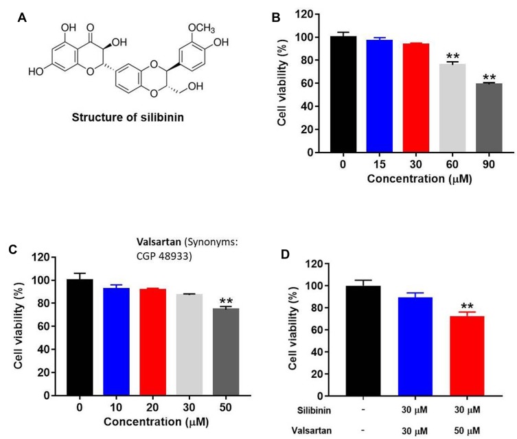
For investigating the cytotoxic effect of silibinin (Figure 1A) or valsartan on HK-2 cells, CCK-8 assay was performed. As showed in Figure 1B, compared to 0 μM group, 15 or 30 μM silibinin had no significant effect on cell viability, while 60 or 90 μM silibinin notably decreased the cell viability. These results indicated that high concentration of silibinin (≥ 60 μM) exhibited significant cytotoxicity. Meanwhile, as demonstrated in Figure 1C, 10, 20 or 30 μM valsartan had very limited effect on cell viability, while 50 μM valsartan markedly decreased cell proliferation. Finally, the combination of silibinin (30 μM) and valsartan (50 μM) could significantly increase cytotoxicity, while silibinin (30 μM) in combination with valsartan (30 μM) did not induce the cytotoxicity (Figure 1D). Therefore, the nontoxic concentration of 30 μM valsartan and 30 μM silibinin were selected for the following experiments.
Figure 1.
Cytotoxicity of silibinin or valsartan in vitro. (A) Chemical structure of silibinin. (B) HK-2 cells were treated with 0, 15, 30, 60 and 90 μM silibinin for 72 hrs, and cell viability was determined by CCK-8 assay. (C) HK-2 cells were treated with 0, 10, 20, 30 and 50 μM valsartan for 72 hrs, and cell viability was determined by CCK-8 assay. (D) HK-2 cells were treated with silibinin plus valsartan for 72 hrs, and cell viability was detected by CCK-8 assay. **P<0.01 vs 0 μM group.
Silibinin Significantly Increased the Anti-Fibrosis Effect of Valsartan in TGF-β1-Treated HK-2 Cells
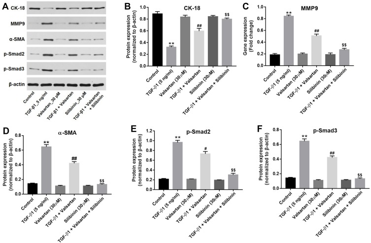
CK-18, MMP9 and α-SMA played critical roles in fibrosis.16 For the purpose of examining the combined effect of silibinin and valsartan on fibrosis in vitro, the expressions of CK-18, MMP9 and α-SMA in HK-2 cells were detected by qRT-PCR. As revealed in Figure 2A–C, TGF-β1 (5 ng/mL) notably increased the expressions of MMP9 and α-SMA and decreased the expression of CK-18 in HK-2 cells. These data indicated that in vitro model of fibrosis was successfully established. Moreover, valsartan (30 μM) significantly increased the expression of CK-18 and inhibited the levels of MMP9 and α-SMA in TGF-β1-treated HK-2 cells. Meanwhile, silibinin significantly enhanced the effect of valsartan on the gene expressions of CK-18, MMP9 and α-SMA in TGF-β1-treated HK-2 cells. In order to verify this result, the expressions of these proteins in HK-2 cells were tested by Western-blot. As showed in Figure 3A–E, silibinin in combination with valsartan significantly upregulated the expressions of CK-18 and downregulated the expression of MMP9, α-SMA, phosphorylated smad2 (p-smad2) and phosphorylated smad3 (p-smad3) in TGF-β1-treated HK-2 cells, compared with valsartan alone or control. Next, we aimed to analyze the functional fibrogenesis in TGF-β1-treated HK-2 cells. The results showed the upregulation of collagen III and fibronectin in TGF-β-treated HK-2 cells was significantly decreased by valsartan, which was further inhibited in the presence of silibinin (Figure 4A–C). All these data revealed that silibinin significantly increased the anti-fibrosis effect of valsartan in TGF-β1-treated HK-2 cells via suppressing TGF-β1 signaling pathway.
Figure 2.
Silibinin in combination with valsartan inhibited TGF-β1-induced renal fibrosis in vitro. HK-2 cells were treated with 5 ng/mL TGF-β1 for 72 hrs. Then, expressions of (A) CK-18, (B) MMP9 and (C) α-SMA in control, TGF-β1 (5 ng/mL), valsartan (30 μM), TGF-β1+valsartan, silibinin (30 μM) or TGF-β1+valsartan+silibinin groups were detected by RT-qPCR. GAPDH was used as an internal control. **P<0.01 vs control group. ##P<0.01 vs TGF-β1 (5 ng/ml); $$PP<0.01 vs TGF-β1+valsartan group.
Figure 3.
Combination of silibinin with valsartan suppressed TGF-β1-induced renal fibrosis via inhibition of TGF-β1 signaling pathway. (A) After 72 hrs of incubation, the protein expressions of CK-18, MMP9, α-SMA, p-smad2 and p-smad3 in control, TGF-β1 (5 ng/mL), valsartan (30 μM), TGF-β1+valsartan, silibinin (30 μM) or TGF-β1+valsartan+silibinin were investigated by Western-blot. β-actin was used as a loading control. The relative protein expression of (B) CK-18, (C) MMP9, (D) α-SMA, (E) p-smad2 and (F) p-smad3 was quantified by normalizing to β-actin. **P<0.01 vs control group; #P<0.05 vs TGF-β1 group; ##P<0.01 vs TGF-β1 group; $$P<0.01 vs TGF-β1+valsartan group.
Figure 4.
Silibinin significantly enhanced the inhibitory effect of valsartan on expression of collagen III and fibronectin. (A) The protein expressions of collagen III and fibronectin in control, TGF-β1 (5 ng/mL), valsartan (30 μM), TGF-β1+valsartan, silibinin (30 μM) or TGF-β1+valsartan+silibinin were investigated by Western-blot. β-actin was used as an internal control. The relative protein expression of (B) collagen III and (C) fibronectin was quantified by normalizing to β-actin. **P<0.01 vs control group; ##P<0.01 vs TGF-β1 group; $$P<0.01 vs TGF-β1+valsartan group.
Silibinin in Combination with Valsartan Exhibited Significant Anti-Renal Fibrosis in vivo
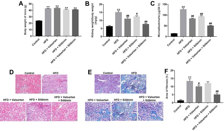
To further investigate the effect of combination treatment (silibinin plus valsartan) on renal fibrosis in vivo, HFD-fed mice model was established. As illustrated in Figure 5A, the body weight of mice was markedly increased in HFD group compared with control, while valsartan, silibinin or combination treatment had a very limited effect on body weight. On the other hand, kidney weight/body weight and microalbuminuria indicated the severity of kidney disease.17,18 In our study, kidney weight/body weight and microalbuminuria of mice analysis indicated that the kidney weight/body weight and microalbuminuria in HFD-induced mice were significantly increased compared with control group, which was markedly reduced by valsartan or silibinin (Figure 5B and C). Moreover, combination treatment exhibited better anti-fibrosis effect compared with valsartan or silibinin (Figure 5B and C). Then, the results of HE staining demonstrated valsartan or silibinin treatment significantly inhibited renal fibrosis in HFD-fed mice. Meanwhile, the renal fibrosis in HFD-fed mice was further alleviated by the combination treatment, compared with valsartan or silibinin alone (Figure 5D). Furthermore, as assayed by masson staining, the symptom of renal fibrosis was significantly increased in HFD mice, which was notably reduced by silibinin, valsartan or combination treatment (Figure 5E). Finally, as illustrated in Figure 5F, the area of fibrosis in kidney was significantly increased in HFD-fed mice, which was partly decreased by valsartan or silibinin treatment. Meanwhile, silibinin further increased the anti-fibrosis effect of valsartan treatment. All these data revealed that combination of silibinin with valsartan exhibited better anti-renal fibrosis effect in vivo, compared with silibinin or valsartan alone treatment.
Figure 5.
Silibinin in combination with valsartan notably attenuated HFD-induced renal fibrosis of mice in vivo. (A) Body weight of mice was determined. (B) Kidney weight/body weight of HFD-induced mice was measured. (C) Microalbuminuria of mice from control, HFD, HFD+valsartan, HFD+silibinin or HFD+valsartan+silibinin groups was measured. (D) H&E staining of mice kidney tissue in control, HFD, HFD+valsartan, HFD+silibinin or HFD+valsartan+silibinin group was detected. (E) Masson staining of mice kidney tissue in control, HFD, HFD+valsartan, HFD+silibinin or HFD+valsartan+silibinin groups was detected. (F) The area of fibrosis in mice was quantified. **P<0.01 vs control group; #P<0.05 vs HFD group; ##P<0.01 vs HFD group.
Silibinin in Combination with Valsartan Attenuated the Process of Renal Fibrosis via Suppression of TGF-β1 Pathway in vivo
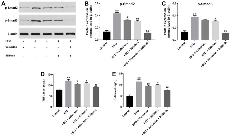
Finally, to further explore the mechanism by which silibinin in combination with valsartan attenuated the renal fibrosis, the expression of p-smad2 and p-smad3 in kidney tissues of mice was investigated. As indicated in Figure 6A–C, valsartan or silibinin partly downregulated the expressions of p-smad2 and p-smad3 in renal tissues, compared with control group; however, the expressions of these two proteins were completely inhibited by combination treatment. Besides, the levels of TNF-α and IL-6 in kidney tissues of mice were significantly increased by HFD, which were notably decreased by valsartan. Additionally, silibinin significantly increased the anti-inflammatory effect of valsartan on HFD-induced renal fibrosis in vivo (Figure 6D and E). Taken together, all these results further demonstrated that combination of silibinin with valsartan suppressed the progression of renal fibrosis via suppressing TGF-β1 signaling pathway.
Figure 6.
Silibinin in combination with valsartan inhibited renal fibrosis in vivo by suppression of TGF-β1 pathway. (A) The protein expressions of p-smad2 and p-smad3 in kidney tissues of mice were measured by Western-blot. Quantification of the ratio of (B) p-smad2 and (C) p-smad3 levels by normalizing to β-actin. The levels of (D) TNF-α and (E) IL-6 in kidney tissues of mice were detected by ELISA kit. **P<0.01 vs control group; #P<0.05 vs HFD group; ##P<0.01 vs HFD group; $$P<0.01 vs TGF-β1+valsartan group.
Discussion
In this study, we aimed to investigate the anti-fibrosis effect of silibinin in combination with valsartan in vitro and in vivo. It has been suggested that valsartan as anti-hypertension agent inhibited nuclear factor erythroid 2-related factor 2 (Nrf2) pathway in renal fibrosis,19 and can be applied in clinical trial as anti-fibrosis agent.20 Our results confirmed that valsartan inhibited cell proliferation in HK-2 cell line with different concentration. On the other hand, the side effect of valsartan seriously reduced the quality of life of patients who receive valsartan as renal fibrosis therapy.21 Silibinin are obtained from the medicinal plant Silybum marianum (milk thistle) and have conventionally been applied for the treatment of liver diseases.22 Previous report has revealed that silibinin was effective in treating neuropathy and hepatopathy.23 In addition, some natural properties of other medicinal plant showed anti-fibrosis ability in vitro.24 The results of our current research were similar to these previous studies, indicating that silibinin could be regarded as another anti-fibrosis agent.
The present data indicated that silibinin notably increased the valsartan-induced anti-fibrosis activities in vitro and in vivo without toxic effects. This finding was similar to the previous study that combination of curcumin, vorinostat and silibinin could reverse nerve cell toxicity.25 Moreover, our results indicated that silibinin, valsartan or combination treatment (silibinin plus valsartan) had a very limited effect on body weight of mice, which suggested that this treatment strategy had no significant systemic toxicity.
Additionally, silibinin enhanced the anti-fibrosis of valsartan in vitro via downregulating the expression of MMP9, α-SMA and upregulating the expression of CK-18. Kosasih et al found that CK-18 played a critical role in progression of liver fibrosis.26 On the other hand, Notch3 ameliorated cardiac fibrosis through inhibiting the expression of MMP9 and α-SMA.27 Similar to these results, combination of silibinin with valsartan could inhibit renal fibrosis via suppression of MMP9, α-SMA and increase of CK-18 in TGF-β1-treated HK-2 cells. In addition, CK-18 and α-SMA played crucial roles in EMT process.28–30 Our results indicated that valsartan could inactivate CK-18 and α-SMA. Silibinin significantly enhanced the inhibitory effect of valsartan on these two proteins. The results were similar to the previous study, indicating that silibinin enhanced the anti-fibrosis effect of valsartan via downregulation of EMT process.
TGF-β1 signaling plays a key role in fibrosis.8,31 It was persistently upregulated in fibrosis of various organs (kidney, lung, and heart).32 It has been reported that TGF-β1 can activate Smad-2/3, and the latter induce transcription of pro-fibrotic genes, factors that can interference this signaling pathway may affect fibrosis.31 In the present study, we found that silibinin further downregulated the expressions of p-smad2 and p-smad3 in TGF-β1-induced HK-2 cells, compared with valsartan alone. Based on these results, the mechanism underlying the anti-fibrosis effects of silibinin in combination with valsartan in vitro and in vivo was associated with the suppression of TGF-β1 signaling pathways. According to Ko et al, silibinin suppressed the fibrotic responses through inhibition of TGF-β1/Smad 2/3 signaling.13 Moreover, silibinin attenuated radiation-induced fibrosis via suppressing TGF-β1/Smad signaling in vitro and in vivo.33 These data were consistent with the results of the present study. Besides, we also found that the expressions of CK-18 and α-SMA were notably upregulated in TGF-β1-treated HK-2 cells. Feng et al have found that upregulation of TGF-β1 signaling could promote the EMT process of melanoma.34 Our data were consistent with these results, suggesting that TGF-β1 signaling could promote EMT process during the fibrosis. Taken together, silibinin has a potential ability for inhibiting fibrosis by suppression of TGF-β1/Smad 2/3 signaling. Otherwise, EGFR and Wnt signaling pathways are involved in the fibrotic process.35,36 However, this study focused only on the effect of silibinin in combination with valsartan on TGF-β1 signaling pathway. Thus, further researches are needed to explore the role of silibinin in combination with valsartan on EGFR or Wnt pathway.
Our study firstly revealed that silibinin increased the anti-fibrosis effect of valsartan in vitro and in vivo via suppression of TGF-β1 signaling. These findings indicated that combination of silibinin with valsartan might serve as an effective strategy for the treatment of renal fibrosis caused by diabetic nephropathy.
Disclosure
The authors declare that they have no competing interests in this work.
References
- 1.Nogueira A, Pires MJ, Oliveira PA. Pathophysiological mechanisms of renal fibrosis: a review of animal models and therapeutic strategies. In Vivo. 2017;31(1):1–22. doi: 10.21873/invivo [DOI] [PMC free article] [PubMed] [Google Scholar]
- 2.Tao Z, Shi A, Zhao J. Epidemiological perspectives of diabetes. Cell Biochem Biophys. 2015;73(1):181–185. doi: 10.1007/s12013-015-0598-4 [DOI] [PubMed] [Google Scholar]
- 3.Bose M, Almas S, Prabhakar S. Wnt signaling and podocyte dysfunction in diabetic nephropathy. J Investig Med. 2017;65(8):1093–1101. doi: 10.1136/jim-2017-000456 [DOI] [PubMed] [Google Scholar]
- 4.Lv W, Fan F, Wang Y, et al. Therapeutic potential of microRNAs for the treatment of renal fibrosis and CKD. Physiol Genomics. 2018;50(1):20–34. doi: 10.1152/physiolgenomics.00039.2017 [DOI] [PMC free article] [PubMed] [Google Scholar]
- 5.Sun YB, Qu X, Caruana G, Li J. The origin of renal fibroblasts/myofibroblasts and the signals that trigger fibrosis. Differentiation. 2016;92(3):102–107. doi: 10.1016/j.diff.2016.05.008 [DOI] [PubMed] [Google Scholar]
- 6.Kim KK, Sheppard D, Chapman HA. TGF-beta1 signaling and tissue fibrosis. Cold Spring Harb Perspect Biol. 2018;10(4):a022293. doi: 10.1101/cshperspect.a022293 [DOI] [PMC free article] [PubMed] [Google Scholar]
- 7.Ma L, Li H, Zhang S, et al. Emodin ameliorates renal fibrosis in rats via TGF-beta1/Smad signaling pathway and function study of Smurf 2. Int Urol Nephrol. 2018;50(2):373–382. doi: 10.1007/s11255-017-1757-x [DOI] [PubMed] [Google Scholar]
- 8.Loboda A, Sobczak M, Jozkowicz A, Dulak J. TGF-beta1/Smads and miR-21 in renal fibrosis and inflammation. Mediators Inflamm. 2016;2016:8319283. doi: 10.1155/2016/8319283 [DOI] [PMC free article] [PubMed] [Google Scholar]
- 9.Higgins SP, Tang Y, Higgins CE, et al. TGF-beta1/p53 signaling in renal fibrogenesis. Cell Signal. 2018;43:1–10. doi: 10.1016/j.cellsig.2017.11.005 [DOI] [PMC free article] [PubMed] [Google Scholar]
- 10.Zhang Q, Li J, Tan XP, Zhao Q. Effects of ME3 on the proliferation, invasion and metastasis of pancreatic cancer cells through epithelial-mesenchymal transition. Neoplasma. 2019;66:896–907. doi: 10.4149/neo_2019_190119N59 [DOI] [PubMed] [Google Scholar]
- 11.Sanajou D, Ghorbani Haghjo A, Argani H, et al. Reduction of renal tubular injury with a RAGE inhibitor FPS-ZM1, valsartan and their combination in streptozotocin-induced diabetes in the rat. Eur J Pharmacol. 2019;842:40–48. doi: 10.1016/j.ejphar.2018.10.035 [DOI] [PubMed] [Google Scholar]
- 12.Sherman B, Hernandez AM, Alhado M, Menge L, Price RS. Silibinin differentially decreases the aggressive cancer phenotype in an in vitro model of obesity and prostate cancer. Nutr Cancer. 2019;72:1–10. [DOI] [PubMed] [Google Scholar]
- 13.Ko JW, Shin NR, Park SH, et al. Silibinin inhibits the fibrotic responses induced by cigarette smoke via suppression of TGF-beta1/Smad 2/3 signaling. Food Chem Toxicol. 2017;106(Pt A):424–429. doi: 10.1016/j.fct.2017.06.016 [DOI] [PubMed] [Google Scholar]
- 14.Cao Y, Zhang L, Wang Y, Fan Q, Cong Y. Astragaloside IV attenuates renal fibrosis through repressing epithelial-to-mesenchymal transition by inhibiting microRNA-192 expression: in vivo and in vitro studies. Am J Transl Res. 2019;11(8):5029–5038. [PMC free article] [PubMed] [Google Scholar]
- 15.Cao T, Xu R, Xu Y, et al. The protective effect of Cordycepin on diabetic nephropathy through autophagy induction in vivo and in vitro. Int Urol Nephrol. 2019;51(10):1883–1892. doi: 10.1007/s11255-019-02241-y [DOI] [PubMed] [Google Scholar]
- 16.Yan T, Chopp M, Ning R, et al. Intracranial aneurysm formation in type-one diabetes rats. PLoS One. 2013;8(7):e67949. doi: 10.1371/journal.pone.0067949 [DOI] [PMC free article] [PubMed] [Google Scholar]
- 17.Wang X, Constans MM, Chebib FT, Torres VE, Pellegrini L. Effect of a vasopressin V2 receptor antagonist on polycystic kidney disease development in a rat model. Am J Nephrol. 2019;49(6):487–493. doi: 10.1159/000500667 [DOI] [PMC free article] [PubMed] [Google Scholar]
- 18.Duborija-Kovacevic N, Tomic Z. Kidney, skeletal muscle and myocardium as potential target sites of pygeum africanum toxicity in wistar rats. Rev Int Androl. 2019;17(1):8–14. doi: 10.1016/j.androl.2017.12.006 [DOI] [PubMed] [Google Scholar]
- 19.Jing W, Vaziri ND, Nunes A, et al. LCZ696 (Sacubitril/valsartan) ameliorates oxidative stress, inflammation, fibrosis and improves renal function beyond angiotensin receptor blockade in CKD. Am J Transl Res. 2017;9(12):5473–5484. [PMC free article] [PubMed] [Google Scholar]
- 20.Zannad F, Ferreira JP. Is sacubitril/valsartan antifibrotic? J Am Coll Cardiol. 2019;73(7):807–809. doi: 10.1016/j.jacc.2018.11.041 [DOI] [PubMed] [Google Scholar]
- 21.Huang QF, Li Y, Wang JG. Overview of clinical use and side effect profile of valsartan in Chinese hypertensive patients. Drug Des Devel Ther. 2013;8:79–86. doi: 10.2147/DDDT.S38617 [DOI] [PMC free article] [PubMed] [Google Scholar]
- 22.Gioti K, Papachristodoulou A, Benaki D, et al. Silymarin enriched extract (Silybum marianum) additive effect on doxorubicin-mediated cytotoxicity in PC-3 prostate cancer cells. Planta Med. 2019;85(11–12):997–1007. [DOI] [PubMed] [Google Scholar]
- 23.Khazim K, Gorin Y, Cavaglieri RC, Abboud HE, Fanti P. The antioxidant silybin prevents high glucose-induced oxidative stress and podocyte injury in vitro and in vivo. Am J Physiol Renal Physiol. 2013;305(5):F691–F700. doi: 10.1152/ajprenal.00028.2013 [DOI] [PMC free article] [PubMed] [Google Scholar]
- 24.Chen DQ, Hu HH, Wang YN, et al. Natural products for the prevention and treatment of kidney disease. Phytomedicine. 2018;50:50–60. doi: 10.1016/j.phymed.2018.09.182 [DOI] [PubMed] [Google Scholar]
- 25.Meng J, Li Y, Zhang M, et al. A combination of curcumin, vorinostat and silibinin reverses Abeta-induced nerve cell toxicity via activation of AKT-MDM2-p53 pathway. PeerJ. 2019;7:e6716. doi: 10.7717/peerj.6716 [DOI] [PMC free article] [PubMed] [Google Scholar]
- 26.Kosasih S, Zhi Qin W, Abdul Rani R, et al. Relationship between serum cytokeratin-18, control attenuation parameter, NAFLD fibrosis score, and liver steatosis in nonalcoholic fatty liver disease. Int J Hepatol. 2018;2018:9252536. doi: 10.1155/2018/9252536 [DOI] [PMC free article] [PubMed] [Google Scholar]
- 27.Zhang M, Pan X, Zou Q, et al. Notch3 ameliorates cardiac fibrosis after myocardial infarction by inhibiting the TGF-beta1/Smad3 pathway. Cardiovasc Toxicol. 2016;16(4):316–324. doi: 10.1007/s12012-015-9341-z [DOI] [PubMed] [Google Scholar]
- 28.Cai Y, Xue Y, An RF. [The expression of EMT associated protein CK-18 and vimentin in gestational trophoblastic disease]. Sichuan Da Xue Xue Bao Yi Xue Ban. 2014;45(6):960–963, 1014. [PubMed] [Google Scholar]
- 29.Kong J, Di C, Piao J, et al. Ezrin contributes to cervical cancer progression through induction of epithelial-mesenchymal transition. Oncotarget. 2016;7(15):19631–19642. doi: 10.18632/oncotarget.7779 [DOI] [PMC free article] [PubMed] [Google Scholar]
- 30.Zhu L, Fu X, Chen X, Han X, Dong P. M2 macrophages induce EMT through the TGF-beta/Smad2 signaling pathway. Cell Biol Int. 2017;41(9):960–968. doi: 10.1002/cbin.10788 [DOI] [PubMed] [Google Scholar]
- 31.Meng XM, Nikolic-Paterson DJ, Lan HY. TGF-beta: the master regulator of fibrosis. Nat Rev Nephrol. 2016;12(6):325–338. doi: 10.1038/nrneph.2016.48 [DOI] [PubMed] [Google Scholar]
- 32.Sisto M, Lorusso L, Ingravallo G, et al. The TGF-beta1 signaling pathway as an attractive target in the fibrosis pathogenesis of sjogren’s syndrome. Mediators Inflamm. 2018;2018:1965935. doi: 10.1155/2018/1965935 [DOI] [PMC free article] [PubMed] [Google Scholar]
- 33.Kim JS, Han NK, Kim SH, Lee HJ. Silibinin attenuates radiation-induced intestinal fibrosis and reverses epithelial-to-mesenchymal transition. Oncotarget. 2017;8(41):69386–69397. doi: 10.18632/oncotarget.20624 [DOI] [PMC free article] [PubMed] [Google Scholar]
- 34.Feng H, Jia XM, Gao NN, et al. Overexpressed VEPH1 inhibits epithelial-mesenchymal transition, invasion, and migration of human cutaneous melanoma cells through inactivating the TGF-beta signaling pathway. Cell Cycle. 2019;18(21):2860–2875. doi: 10.1080/15384101.2019.1638191 [DOI] [PMC free article] [PubMed] [Google Scholar] [Retracted]
- 35.Liang D, Chen H, Zhao L, et al. Inhibition of EGFR attenuates fibrosis and stellate cell activation in diet-induced model of nonalcoholic fatty liver disease. Biochim Biophys Acta Mol Basis Dis. 2018;1864(1):133–142. doi: 10.1016/j.bbadis.2017.10.016 [DOI] [PubMed] [Google Scholar]
- 36.Okazaki H, Sato S, Koyama K, et al. The novel inhibitor PRI-724 for Wnt/beta-catenin/CBP signaling ameliorates bleomycin-induced pulmonary fibrosis in mice. Exp Lung Res. 2019;45:1–12. [DOI] [PubMed] [Google Scholar]


