Abstract
目前牙种植体的生物力学研究主要集中于种植体-骨结合界面及种植牙内各个部件连接界面的力学损伤及增强机制,以及如何获得更好的牙种植体-基台复合体的整体机械强度和服役寿命等方面的研究。目前有关生物力学的研究,主要通过电阻应力测试方法、光弹应力分析法、数字图像相关分析法、有限元分析法、种植体-骨结合强度力学测试和机械性能测试法对实验样本进行综合评价。目前有限元分析法是最常见的牙种植体应力分布研究法,而静态压力实验和疲劳实验是最常见的机械强度测试研究方法。本文通过对这些研究方法的原理、应用范围及特点的介绍和对实验结果影响因素的梳理,给相关领域研究者们提供方法学的指导,并可拓展种植体的设计思路。
Keywords: 牙种植体, 生物力学, 应力, 机械强度
Abstract
Current biomechanical research of dental implants focuses on the mechanical damage and enhancement mechanism of the implant-abutment interface as well as how to obtain better mechanical strength and longer fatigue life of dental implants. The mechanical properties of implants can be comprehensively evaluated by strain gauge analysis, photo elastic stress analysis, digital image correlation, finite element analysis, implant bone bonding strength test, and measurement of mechanical properties. Finite element analysis is the most common method for evaluating stress distribution in dental implants, and static pressure and fatigue tests are commonly used in mechanical strength test. This article reviews biomechanical research methods and evaluation indices of dental implants. Results provide methodology guidelines in the field of biomechanics by introducing principles, ranges of application, advantages, and limitations, thereby benefitting researchers in selecting suitable methods. The influencing factors of the experimental results are presented and discussed to provide implant design ideas for researchers.
Keywords: dental implant, biomechanics, stress, mechanical strength
随着种植体-骨界面骨结合理论的提出,口腔种植学进入飞速发展时期,越来越多缺牙患者选择种植治疗为首选治疗方案。在临床应用中,也频频有种植修复的失败病例,这与生物力学因素紧密相关。近年来许多学者将研究重点致力于此,本文将近年来牙种植体生物力学相关研究的方法与结果进行总结,以便为临床应用提供参考。
1. 种植体生物力学研究方法
1.1. 电阻应力测试方法
电阻应力测试方法(简称电测法)是以电阻应变片为传感器,通过将构件的应变转为连接电桥中电阻应变片的电阻变化,测量电桥输出端电压的变化,通过相应的换算关系,最终获得测量构件的应力变化值(图1)。电测法灵敏度与精确度较高,且可现场测定,故被用于各种复杂环境下测量多种力学参数。
图 1. 电测法测量系统简易图.
Fig 1 Simple diagram of stain electronic measurement system
电测法的缺点在于:1)条件恶劣时误差较大;2)只能通过单点测量物件表面的应变,获得应变片检测位置的应变平均值,不能直观得出构件整体的应力分布;3)在测量复杂构件表面应力梯度较大点时,对粘贴应变片的准确度要求较高,使得测量过程比较复杂。
电测法是分析牙种植体应力的基础检测方法,可测定牙种植体内部结构的应力分布情况[1],也可用于对多单位的牙种植体进行应力分析[2],便于直接测定力学数值。
1.2. 光弹应力分析法
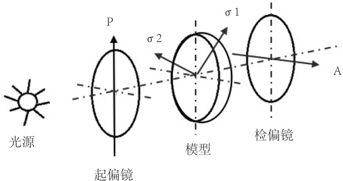
光弹应力分析是利用光敏材料(如环氧树脂等)制成和实物几何形状相似的模型,模拟实际受力后,以偏振光透过模型,由于应力的存在,产生光的暂时双折射现象,再透过分析镜后产生光的干涉,得到等倾线和等差线的图像,据此推算出构件内的应力大小、方向及分布情况(图2)。
图 2. 光弹应力分析法原理.
Fig 2 Principle of photoelastic method
P:起偏镜,A:检偏镜,σ1、σ2:光沿着两个主应力方向分解的偏振光。
光弹法测量是全域性的,故直观性强,可靠性高,适应性广,能求出在各种复杂条件下的全部应力状态,但不能计算出模型内任意处的应力值和位移值。
光弹应力分析法具有直观性强和能分析复杂构件的优点,故成为分析牙种植体应力时常用的方法。目前光弹应力分析法主要用在种植覆盖义齿应力分析[3]和牙种植体与基台界面应力分析中[4]。
1.3. 数字图像相关(digital image correlation,DIC)分析法
20世纪70年代以前,生物力学研究和应力分布的检测多采用电测法和光弹应力分析法。随着计算机应用的推广,DIC分析法的应用更加广泛,是当前实验力学领域最活跃也最受关注的光测力学方法之一。
DIC分析法是应用计算机视觉技术的一种图像测量方法,是一种非接触的、用于全场形状、变形、运动测量的方法(图3)。该方法基于数字图像处理和数值计算的非干涉变形,与其他基于相干光波干涉原理的光测方法(如电子散斑干涉、云纹干涉法)相比,DIC分析法具有明显且独特的优势:1)仅需要1个数字相机(二维数字图像相关技术,two-dimensional digital image correlation,2D DIC)或两个数字相机(三维数字图像相关技术,three-dimensional digital image correlation,3D DIC)拍摄变形前后被测物体表面的数字图像,其光路布置、测量过程和试样准备简单;2)无需激光照明和隔振,对测量环境要求较低;3)可与不同时间分辨率和空间分辨率的数字成像设备(如高速摄像机、光学显微镜、扫描电子显微镜)直接结合,适用测量范围广泛,可用于固体材料和结构表面位移、变形和形貌测量。
图 3. DIC分析法流程图.
Fig 3 Flowchart of DIC analysis
在牙种植体生物力学研究领域,DIC分析法作为一种灵活、有效和功能强大的变形测量手段,不仅能够显示种植体表面的细微变化[5],而且可实现应力场传递的完整测量[6]–[7];但有学者[8]发现,DIC分析法由于离面位移和摄像机的噪声会产生较大的误差。
DIC分析法可以进行种植体与基台界面应力分析[9],也可研究种植体支持组织的应力传递情况[10],还可分析冠修复方式 [11]。
1.4. 有限元分析法(finite element analysis,FEA)
FEA是一种与计算机技术结合的数值分析方法,其基本原理是:将物体划分为有限数量的单元,后者相互连接形成几何实体,将单元顶点作为力在单元间传导的途径,每个单元的力学效应的总体效果反映出物体的整体力学特征[12]。FEA可对复杂的构件进行应力分析,能模拟口腔内多变的生物力学过程,通过计算机软件ABAQUS、ANSYS、COMSOL等进行高效求解,代替实验测试,节省成本,已被广泛应用于口腔生物力学的研究中[13]。
FEA技术应用于牙种植体的研究己有40余年历史,但其分析结果的准确性和科学价值在很大程度上取决于模型的几何相似性和力学相似性[14]。由于牙种植体及牙颌组织形态结构的复杂性及计算条件限制,早期的大部分研究将其简化为二维对称平面模型,并将各材料均假设为均质性、各向同性的线弹性材料,分析了一些特定问题,对早期牙种植体的研究和设计做出了贡献[15],但结果与实际情况差异较大。目前,应用于牙种植领域中的FEA已由二维转化为三维,基于CT通过计算机软件构建更为符合解剖学特征的三维模型已成为比较成熟的技术。在力学相似性方面,由于牙槽骨及颌骨表现为明显的非均质量性和各向异性,目前较多采用线弹性的各向异性本构模型进行表述[16]。
目前有限元法在牙种植体领域中的应用主要包括:指导种植体材料的选择、种植体表面处理、上部结构的合理修复;种植体形状与结构的力学分析;仿生种植牙的研究等[12],[17]。FEA能够对牙种植体的力学性能进行检验,模拟各种载荷变化,指导种植体的优化设计,进而提高种植义齿修复的远期效果,是一种研究复杂应力的有效方法。
1.5. 种植体-骨结合强度力学测试
种植体初期稳定性是获得种植成功的必要条件,也是种植体在骨内顺利渡过愈合期的关键因素。客观、无损地评价种植体的初期稳定性具有非常重要的临床意义,是指导修复时机的重要指标。
种植体与骨形成良好的骨结合是能够进行上部结构修复和恢复咬合功能的前提,目前评价种植体-骨结合强度的力学测试方法主要包括:侵袭性测量方法(即推出实验、推入实验、植入扭矩实验及旋出扭矩实验等)和非侵袭性测量方法(即Periotest法以及共振频率)分析。
1.5.1. 推出实验
推出实验是用力学设备将已经植入骨内的种植体推出以测定种植体-骨结合强度。运用实验机将种植体样本推出,根据负荷位移曲线求得最大作用力(Fmax);测量种植体骨接触的平均长度和直径,计算界面最大抗剪切强度(σ)=Fmax/πDT,其中D为种植体直径,T为骨接触厚度。
1.5.2. 推入实验
推入实验是使用特殊仪器将与骨结合的种植体推入骨内,以推入过程中位移-载荷曲线最大值作为种植体-骨结合强度值。有学者[18]提出,推入实验比推出实验敏感度更高,且更接近临床中牙种植体承受功能性负荷的状态,能够更好地反映种植体-骨生物力学性能。
1.5.3. 植入扭矩实验
植入扭矩实验是指种植手术时使用专用的扭矩仪测量其植入时的最大扭矩。影响植入扭矩最主要的因素是种植区骨质的密度,骨密度和植入扭矩呈正相关[19]。此外,手术方式也会影响植入扭矩,如采用极差备洞可使植入扭矩增大,从而增加种植体初期稳定性;反之,若种植手术制备的种植窝直径大于种植体直径,植入扭矩则会下降,导致种植体初期稳定性降低。
该方法测量结果可作为即刻负重的依据,但因该方法的局限性为其测定时机,故不能用于连续性监测,也不能用于术前种植体稳定性的评估。
1.5.4. 旋出扭矩实验
旋出扭矩实验是指用专用仪器测试得到的使种植体松动过程中,种植体-骨界面破坏的扭矩值,也被认为是种植体骨界面抵抗剪切破坏的最大强度。该测量值属于破坏性的测量方法,故无法连续测量。种植体与骨组织接触面积越大,旋出扭矩值越大,说明种植体与骨结合力越强。这种方法主要适用于评价螺纹状种植体经不同表面处理后骨结合界面的生物力学性能[20]。
1.5.5. Periotest法
Periotest法是通过反复叩击牙齿,采用仪器捕捉叩击头被弹回的信号,经换算即可以得到Periotest value(PTV)。PTV最早用来评价牙齿的松动度,同样可以用来评价种植体的松动度。PTV越小,种植体稳定性越好。有学者[21]通过测量种植体软硬组织界面的稳定性,证明Osstell系统和Periotest系统都具有较高敏感度。
Periotest法虽然操作简便、无创,但是存在以下争议。1)可重复性差:由于叩击的位置重复性差,而叩击位置对实验结果影响较大,故测试结果不稳定;2)特异性差;3)对骨吸收的灵敏度差;4)实验结果受种植体本身影响与否还待商榷[21]–[22]。
1.5.6. 共振频率分析(resonance frequency analysis,RFA)
从20世纪90年代起,口腔种植界开始关注与探究RFA在牙种植稳定性判断中的应用。这种方法利用共振原理,激发种植体-悬臂梁自由振荡,以共振频率来数字化评估种植体组织界面的稳定性,结果以种植体的稳定系数(implant stability quotient,ISQ)表示。ISQ值越高,种植体稳定性越好[22]。影响RFA值的因素主要包括有效长度(测量用基台长度+种植体颈部骨吸收的高度)和骨组织硬度[23],因此RFA法能够定量测量种植体与骨组织在组织测量学层次上的结合情况。
RFA法可无创、连续性监测种植体稳定性的动态变化,可重复性好;但也存在一定缺陷,例如其检测值不能完全说明骨整合情况,测量部位不包含固定修复体,传感器不能通用于各种植体系统等等。
1.6. 机械性能测试方法
1.6.1. 压缩实验
压缩实验又称压力实验,是测定材料在轴向静压力作用下的力学性能的试验,是材料机械性能测试的基本方法之一。因牙种植体在口腔中主要受的咬合力为压力,可初步评测不同设计的牙种植体的机械性能,比较牙种植体在压力条件下的最大位移、极限强度和弹性模量等[24]–[25]。
1.6.2. 疲劳实验
牙种植体颈部较窄,在其受力时易导致应力集中而使周围皮质骨吸收,最终引起种植体折断,修复失败,故大多数牙种植体体外实验采用疲劳实验。疲劳实验可通过不同形式的力(如拉、压和扭转)测定牙种植体样本的疲劳断裂需要的循环次数和最大载荷,以对实验研究的不同条件下的样本进行抗疲劳能力的评价[26]。
2. 种植体应力分布影响因素的研究
2.1. 种植体材料
纯钛及其合金具有优异的物理性能,并且具有较好的生物相容性,已被广泛地应用在牙种植体上。此外,钛还具有耐腐蚀好、弹性模量高等优点。新式的β型钛合金具有良好的延展性和机械性能,电阻高且毒性低,与其他类型的合金相比更接近人体骨组织的弹性模量,因此被广泛应用。
基台的材料选择较为广泛,目前常用的有氧化锆和钛及钛合金基台。在同等角度下,氧化锆基台的Von Mises应力最大值要大于钛及钛合金基台[27]。冠部使用不同的材料会影响应力的分布和传递,使用硬度较低的材料会减少施加在颌骨的应力,会吸收更多的外加负荷产生的能量,转移较少的能量到种植体-基台复合体和骨界面上[28]。
2.2. 种植体形态结构
目前,种植体的外形主要为圆柱形和圆锥形,种植体体部基本上趋向于螺纹状设计。张永丽[29]比较了4种不同形状的种植体(光滑圆柱形种植体、螺纹圆柱形种植体、光滑圆锥形种植体、螺纹圆锥形种植体)的应力分布差异,发现无论是否有螺纹,柱形种植体颈部的最大Von Mises应力均大于锥形种植体,而根端部位的最小Von Mises应力均小于锥形种植体,即锥形种植体的应力分布情况较柱形更合理。对柱形种植体来说,有无螺纹对应力影响不大;而对于锥形种植体,有螺纹者比无螺纹者的应力分布更为合理[29]。
研究[30]表明,相比于颈部光滑设计,螺纹结构对于保存骨水平更有利。从结构上看,螺纹单元的参数包括:螺纹形态、螺距、螺纹深度。通过有限元分析,Oswal等[31]评价了3种螺纹形态(V形、偏梯形、反偏梯形)的应力分布,结果提示,反偏梯形螺纹更有利于保存骨组织。对于V形螺纹种植体模型的即刻负载情况,马洁等[32]发现,在0.4、0.6、0.8、1.0、1.2 mm的颈部螺距中,螺距为0.8、1.0 mm时种植体-骨界面综合力学性能较好。王维丽等[33]在即刻负载情况下进行研究,发现螺纹深度为0.3~0.5 mm而宽度不变时,颌骨产生的应力峰值相对较小;螺纹宽度为0.1~0.3 mm而深度不变时,颌骨产生的应力峰值相对较小。较大的颈部螺纹结构(螺距宽度和深度分别为0.6 mm和0.35 mm)与较小的颈部螺纹结构(螺距宽度和深度分别为0.3 mm和0.15 mm)在功能性负载1年后,平均边缘骨吸收量的差异没有统计学意义[34]。目前多采用颈部螺纹结构增加种植体-骨界面接触面积,以减小种植体颈部应力,降低周围蝶形骨吸收,但螺纹的具体几何参数对种植体周围组织的影响尚存在争议。
2.3. 种植体长度和直径
关于种植体长度和直径对应力的影响,目前尚未达成一致的意见,其中种植体长度对应力的影响存在较大争议。有研究者[35]认为,增加种植体长度能减少种植体周围的应力。种植体长度增加可以使应力减少,并减少牙槽骨的变形[36],故建议在低密度骨质环境下使用长种植体[37]。也有研究者[38]认为种植体长度对周围应力影响不大,减少种植体长度对周围应力分布无显著影响。还有研究者[39]认为,增加种植体长度能有效地增强种植体承受侧向力的能力,而对种植体承受轴向力的影响不大。
相较于种植体长度,普遍认为增加种植体直径能显著降低种植体骨界面的应力值和应力集中。加载同等大小力的情况下,增加种植体直径能明显降低种植体内部以及种植体周围的应力[36]。
2.4. 种植体上部结构的连接装置
目前市场上有超过20种种植体,包含了不同材料和不同的种植体-基台界面设计方法[40]。虽连接方式对作用在周围骨上的压力值有影响,但相对种植体的位置和种植环境而言,是次要的影响因素[41]。
种植体连接的几何形态包括了外六边形、内六边形和莫尔斯锥度[13]。应力的最大集中区是在基台部位,平台转移使得种植体与基台间的间隙向种植体的中央移动,让间隙变小从而改善局部的应力集中。平台转移后的模型应力集中程度较小,内六边形的设计方法是上述3种方法中应力值最小的,外六边形设计则产生较大应力[18]。
在有些种植体设计中使用了角度基台。角度基台改变了局部的应力分布状况,但是同时也让应力分析复杂化,使得骨界面的应力从颈部转移向底部,变为损害性更大的剪切力,极大影响了骨的再生能力,并破坏了骨的结构。超过15°的氧化锆角度基台,基台和种植体周围的骨内最大Von mises应力显著增高[42]。
2.5. 种植体数量及排列
传统全口义齿常有咀嚼效率低、固位稳定性差、适应期长等缺点,难以满足患者的需求。全牙列种植修复给患者造成较大的经济负担,因此种植覆盖义齿成为一种经济、有效的修复方案。下颌覆盖种植义齿一般需植入2~4枚种植体[43],临床随访结果显示,2枚与4枚种植体支持的覆盖义齿都具有良好的固位和稳定性,没有显著的差异。通过FEA研究可知,植入1枚种植体时,种植体周围无应力集中,但义齿可围绕种植体旋转;2枚种植体支持时可形成支点线,种植体周围骨组织上显示应力集中,并且在黏膜上有较大应力;当有3、4枚种植体支持时,种植体周围应力均较小[44]。
种植体植入部位的改变也可以降低种植体周围的应力并减少上部结构的应力集中。在磨牙区的应力值显著小于前牙区[45]。后牙的游离距越长,种植体骨界面的应力越小,黏膜承受的应力越大[46]。随着种植体植入倾斜角度的增加,种植体周围骨应力增大,而下颌尖牙区颊向植入两枚种植体时,种植体周围应力最小[47]。
3. 种植体机械强度影响因素的研究
国内外不少学者对种植体机械强度的影响因素进行研究,实验条件对结果影响较大,其中包括种植体的形状和尺寸设计、表面粗化处理、种植体与基台连接方式等,现将影响实验室种植体机械强度的因素总结如下。
3.1. 种植体尺寸和形状
目前,种植体的主导外形为圆柱形和圆锥形,其体部基本上均趋向于螺纹状设计。锥形种植体比柱形种植体的抗折强度更高。比较0.35、0.6、0.85、1.0 mm四种种植体的螺纹深度,螺纹深度越大,种植体与骨整合的稳定性越好,机械强度也越高[24]。带有微螺纹的牙种植体[48]、直径更大的牙种植体[49]表现出更高的机械强度,其机制可能与应力的分布有关。
3.2. 种植体表面处理
目前临床应用最广泛的种植体由金属钛制成。钛金属因其机械强度高、抗腐蚀、易加工的优点被作为牙种植体制造常用金属之一。不过,钛金属的机械强度小于某些生物材料,为提高钛金属制造的牙种植体在口腔中的抗压性能,部分学者将关注点集中于种植体表面的改性,这些处理包括纳米级粗糙度、纹理、孔隙等。
近年来发现,种植体表面处理对种植体骨整合、抗菌性能以及机械性能均有影响。研究表明,喷砂-常温酸蚀的种植体强度大于未经过表面粗化的种植体[25],超细晶纯钛微种植体的最大破坏强度显著大于普通纯钛微种植体[50],应用微弧氧化技术也能对牙种植体的机械强度进行提升[51]。羟磷灰石-生物玻璃复合涂层具有一定的塑性变形能力,具备添加生物玻璃过渡层涂层的种植体的临界载荷高于单一HA涂层,两者的临界载荷均高于无涂层的种植体[52]。近年来,纳米材料体现出越来越大的优势,学者们提出纳米级二氧化钛涂层的改性方法[53]。由此可见,种植体表面处理也是提高种植体机械稳定性的必要手段。
3.3. 种植体基台连接方式
种植体的设计,基台连接的几何形态和类型,是建立和稳定种植体骨整合的一个重要因素,并且在很大程度上影响着力在种植体上的传递及周围骨的吸收[54]。种植体连接的几何形态包括了外六边形、内六边形和莫尔斯锥度[13]。应用最广泛的是由单独的基台螺丝和六边形的上部平台结构构成的外六边形种植体。内六边形种植体可以很好地消除水平载荷并且保护基台螺丝免受更多的压力。关于内六边形的种植体设计是否优于外六边形的种植体设计现在仍存争议[40]。莫尔斯锥度种植体是由一个长锥度螺丝和恰当贴合的基台构成,会聚角一般在4°~16°,几何锁合锥度的摩擦力是基台载荷的基础[55]。莫尔斯锥度最大的优势在于种植体和基台间的封闭,这种应用几乎杜绝了基台的断裂和松动的发生。
有限元分析[56]证明内连接界面具有更广泛的应力分布:在侧向载荷下,采用外连接方式的基台应力集中于基台螺丝,而内连接锥形基台的载荷通过锥体转移,从而降低了螺纹部分的应力。实验室牙种植体断裂发生的位置[57]–[58]及临床断裂发生的位置与上述实验的结果一致[59]。
3.4. 基台材料
基台是连接种植体和上部修复体的重要结构,目前临床上应用的纯钛基台有着良好的生物相容性和与种植体的摩擦配副,但纯钛基台在种植体肩部位置较高或牙龈较薄的前牙区的患者中,基台金属颜色暴露严重影响美观,降低患者满意度。氧化锆基台因其优秀的生物相容性和美学性能更能满足患者和修复医生的要求,然而该基台的强度较低且脆性较高,易于折裂崩断。因此,针对纯钛基台和氧化锆基台在美观性和机械性能方面的缺陷,研究者使用了新型的材料制作基台。
基台材料的强度是牙种植体-基台复合体机械强度重要的影响因素,建议采用较高强度的材料[58]。氧化铝基台与氧化锆基台机械强度不同,断裂方式也有区别[60]。(Y, Nb)-TZP/Al2O3复相陶瓷具有相对较高的韧性和较低的磨损率,运用此材料加工制作的基台表现出较高的断裂强度,且随着Al2O3含量增大,机械强度更高[61]。Ti-Zr合金制作的基台,合金中Zr含量为质量分数为30%时,机械强度最大[62]。
3.5. 种植体中央螺丝(基台螺丝)
种植体最常见的失败原因常常源于种植体-基台的连接界面。种植体-基台连接和中央螺丝的失效常见于中央螺丝折断和松动的情况,因此种植体中央螺丝对种植体的机械强度有很大的影响。30%~50%的种植体断裂样本最先出现中央螺丝弯曲断裂[56],[63]。
为了加强种植体-基台连接界面的稳定性,增加种植体-基台复合体的机械稳定性,提高种植修复的成功率,国内外学者进行了大量的实验研究。对中央螺丝的改性研究大部分着眼于中央螺丝螺纹处的表面处理,其机制可能是涂层材料对中央螺丝螺纹部分具有保护作用。具有类金刚石涂层的中央螺丝可以提升种植体-基台复合体的机械强度[64]。与装配传统钛合金中央螺丝的种植体相比,装配微晶金刚石[65]和纳米金刚石涂层[66]的中央螺丝具有更高的机械强度。
综上所述,不同的生物力学测试方法在不同的研究层面中各有优势,围绕种植体-骨界面,采用不同方法从不同角度探讨牙种植体生物力学问题,必将丰富和发展对牙种植系统界面理想特征的认识。
Funding Statement
[基金项目] 十三五国家重点研发项目(2016YFC1102704)
Supported by: The National Key Research and Development Program of China (2016YFC1102704).
References
- 1.Asvanund P. A strain gauge analysis comparing external and internal implant-abutment connections[J] Implant Dent. 2014;23(2):206–211. doi: 10.1097/ID.0000000000000063. [DOI] [PubMed] [Google Scholar]
- 2.Waly KM, Elwakad MT, Eldosoky MAA, et al. Effect of bone height on stresses developed around dental implant using strain gauge (in-vitro study)[J] Int J Eng Res Develop. 2014;10(9):20–26. [Google Scholar]
- 3.Figueirêdo EP, Sigua-Rodriguez EA, Pimentel MJ, et al. Photoelastic analysis of fixed partial prosthesis crown height and implant length on distribution of stress in two dental implant systems[J] Int J Dent. 2014:206723. doi: 10.1155/2014/206723. [DOI] [PMC free article] [PubMed] [Google Scholar]
- 4.Geramizadeh M, Katoozian H, Amid R, et al. Comparison of finite element results with photoelastic stress analysis around dental implants with different threads[J] Dent Med Probl. 2018;55(1):17–22. doi: 10.17219/dmp/85077. [DOI] [PubMed] [Google Scholar]
- 5.Tiossi R, Lin LS, Rodrigues RC, et al. Digital image correlation analysis of the load transfer by implant-supported restorations[J] J Biomech. 2011;44(6):1008–1013. doi: 10.1016/j.jbiomech.2011.02.015. [DOI] [PubMed] [Google Scholar]
- 6.Li JY, Fok ASL, Satterthwaite J, et al. Measurement of the full-field polymerization shrinkage and depth of cure of dental composites using digital image correlation[J] Dent Mater. 2009;25(5):582–588. doi: 10.1016/j.dental.2008.11.001. [DOI] [PubMed] [Google Scholar]
- 7.Tung SH, Shih MH, Kuo JC. Application of digital image correlation for anisotropic plastic deformation during tension testing[J] Opt Lasers Eng. 2010;48(5):636–641. [Google Scholar]
- 8.王 楠, 李 恩普, 汤 忠斌, et al. 二维数字图像相关方法的拉伸实验误差分析[J] 光学仪器. 2012;34(3):5–12. [Google Scholar]; Wang N, Li EP, Tang ZB, et al. An investigation of the experimental error of 2-D DIC method applied to tensile strain measurement[J] Opt Instru. 2012;34(3):5–12. [Google Scholar]
- 9.Peixoto RF, Macedo AP, Martinelli J, et al. A digital image correlation analysis of strain generated by 3-unit implant-supported fixed dental prosthesis[J] Implant Dent. 2017;26(4):567–573. doi: 10.1097/ID.0000000000000588. [DOI] [PubMed] [Google Scholar]
- 10.Nenad M, Ivan T, Dušan Š, et al. Analysis of the effect of implant distance from the surrounding structure, in the PMMA block model[J] Procedia Struct Integr. 2016(2):1260–1265. [Google Scholar]
- 11.Clelland NL, Yilmaz B, Seidt JD. Three-dimensional image correlation analyses for strains generated by cement and screw-retained implant prostheses[J] Clin Implant Dent Relat Res. 2013;15(2):271–282. doi: 10.1111/j.1708-8208.2011.00411.x. [DOI] [PubMed] [Google Scholar]
- 12.刘 宝林. 口腔种植学[M] 北京: 人民卫生出版社; 2011. pp. 87–92. [Google Scholar]; Liu BL. Dental implantology[M] Beijing: People's Medical Publishing House; 2011. pp. 87–92. [Google Scholar]
- 13.王 姝, 李 琼, 金 武龙. 有限元法在口腔种植领域的研究进展[J] 中华老年口腔医学杂志. 2018;16(2):125–128. [Google Scholar]; Wang S, Li Q, Jin WL. The research progress of finite element method in dental implants[J] Chin J Geriatric Dent. 2018;16(2):125–128. [Google Scholar]
- 14.王 晓洁. 牙种植体的三维有限元分析和生物密封研究[D] 成都: 四川大学; 2005. [Google Scholar]; Wang XJ. Three-dimensional finite element analysis and bioseal study of dental implants[D] Chengdu: Sichuan University; 2005. [Google Scholar]
- 15.Holmgren EP, Seckinger RJ, Kilgren LM, et al. Evaluating parameters of osseointegrated dental implants using finite element analysis—a two-dimensional comparative study examining the effects of implant diameter, implant shape, and load direction[J] J Oral Implantol. 1998;24(2):80–88. doi: 10.1563/1548-1336(1998)024<0080:EPOODI>2.3.CO;2. [DOI] [PubMed] [Google Scholar]
- 16.陈 新民. 口腔生物力学[M] 北京: 科学出版社; 2010. pp. 335–336. [Google Scholar]; Chen XM. Oral biomechanics[M] Beijing: Science Press; 2010. pp. 335–336. [Google Scholar]
- 17.王 宗篪, 谢 培庆, 庄 国铭, et al. 仿生种植牙三维有限元模型[J] 三明学院学报. 2006;23(2):137–140. [Google Scholar]; Wang ZC, Xie PQ, Zhuang GM, et al. The three-dimensional finite element model of bionic dental implant[J] J Sanming Univ. 2006;23(2):137–140. [Google Scholar]
- 18.Seong WJ, Grami S, Jeong SC, et al. Comparison of push-in versus pull-out tests on bone-implant interfaces of rabbit tibia dental implant healing model[J] Clin Implant Dent Relat Res. 2013;15(3):460–469. doi: 10.1111/j.1708-8208.2011.00357.x. [DOI] [PubMed] [Google Scholar]
- 19.Beer A, Gahleitner A, Holm A, et al. Correlation of insertion torques with bone mineral density from dental quantitative CT in the mandible[J] Clin Oral Implants Res. 2003;14(5):616–620. doi: 10.1034/j.1600-0501.2003.00932.x. [DOI] [PubMed] [Google Scholar]
- 20.栗 兴超, 董 福生, 李 向军, et al. 不同颈部结构钛人工牙种植体的骨结合性能研究[J] 现代口腔医学杂志. 2017;31(3):129–132. [Google Scholar]; Li XC, Dong FS, Li XJ, et al. Study on the osseointegration of titanium dental implant with different neck structure[J] J Modern Stomatol. 2017;31(3):129–132. [Google Scholar]
- 21.Li DH, Ferguson SJ, Beutler T, et al. Biomechanical comparison of the sandblasted and acid-etched and the machined and acid-etched titanium surface for dental implants[J] J Biomed Mater Res A. 2002;60(2):325–332. doi: 10.1002/jbm.10063. [DOI] [PubMed] [Google Scholar]
- 22.Huang HM, Lee SY, Yeh CY, et al. Resonance frequency assessment of dental implant stability with various bone qualities: a numerical approach[J] Clin Oral Implants Res. 2002;13(1):65–74. doi: 10.1034/j.1600-0501.2002.130108.x. [DOI] [PubMed] [Google Scholar]
- 23.Nkenke E, Hahn M, Weinzierl K, et al. Implant stability and histomorphometry: a correlation study in human cadavers using stepped cylinder implants[J] Clin Oral Implants Res. 2003;14(5):601–609. doi: 10.1034/j.1600-0501.2003.00937.x. [DOI] [PubMed] [Google Scholar]
- 24.Lee SY, Kim SJ, An HW, et al. The effect of the thread depth on the mechanical properties of the dental implant[J] J Adv Prosthodont. 2015;7(2):115–121. doi: 10.4047/jap.2015.7.2.115. [DOI] [PMC free article] [PubMed] [Google Scholar]
- 25.李 璁. 纯钛种植体表面不同粗化处理对种植体强度的影响[D] 石家庄: 河北医科大学; 2016. [Google Scholar]; Li C. Effects of different roughening treatments on the strength of pure titanium implants[D] Shijiazhuang: Hebei Medical University; 2016. [Google Scholar]
- 26.栗 兴超, 董 福生, 李 向军, et al. 表面粗化处理不均衡螺纹钛人工牙种植体的疲劳性能研究[J] 现代口腔医学杂志. 2017;31(4):202–205, 234. [Google Scholar]; Li XC, Dong FS, Li XJ, et al. Study on the fatigue performance of the uneven-threaded titanium dental implants treated by surface roughening[J] J Modern Stomatol. 2017;31(4):202–205, 234. [Google Scholar]
- 27.张 波. 牙种植体生物力学评价研究进展[J] 中国实用口腔科杂志. 2014;7(9):566–571. [Google Scholar]; Zhang B. Advances on the biomechanical evaluation methods of dental implant[J] Chin J Pract Stomatol. 2014;7(9):566–571. [Google Scholar]
- 28.Soliman TA, Tamam RA, Yousief SA, et al. Assessment of stress distribution around implant fixture with three different crown materials[J] Tanta Dent J. 2015;12(4):249–258. [Google Scholar]
- 29.张 永丽. 四种不同形状种植体应力分布比较的三维有限元分析[D] 济南: 山东大学; 2003. [Google Scholar]; Zhang YL. Three-dimensional finite element analysis of stress distribution of implants with four different shapes[D] Jinan: Shandong University; 2003. [Google Scholar]
- 30.Lee DW, Choi YS, Park KH, et al. Effect of microthread on the maintenance of marginal bone level: a 3-year prospective study[J] Clin Oral Implants Res. 2007;18(4):465–470. doi: 10.1111/j.1600-0501.2007.01302.x. [DOI] [PubMed] [Google Scholar]
- 31.Oswal M, Amasi U, Oswal M, et al. Influence of three different implant thread designs on stress distribution: a three-dimensional finite element analysis[J] J Indian Prosthodon Soc. 2016;16(4):359–365. doi: 10.4103/0972-4052.191283. [DOI] [PMC free article] [PubMed] [Google Scholar]
- 32.马 洁, 王 维丽, 雷 蕾, et al. 种植体颈部不同螺距动态负载的生物力学分析[J] 解放军医学院学报. 2016;37(7):784–787, 797. [Google Scholar]; Ma J, Wang WL, Lei L, et al. Biomechanical analysis of stress distribution of dental implant with different neck thread pitch design under dynamic loading[J] Acad J Chin PLA Med School. 2016;37(7):784–787, 797. [Google Scholar]
- 33.王 维丽, 马 洁, 李 鑫, et al. 反支撑形螺纹种植体即刻负载时应力分布的三维有限元分析[J] 口腔颌面修复学杂志. 2016;17(4):211–215. [Google Scholar]; Wang WL, Ma J, Li X, et al. Analysis of the biomechanics stress distribution of the reverse buttress thread implant under immediate loading: a three dimensional finite element study[J] Chin J Prosthodont. 2016;17(4):211–215. [Google Scholar]
- 34.Kang YI, Lee DW, Park KH, et al. Effect of thread size on the implant neck area: preliminary results at 1 year of function[J] Clin Oral Implants Res. 2012;23(10):1147–1151. doi: 10.1111/j.1600-0501.2011.02298.x. [DOI] [PubMed] [Google Scholar]
- 35.Rubo JH, Capello Souza EA. Finite-element analysis of stress on dental implant prosthesis[J] Clin Implant Dent Relat Resh. 2009;12(2):105–113. doi: 10.1111/j.1708-8208.2008.00142.x. [DOI] [PubMed] [Google Scholar]
- 36.Petrie CS, Williams JL. Comparative evaluation of implant designs: influence of diameter, length, and taper on strains in the alveolar crest[J] Clin Oral Implants Res. 2005;16(4):486–494. doi: 10.1111/j.1600-0501.2005.01132.x. [DOI] [PubMed] [Google Scholar]
- 37.Tada S, Stegaroiu R, Kitamura E, et al. Influence of implant design and bone quality on stress/strain distribution in bone around implants: a 3-dimensional finite element analysis[J] Int J Oral Maxillofac Implants. 2003;18(3):357–368. [PubMed] [Google Scholar]
- 38.Meijer HJ, Kuiper JH, Starmans FJ, et al. Stress distribution around dental implants: influence of superstructure, length of implants, and height of mandible[J] J Prosthet Dent. 1992;68(1):96–102. doi: 10.1016/0022-3913(92)90293-j. [DOI] [PubMed] [Google Scholar]
- 39.董 福生, 董 玉英, 邢 汝东, et al. 种植体长度对骨界面应力分布影响的三维有限元分析[J] 中国口腔种植学杂志. 2001;6(3):106–108. [Google Scholar]; Dong FS, Dong YY, Xing RD, et al. Three-dimensional finite element method analysis of stress distribution around dental implants to estimate the influence of the length[J] Chin J Oral Implant. 2001;6(3):106–108. [Google Scholar]
- 40.Borie E, Orsi IA, de Araujo CP. The influence of the connection, length and diameter of an implant on bone biomechanics[J] Acta Odontol Scand. 2015;73(5):321–329. doi: 10.3109/00016357.2014.961957. [DOI] [PubMed] [Google Scholar]
- 41.Lin CL, Chang SH, Chang WJ, et al. Factorial analysis of variables influencing mechanical characteristics of a single tooth implant placed in the maxilla using finite element analysis and the statistics-based Taguchi method[J] Eur J Oral Sci. 2007;115(5):408–416. doi: 10.1111/j.1600-0722.2007.00473.x. [DOI] [PubMed] [Google Scholar]
- 42.陈 轶珺. 氧化锆全瓷角度基台的三维有限元应力分析[J] 口腔医学研究. 2008;24(3):317–319. [Google Scholar]; Chen YJ. Finite element analysis of zirconia angled ceramic abutment[J] J Oral Sci Res. 2008;24(3):317–319. [Google Scholar]
- 43.Visser A, Raghoebar GM, Meijer HJ, et al. Mandibular overdentures supported by two or four endosseous implants. A 5-year prospective study[J] Clin Oral Implants Res. 2005;16(1):19–25. doi: 10.1111/j.1600-0501.2004.01085.x. [DOI] [PubMed] [Google Scholar]
- 44.Liu J, Pan S, Dong J, et al. Influence of implant number on the biomechanical behaviour of mandibular implant-retained/supported overdentures: a three-dimensional finite element analysis[J] J Dent. 2013;41(3):241–249. doi: 10.1016/j.jdent.2012.11.008. [DOI] [PubMed] [Google Scholar]
- 45.简 波, 宋 应亮, 李 德华, et al. 下无牙颌覆盖义齿四颗种植体套简冠固位位置的优化设计及力学分析[J] 中国口腔种植学杂志. 2009;14(2):98–99. [Google Scholar]; Jian B, Song YL, Li DH, et al. The optimizing designs and mechanics analyses of implants location for the edentulous mandibular overdenture which be retained by four implants and telescopic crowns[J] Chin J Oral Implant. 2009;14(2):98–99. [Google Scholar]
- 46.张 少锋, 马 轩祥, 欧 阳官, et al. 加载部位对种植全口义齿应力的影响[J] 实用口腔医学杂志. 1997;13(3):37–39. [Google Scholar]; Zhang SF, Ma XX, Ouyang G, et al. Effect of loading position on stress of implant complete denture[J] J Pract Stomatol. 1997;13(3):37–39. [Google Scholar]
- 47.Hong HR, Pae A, Kim Y, et al. Effect of implant position, angulation, and attachment height on peri-implant bone stress associated with mandibular two-implant overdentures: a finite element analysis[J] Int J Oral Maxillofac Implants. 2012;27(5):e69–e76. [PubMed] [Google Scholar]
- 48.Elias CN, Rocha FA, Nascimento AL, et al. Influence of implant shape, surface morphology, surgical technique and bone quality on the primary stability of dental implants[J] J Mechan Behav Biomed Mater. 2012;16(1):169–180. doi: 10.1016/j.jmbbm.2012.10.010. [DOI] [PubMed] [Google Scholar]
- 49.Kong L, Sun YY, Hu KJ, et al. Selections of the cylinder implant neck taper and implant end fillet for optimal biomechanical properties: a three-dimensional finite element analysis[J] J Biomech. 2008;41(5):1124–1130. doi: 10.1016/j.jbiomech.2007.12.013. [DOI] [PubMed] [Google Scholar]
- 50.贾 晓瑞, 辛 海涛, 张 强, et al. 超细晶纯钛种植体表面微弧氧化处理的实验研究[J] 牙体牙髓牙周病学杂志. 2015;25(3):158–161, 172. [Google Scholar]; Jia XR, Xin HT, Zhang Q, et al. Surface treatment of ultrafine-grained titanium implant by micro-arc oxidation[J] Chin J Conserv Dent. 2015;25(3):158–161, 172. [Google Scholar]
- 51.丁 鹏飞, 徐 欣. 微弧氧化表面处理后的种植体性能改良[J] 滨州医学院学报. 2015(3):224–225. [Google Scholar]; Ding PF, Xu X. Improvement of implant properties after micro-arc oxidation surface treatment[J] J Binzhou Med Univ. 2015(3):224–225. [Google Scholar]
- 52.Jemat A, Ghazali MJ, Razali M, et al. Surface modifications and their effects on titanium dental implants[J] Biomed Res Int. 2015;2015(6):791725. doi: 10.1155/2015/791725. [DOI] [PMC free article] [PubMed] [Google Scholar]
- 53.贺 刚, 陈 治清, 盛 祖立. 纯钛种植体表面纳米TiO2生物活性涂层的构建[J] 中国口腔种植学杂志. 2008;13(3):101–104, 135. [Google Scholar]; He G, Chen ZQ, Sheng ZL. The construction of nano-TiO2 biocoating on titanium surface[J] Chin J Oral Implant. 2008;13(3):101–104, 135. [Google Scholar]
- 54.Bozkaya D, Muftu S, Muftu A. Evaluation of load transfer characteristics of five different implants in compact bone at different load levels by finite elements analysis[J] J Prosthet Dent. 2004;92(6):523–530. doi: 10.1016/j.prosdent.2004.07.024. [DOI] [PubMed] [Google Scholar]
- 55.Pita MS, Anchieta RB, Barão VAR, et al. Prosthetic platforms in implant dentistry[J] J Craniofac Surg. 2011;22(6):2327–2331. doi: 10.1097/SCS.0b013e318232a706. [DOI] [PubMed] [Google Scholar]
- 56.Gehrke P, Dhom G, Brunner J, et al. Zirconium implant abutments: fracture strength and influence of cyclic loading on retaining-screw loosening[J] Quintessence Int. 2006;37(1):19–26. [PubMed] [Google Scholar]
- 57.Adatia ND, Bayne SC, Cooper LF, et al. Fracture resistance of yttria-stabilized zirconia dental implant abutments[J] J Prosthodont. 2009;18(1):17–22. doi: 10.1111/j.1532-849X.2008.00378.x. [DOI] [PubMed] [Google Scholar]
- 58.Elsayed A, Wille S, Al-Akhali M, et al. Comparison of fracture strength and failure mode of different ceramic implant abutments[J] J Prosthet Dent. 2017;117(4):499–506. doi: 10.1016/j.prosdent.2016.06.018. [DOI] [PubMed] [Google Scholar]
- 59.Nguyen HQ, Tan KB, Nicholls JI. Load fatigue performance of implant-ceramic abutment combinations[J] Int J Oral Maxillofac Implants. 2009;24(4):636–646. [PubMed] [Google Scholar]
- 60.Jo JY, Yang DS, Huh JB, et al. Influence of abutment materials on the implant-abutment joint stability in internal conical connection type implant systems[J] J Adv Prosthodont. 2014;6(6):491–497. doi: 10.4047/jap.2014.6.6.491. [DOI] [PMC free article] [PubMed] [Google Scholar]
- 61.Kim DJ, Lee MH, Lee DY, et al. Mechanical properties, phase stability, and biocompatibility of (Y, Nb)-TZP/Al2O3 composite abutments for dental implant[J] J Biomed Mater Res. 2000;53(4):438–443. doi: 10.1002/1097-4636(2000)53:4<438::aid-jbm19>3.0.co;2-3. [DOI] [PubMed] [Google Scholar]
- 62.Lee T, Ueno T, Nomura N, et al. Titanium-zirconium binary alloy as dental implant material: analysis of the influence of compositional change on mechanical properties and in vitro biologic response[J] Int J Oral Maxillofac Implants. 2016;31(3):547–554. doi: 10.11607/jomi.4349. [DOI] [PubMed] [Google Scholar]
- 63.Basílio MA, Delben JA, Cesar PF, et al. Failure modes of Y-TZP abutments with external hex implant-abutment connection determined by fractographic analysis[J] J Mech Behav Biomed Mater. 2016(60):187–194. doi: 10.1016/j.jmbbm.2015.12.042. [DOI] [PubMed] [Google Scholar]
- 64.Bordin D, Coelho PG, Bergamo ETP, et al. The effect of DLC-coating deposition method on the reliability and mechanical properties of abutment's screws[J] Dent Mater. 2018;34(6):e128–e137. doi: 10.1016/j.dental.2018.03.005. [DOI] [PubMed] [Google Scholar]
- 65.Prado CJ, Neves FD, Soares CJ, et al. Influence of abutment screw design and surface coating on the bending flexural strength of the implant set[J] J Oral Implantol. 2014;40(2):123–128. doi: 10.1563/AAID-JOI-D-11-00116. [DOI] [PubMed] [Google Scholar]
- 66.Xie YN, Zhou J, Wei QP, et al. Improving the long-term stability of Ti6Al4V abutment screw by coating micro/nano-crystalline diamond films[J] J Mech Behav Biomed Mater. 2016(63):174–182. doi: 10.1016/j.jmbbm.2016.06.018. [DOI] [PubMed] [Google Scholar]


