Abstract
目的
对比研究美学区改良盾构术与传统即刻种植术的临床效果。
方法
选取24例行上颌前牙区单颗牙即刻种植的患者,12例行改良盾构术(试验组),12例行传统即刻种植术(对照组),随访1年,比较两组患者的种植成功率、红色美学指数(PES)、唇侧骨板吸收量以及患者满意度。采用SPSS 21.0软件进行统计学分析。
结果
术后1年,两组患者的种植成功率均为100%;试验组PES评分以及患者满意度均高于对照组,唇侧骨板吸收量低于对照组,差异均有统计学意义(P<0.05)。
结论
改良盾构术有利于维持唇侧骨量,从而获得更好的软组织美学效果。
Keywords: 即刻种植, 盾构术, 美学区, 唇侧骨板, 美学效果
Abstract
Objective
To compare the clinical effects of a modified socket shield technique (SST) and the conventional immediate implant placement in the esthetic zone.
Methods
Twenty-four patients with one hopeless upper incisor were consecutively enrolled in this study. All patients were scheduled for immediate implant placement. Twelve patients underwent a modified SST (experimental group) whereas the others underwent conventional procedures (control group). Radiographic and clinical data were obtained preoperatively, immediately after surgery, and 12 months after surgery. The success rate of the implants, the pink esthetic score (PES) and patient satisfaction were analyzed. Statistical analysis was conducted using SPSS 21.0 software.
Results
One year after operation, the success rate of the implants was 100% in both groups. The PES and patient satisfaction of the experimental group were significantly higher than those of the control group (P<0.05). The buccal bone plate resorbed less in the experimental group than in the control group (P<0.05).
Conclusion
The modified SST is advantageous over the conventional immediate implant placement in terms of buccal bone and soft tissue maintenance.
Keywords: immediate implant placement, socket shield technique, esthetic zone, buccal bone plate, esthetic outcomes
随着社会经济的飞速发展,人们对前牙缺失后种植修复的美学效果提出了越来越高的要求,如何为患者提供更理想的治疗方案成为种植医生的挑战。即刻种植与延期种植相比,具有减少患者就诊次数,缩短治疗周期,减轻患者痛苦等优点,因而受到临床医生的广泛关注[1]。动物实验和临床研究[2]–[4]均显示,即刻种植并不能防止拔牙后牙槽骨的吸收,且唇侧的吸收较舌侧尤为明显,即刻种植后龈缘退缩的美学风险增大。因此,如何在利用即刻种植优势的同时减少美学并发症、维持颊侧软硬组织稳定,已经成为种植医学亟待解决的问题。
盾构术(socket shield technique,SST)是Hürzeler等[5]提出的保留部分颊侧牙根的即刻种植技术,其核心理念是在拔除天然牙时保留部分唇侧牙片,最终实现牙槽嵴轮廓的长期维持[5]。该方法在动物实验和临床试验中均获得了成功[6]–[8]。传统SST技术敏感性较高,在种植备洞与种植体植入时可能引起唇侧牙片松动及菲薄的唇侧骨板发生骨折。在此基础上,本研究设计了改良SST,即种植体与唇侧牙片之间留有间隙,在保障唇侧牙片盾稳定的同时植入种植体;同时引入位点保存的概念,即在间隙中植入异种骨替代材料Bio-Oss骨粉,进一步支撑唇侧牙片盾与骨板。本研究拟通过前瞻性非随机对照研究,对比美学区改良SST与传统即刻种植术的临床效果。
1. 材料和方法
1.1. 临床资料
于2016年9月至2017年9月在重庆医科大学附属口腔医院种植科就诊患者中选取因外伤或牙体牙髓病变导致上前牙无法保留而要求种植修复的患者24例。12例行改良SST(试验组),男6例,女6例,年龄25~52岁,平均年龄(38.08±10.5)岁,其中中切牙11例,侧切牙1例;5例采用瑞典Nobel Active种植体系统,7例采用韩国Osstem系统,种植体的直径平均为(3.53±0.08) mm,长度平均为(12.75±1.42) mm。12例行传统即刻种植术(对照组),男6例,女6例,年龄27~53岁,平均年龄为(40.0±9.0)岁,其中中切牙9例,侧切牙3例;7例采用瑞典Nobel Active种植体系统,5例采用韩国Osstem系统,种植体的直径平均为(3.73±0.33) mm,长度平均为(12.75±0.73) mm。两组各基线水平比较,差异无统计学意义(P>0.05),具有可比性。
对照组纳入标准:单颗上颌前牙;唇侧骨板完整,且厚度≥1 mm;牙龈生物型为厚龈型;牙周组织健康,根尖无明显炎症;骨量充足,初期稳定性可达到35 N·cm;不吸烟且口腔卫生良好;无手术禁忌的全身疾患。试验组的纳入标准在此基础上还应满足:牙根颊侧没有横向或纵向的折裂。两组的排除标准一致:牙根有病变,如牙根内吸收外吸收等;唇侧附着丧失大于3 mm;治疗依从性差。患者均签署了知情同意书,并经伦理委员会批准。
1.2. 治疗方法
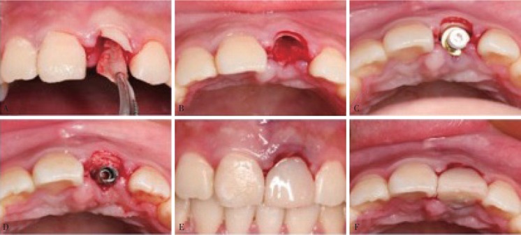
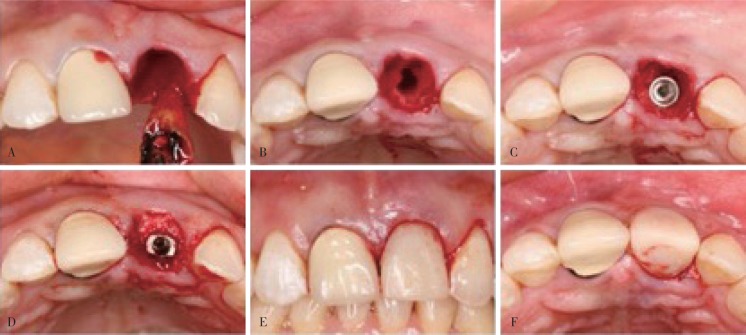
完善术前口腔检查,并进行牙周洁治术。术前0.12%氯己定含漱。局部麻醉下采用微创方法不翻瓣拔牙。试验组:显微镜下约龈缘上方1 mm磨除牙冠,近远中向分根,小心拔除牙根的腭侧部分(图1A);修整剩余唇侧牙片盾的厚度约为1 mm,高度位于牙槽嵴顶上方1 mm,形成盾牌状结构(图1B);探查唇侧牙片盾的松动度以确保其稳定无松动;按即刻种植位点植入种植体,与唇侧牙片盾之间保留约2 mm的间隙(图1C),在间隙内严密充填Bio-Oss骨粉(Geistlich公司,瑞士)(图1D)。对照组:完整拔除患牙(图2A),偏腭侧备洞(图2B),植入种植体(图2C),唇侧跳跃间隙填入Bio-Oss骨粉(图2D)。术后两组患者均采用即刻修复(图1E、1F,图2E、2F)。术后口服抗生素3 d,0.12%氯己定含漱7 d。术后6个月复诊,取终模,行最终修复体修复。术后12个月复诊。
图 1. 试验组(改良SST法)治疗过程.
Fig 1 Treatment of experiment group (the modified SST)
A:近远中向分根,拔除牙根腭侧部分;B:保留并修整唇侧牙片盾;C:植入种植体,与唇侧牙片盾之间保留约2 mm的间隙;D:在间隙内严密充填 Bio-Oss骨粉;E:即刻修复正面照;F:即刻修复 面照。
面照。
图 2. 对照组(传统即刻种植法)治疗过程.
Fig 2 Treatment of the control group (conventional immediate implant placement)
A:完整拔除患牙;B:偏腭侧备洞;C:植入种植体;D:唇侧跳跃间隙填入Bio-Oss骨粉;E:即刻修复正面照;F:即刻修复 面照。
面照。
1.3. 评价指标
1.3.1. 种植成功率
根据Albrektsson提出的种植成功标准判断种植成功率。成功标准为:临床检查种植体无松动;影像学检查种植体周围无透射区;种植体无持续性或不可逆的症状,如疼痛、感染、麻木等;负载1年,垂直方向种植体骨吸收第1年<2 mm,以后每年<0.2 mm[9]。
1.3.2. 红色美学指数(pink esthetic score,PES)
在术前(T0)和术后1年(T2),采用PES评价种植体周围软组织美学效果。评价指标包括:近中龈乳头、远中龈乳头、唇侧牙龈曲线、唇侧牙龈高度、牙槽骨外形、软组织颜色、软组织质地,每个项目评分为0、1、2分,总分14分[10]。
1.3.3. 唇侧骨板吸收情况
患者在术后即刻(T1)及术后1年(T2)拍摄锥形束CT,用自带软件Kavo eXam Vision测量种植体肩部(implant shoulder,IS)根方1、3、5 mm的多水平唇侧骨板厚度,分别记为1 mm-IS、3 mm-IS、5 mm-IS。每处测量3次,取平均值。测量精度是0.01 mm,测量图像均穿过种植体中心。
1.3.4. 患者满意度调查
使用视觉模拟评分法(visual analogue scale,VAS)调查患者对种植义齿周围软组织美学效果的满意度。基本方法是使用一条长约10 cm的游动标尺,"0”代表完全不满意,“10”代表完全满意,患者根据自己的满意度在线段上作标注,医师根据患者标出的位置为其评分。
1.4. 统计学处理
采用SPSS 21.0软件进行统计分析。计数资料采用均值±标准差表示。同组不同时间点的比较,满足正态性和方差齐性采用配对资料的t检验,不满足则采用Wilcoxon符号秩和检验;同时间点组间比较,满足正态性和方差齐性采用独立样本t检验,不满足则采用两独立样本非参数检验;计量资料采用率值表示,组间比较采用卡方检验。检验水准为双侧α=0.05。
2. 结果
2.1. 术后1年两组患者种植成功率对比
试验组与对照组的12例种植体均取得成功,成功率均为100%,组间对比无差异。
2.2. 术后1年两组患者PES评分对比
试验组与对照组术后1年的软组织情况见图3,术前术后的PES评分见表1。拔牙前(T0)两组的PES分别为13.17±0.83和13.08±0.79,差异无统计学意义(P>0.05)。试验组术后1年(T2)的评分为13.25±0.75,明显高于对照组11.83±0.94(P<0.05)。试验组拔牙前和术后1年分值变化(T2-T0)为0.08±1.08,对照组为-1.25±1.22,两组的差异有统计学意义(P<0.05)。
图 3. 术后1年两组患者软组织情况对比.
Fig 3 Comparison of soft tissue condition in two groups one year after surgery
A1、A2:试验组拔牙前;B1、B2:试验组术后1年,唇侧轮廓得以维持;C1、C2:对照组拔牙前;D1、D2:对照组术后1年,唇侧轮廓发生塌陷。上:正面照;下: 面照。
面照。
表 1. 两组患者术后1年PES评分对比.
Tab 1 Comparison of PES in two groups one year after surgery
| 组别 | 例数 | T0 | T2 | T2-T0 |
| 试验组 | 12 | 13.17±0.83 | 13.25±0.75 | 0.08±1.08 |
| 对照组 | 12 | 13.08±0.79 | 11.83±0.94 | -1.25±1.22 |
| P值 | - | 0.782 | 0.001* | 0.015* |
注:T0为拔牙前,T2为术后1年;*为P<0.05。
x±s
2.3. 术后1年两组患者种植体唇侧骨量情况
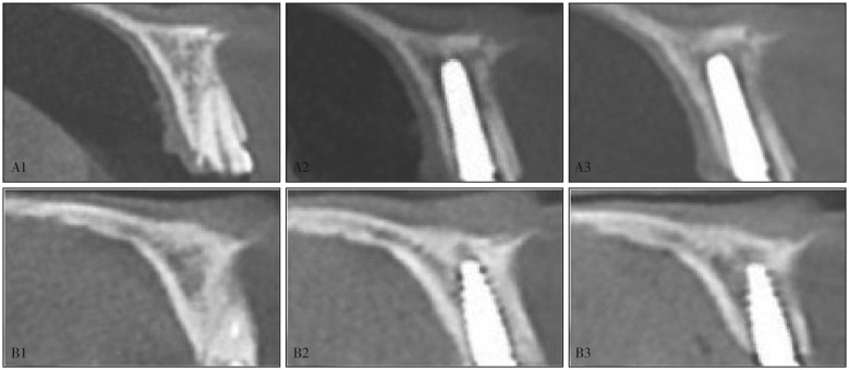
两组患者在术后即刻(T1)和术后1年(T2)唇侧骨量情况见图4,各水平唇侧骨板厚度的测量值见表2。术后即刻(T1)各水平唇侧骨板厚度无明显差异(P>0.05);试验组各水平唇侧骨板厚度在术后1年(T2)较术后即刻(T1)变化较小,差异无统计学意义(P>0.05);而对照组在术后1年(T2)的变化比较明显,差异有统计学意义(P<0.05)。
图 4. 两组术前和术后的唇侧骨量情况.
Fig 4 Labial bone thickness in two groups before and after operation
A1、A2、A3:试验组拔牙前、术后即刻、术后1年的锥形束CT片,唇侧骨板厚度得以维持;B1、B2、B3:对照组拔牙前、术后即刻、术后1年的锥形束CT片,唇侧骨板出现明显吸收。
表 2. 两组患者术后唇侧骨量对比.
Tab 2 Comparison of postoperative labial bone thickness in two groups after operation
| 组别 | 1 mm-IS |
P值 | 3 mm-IS |
P值 | 5 mm-IS |
P值 | |||
| T1 | T2 | T1 | T2 | T1 | T2 | ||||
| 试验组 | 2.94±0.68 | 2.90±0.64 | 0.256 | 3.00±0.76 | 2.94±0.71 | 0.179 | 2.84±0.64 | 2.73±0.57 | 0.185 |
| 对照组 | 3.35±0.64 | 2.51±0.69 | 0.000* | 3.08±0.66 | 2.33±0.78 | 0.002* | 2.78±0.67 | 2.13±0.72 | 0.005* |
注:1 mm-IS、3 mm-IS、5 mm-IS分别为种植体肩部根方1、3、5 mm的唇侧骨厚度;*表示与T1相比,P<0.05。
mm, x±s
2.4. 术后1年两组患者唇侧骨板的吸收量
两组患者术后1年各水平唇侧骨板的吸收量见表3。试验组术后1年水平唇侧骨板吸收量(T1-T2)均小于对照组,差异有统计学意义(P<0.05)。
表 3. 术后1年两组患者唇侧骨板吸收量对比.
Tab 3 Comparison of postoperative labial bone thickness in two groups one year after operation
| 组别 | 例数 | T1-T2 |
||
| 1 mm-IS | 3 mm-IS | 5 mm-IS | ||
| 试验组 | 12 | 0.04±0.13 | 0.05±0.13 | 0.11±0.26 |
| 对照组 | 12 | 0.85±0.45 | 0.75±0.37 | 0.65±0.35 |
| P值 | - | 0.000* | 0.000* | 0.005* |
注:T1-T2为术后1年较术后即刻唇侧骨板吸收量;*为P<0.05。
mm, x±s
2.5. 术后1年两组患者的满意度
试验组的满意度(VAS评分)为9.08±0.29,对照组满意度为8.77±0.45,两组差异有统计学意义(P<0.05)。
3. 讨论
上颌前牙区对个体美观具有极其重要的意义。研究[11]–[13]表明,当严格选择病例、谨慎操作、合理设计时,即刻种植即刻修复的成功率与传统种植相比无明显差异。本研究采用即刻种植即刻修复的方法,治疗时严格掌握适应证,结果显示,试验组与对照组的种植成功率均为100%,提示改良SST不会影响种植成功率。
上颌前牙区牙槽窝唇侧骨板菲薄,主要由固有牙槽骨构成。天然牙丧失后,由于失去牙周膜的滋养,固有牙槽骨常发生不可逆吸收[14]。Lee等[15]通过Meta分析表明,即刻种植术后1年水平向骨吸收可达到1.07 mm;并且即刻种植术后常常伴发唇侧轮廓塌陷和牙龈退缩的美学并发症[16]。Hürzeler等[5]和Bäumer等[6]通过动物研究发现,在保留唇侧牙片盾的颊侧,健康的牙周膜和唇侧骨板得到很好的保留。Bäumer等[8]进一步通过5年回顾性研究,定量分析种植体唇侧轮廓改变和牙龈退缩情况,结果显示,唇侧轮廓平均吸收量仅为(-0.37±0.18) mm,牙龈退缩量与邻牙相当。目前,SST的前瞻性对照研究尚少,本研究通过纳入对照组患者,设计前瞻性对照研究,结果显示,试验组的PES评分以及患者满意度均优于对照组,而唇侧骨板吸收量低于对照组,进一步证实SST通过保留唇侧牙片盾可较好地维持唇侧骨板,达到更好的软组织美学效果。
传统的SST在种植体植入过程中容易导致唇侧牙片盾的松动而使牙片盾不能保留,并且有并发症的报道。Siormpas等[7]对46例SST患者随访了24~60个月,有1例患者的牙片盾在根方出现了吸收。Lagas等[17]报道了16例系列病例,发现仅有1例患者的牙片盾根方由于感染导致骨板吸收。近年传统SST术得到了改良。Kan等[18]和Cherel等[19]的病例报道中,保留邻面牙片,在牙片与种植体之间的间隙填入骨粉,用于保存种植体间的牙间乳头,效果满意。基于此,本研究在唇侧牙片盾与种植体之间保留约2 mm的间隙,在间隙内严密充填骨粉。这种改良一方面防止了种植术中对牙片盾以及唇侧骨板的挤压损伤,另一方面避免了种植体与牙片盾直接接触导致的并发症,降低了传统SST的技术敏感性。
2013年国际牙种植学会共识会议提出即刻种植适应证为:满足唇侧骨壁完整且厚度≥1 mm[20]。50%以上的中国人前牙唇侧骨板厚度不足1 mm,使得即刻种植在前牙的应用受到很大的限制[21]–[22]。扩大即刻种植的应用范围,同时避免即刻种植的美学并发症,是本研究设计的初衷。本研究中,为了实现试验组和对照组的可比性,将两组患者唇侧骨板厚度的纳入标准均设定为≥1 mm,结果显示,改良SST由于保留牙片盾增强了唇侧骨板对外力的抵抗性,保留牙周膜维持了唇侧骨板的血供,其美学效果优于传统即刻种植法。在此基础上,笔者推测,当唇侧骨板不足1 mm时,通过改良SST可实现即刻种植,扩大即刻种植的适应证,本课题组将在后续研究中对此进行进一步验证。
Funding Statement
[基金项目] 重庆市医学科研计划(2012-2-140);2016年重庆市高校创新团队建设计划(CXTDG-201602006);重庆市高校市级口腔生物医学工程重点实验室资助项目[渝教科(2014)55号]
Supported by: Chongqing Medical Research Project (2012-2-140); Program for Innovation Team Building at Institutions of Higher Education in Chongqing in 2016 (CXTDG-201602006); Project Supported by Chongqing Municipal Key Laboratory of Oral Biomedical Engineering of Higher Education [(2014)55].
Footnotes
作者声明本文无利益冲突。
References
- 1.Bhola M, Neely AL, Kolhatkar S. Immediate implant placement: clinical decisions, advantages, and disadvantages[J] J Prosthodont. 2008;17(7):576–581. doi: 10.1111/j.1532-849X.2008.00359.x. [DOI] [PubMed] [Google Scholar]
- 2.Roe P, Kan JY, Rungcharassaeng K, et al. Horizontal and vertical dimensional changes of peri-implant facial bone following immediate placement and provisionalization of maxillary anterior single implants: a 1-year cone beam computed tomography study[J] Int J Oral Maxillofac Implants. 2012;27(2):393–400. [PubMed] [Google Scholar]
- 3.Botticelli D, Berglundh T, Lindhe J. Hard-tissue alterations following immediate implant placement in extraction sites[J] J Clin Periodontol. 2004;31(10):820–828. doi: 10.1111/j.1600-051X.2004.00565.x. [DOI] [PubMed] [Google Scholar]
- 4.Nisapakultorn K, Suphanantachat S, Silkosessak O, et al. Factors affecting soft tissue level around anterior maxillary single-tooth implants[J] Clin Oral Implants Res. 2010;21(6):662–670. doi: 10.1111/j.1600-0501.2009.01887.x. [DOI] [PubMed] [Google Scholar]
- 5.Hürzeler MB, Zuhr O, Schupbach P, et al. The socket-shield technique: a proof-of-principle report[J] J Clin Periodontol. 2010;37(9):855–862. doi: 10.1111/j.1600-051X.2010.01595.x. [DOI] [PubMed] [Google Scholar]
- 6.Bäumer D, Zuhr O, Rebele S, et al. The socket-shield technique: first histological, clinical, and volumetrical observations after separation of the buccal tooth segment—a pilot study[J] Clin Implant Dent Relat Res. 2015;17(1):71–82. doi: 10.1111/cid.12076. [DOI] [PubMed] [Google Scholar]
- 7.Siormpas KD, Mitsias ME, Kontsiotou-Siormpa E, et al. Immediate implant placement in the esthetic zone utilizing the “root-membrane” technique: clinical results up to 5 years postloading[J] Int J Oral Maxillofac Implants. 2014;29(6):1397–1405. doi: 10.11607/jomi.3707. [DOI] [PubMed] [Google Scholar]
- 8.Bäumer D, Zuhr O, Rebele S, et al. Socket shield technique for immediate implant placement—clinical, radiographic and volumetric data after 5 years[J] Clin Oral Implants Res. 2017;28(11):1450–1458. doi: 10.1111/clr.13012. [DOI] [PubMed] [Google Scholar]
- 9.Albrektsson T, Zarb G, Worthington P, et al. The long-term efficacy of currently used dental implants: a review and proposed criteria of success[J] Int J Oral Maxillofac Implants. 1986;1(1):11–25. [PubMed] [Google Scholar]
- 10.Fürhauser R, Florescu D, Benesch T, et al. Evaluation of soft tissue around single-tooth implant crowns: the pink esthetic score[J] Clin Oral Implants Res. 2010;16(6):639–644. doi: 10.1111/j.1600-0501.2005.01193.x. [DOI] [PubMed] [Google Scholar]
- 11.Chen ST, Wilson TG, Jr, Hämmerle CH. Immediate or early placement of implants following tooth extraction: review of biologic basis, clinical procedures, and outcomes[J] Int J Oral Maxillofac Implants. 2004;19(Suppl):12–25. [PubMed] [Google Scholar]
- 12.Atieh MA, Payne AG, Duncan WJ, et al. Immediate placement or immediate restoration/loading of single implants for molar tooth replacement: a systematic review and meta-analysis[J] Int J Oral Maxillofac Implants. 2010;25(2):401–415. [PubMed] [Google Scholar]
- 13.Grandi T, Guazzi P, Samarani R, et al. Immediate provisionalisation of single post-extractive implants versus implants placed in healed sites in the anterior maxilla: 1-year results from a multicentre controlled cohort study[J] Eur J Oral Implantol. 2013;6(3):285–295. [PubMed] [Google Scholar]
- 14.Huynh-Ba G, Pjetursson BE, Sanz M, et al. Analysis of the socket bone wall dimensions in the upper maxilla in relation to immediate implant placement[J] Clin Oral Implants Res. 2010;21(1):37–42. doi: 10.1111/j.1600-0501.2009.01870.x. [DOI] [PubMed] [Google Scholar]
- 15.Lee CT, Chiu TS, Chuang SK, et al. Alterations of the bone dimension following immediate implant placement into extraction socket: systematic review and meta-analysis[J] J Clin Periodontol. 2014;41(9):914–926. doi: 10.1111/jcpe.12276. [DOI] [PubMed] [Google Scholar]
- 16.Chen ST, Buser D. Esthetic outcomes following immediate and early implant placement in the anterior maxilla: a systematic review[J] Int J Oral Maxillofac Implants. 2014;29(Suppl):186–215. doi: 10.11607/jomi.2014suppl.g3.3. [DOI] [PubMed] [Google Scholar]
- 17.Lagas LJ, Pepplinkhuizen JJ, Bergé SJ, et al. Implant placement in the aesthetic zone: the socket-shield-technique[J] Ned Tijdschr Tandheelkd. 2015;122(1):33–36. doi: 10.5177/ntvt.2015.01.14180. [DOI] [PubMed] [Google Scholar]
- 18.Kan JY, Rungcharassaeng K. Proximal socket shield for interimplant papilla preservation in the esthetic zone[J] Int J Periodontics Restorative Dent. 2013;33(1):e24–e31. doi: 10.11607/prd.1346. [DOI] [PubMed] [Google Scholar]
- 19.Cherel F, Etienne D. Papilla preservation between two implants: a modified socket-shield technique to maintain the scalloped anatomy? A case report[J] Quintessence Int. 2014;45(1):23–30. doi: 10.3290/j.qi.a30765. [DOI] [PubMed] [Google Scholar]
- 20.Morton D, Chen ST, Martin WC, et al. Consensus statements and recommended clinical procedures regarding optimizing esthetic outcomes in implant dentistry[J] Int J Oral Maxillofac Implants. 2014;29(Suppl):216–220. doi: 10.11607/jomi.2013.g3. [DOI] [PubMed] [Google Scholar]
- 21.沈 建伟, 何 福明, 江 巧红, et al. 上颌前牙与前磨牙唇侧骨壁厚度的CBCT测量分析[J] 浙江大学学报(医学版) 2012;41(3):234–238. [PubMed] [Google Scholar]; Shen JW, He FM, Jiang QH, et al. Measurement of facial bone wall thickness of maxillary anterior teeth and premolars on cone beam computed tomography images[J] J Zhejiang Univ (Med Sci) 2012;41(3):234–238. [PubMed] [Google Scholar]
- 22.蒙 萌, 陶 荣, 夏 雨凝, et al. 中西部人群健康上颌前牙唇侧骨板厚度的CBCT测量[J] 牙体牙髓牙周病学杂志. 2018;28(2):73–77, 102. [Google Scholar]; Meng M, Tao R, Xia YN, et al. Facial alveolar bone thickness of maxillary anterior teeth in the subjects from central and west China with normal occlusion measured by CBCT[J] Chin J Conserv Dent. 2018;28(2):73–77, 102. [Google Scholar]


