Abstract
Background
Inflammation is a necessary component of chronic kidney disease (CKD) that can be attributed to an accumulation of toxins and a reduced clearance of proinflammatory cytokines. Procalcitonin (PCT) is a widely applied biomarker in the diagnosis of infection, and considering the presence of pre‐existing inflammation in CKD patients, the PCT level could be high in such a population; however, no reference value for PCT in CKD patients has been available to date.
Methods
During the present study period, 361 CKD patients and 119 healthy controls were included. The PCT level and other biochemistry parameters were assayed by using a COBAS system. Statistical analysis was conducted to compare the differences in PCT levels and other biochemistry parameters between the two groups, and linear regression was used to assess the correlation between two variables. Receiver operating characteristic (ROC) curve analysis was performed to evaluate the performance of PCT and the optimal cutoff value to differentiate between CKD patients and healthy controls.
Results
The PCT level in CKD patients was significantly higher than that in healthy controls, and among the CKD patients, the PCT level was increased with advanced clinical stage. Moreover, PCT was moderately correlated with CysC. The optimal off‐value was 0.075 with a sensitivity of 94.7% and specificity of 90.8%.
Conclusion
The PCT level was significantly higher in CKD patients than in healthy controls, and the reference value for CKD patients should be adjusted to avoid unnecessary antibiotic treatments which may pose a negative impact on residual renal function.
Keywords: chronic kidney disease, diagnostic performance, infection, procalcitonin
1. INTRODUCTION
Chronic kidney disease (CKD) is a kidney disease in which there is gradual loss of renal function over a period of years or decades. During the early stages of CKD, due to the kidney's significant compensation mechanism, patients with CKD can be asymptotic since the remaining renal nephrons are capable of removing toxins and maintaining homeostasis.1 Therefore, symptoms of CKD only appear when the kidneys are significantly impaired. The main challenge faced by the public health system is the accurate diagnosis of CKD; without regular surveillance of renal function, most CKD patients have progressed to the advanced stage when diagnosed. Under such circumstances, patients with CKD might need to receive regular dialysis or a kidney transplant to survive. According to statistics, in 2013, there were 956 000 deaths attributed to CKD worldwide2; therefore, CKD has been considered to have a major impact on the quality of life, especially in the elderly population. Among the general population, the prevalence of CKD including all five stages is approximately 13.4%.3 Despite this astonishing high prevalence of CKD, the trend of this chronic disease is expected to grow in coming decades. The high prevalence of diabetes, hypertension,4 and tobacco abuse5 is believed to be responsible for the increasing trend of CKD. With the facts stated above, we can conclude that CKD is a pressing public health issue affecting the health and quality of life of the general population.
Inflammation has been recognized as an essential part of CKD and is associated with cardiovascular disease, protein‐energy wasting, and mortality among patients with CKD.6 The origin of inflammation in CKD patients can be attributed to uremia, which is the direct consequence of impaired renal function. Accumulated toxins in the circulation produce oxidative stress and carbonyl stress, which are highly proinflammatory states.7 Moreover, impaired renal function leads to the decreased clearance of several kinds of proinflammatory cytokines,8 and this impaired function is also responsible for the chronic inflammation observed in CKD patients. Most CKD patients are at high risk of some infectious events, such as catheter‐related infections, access site infections, and peritonitis, in addition to endogenous factors, in patients receiving peritoneal dialysis (PD) and hemodialysis (HD).9 Therefore, the management of infection in CKD patients is of critical importance in improving the medical condition and outcome.
Procalcitonin (PCT) is a common biomarker for diagnosing infection, especially bacterial infection.10 The PCT level in healthy individuals without infection is below the limit of detection (0.01 ng/mL), and it is significantly elevated under the stimulation of pathogens. However, due to the pre‐existing endogenous inflammation that occurs in CKD patients and the impaired kidney clearance, the reference range that applies to the general population may not be appropriate for diagnosing infections in CKD patients. More recently, debate has continued regarding whether the PCT level is increased in CKD patients without infection, and the optimal reference for CKD patients remains undetermined. This study therefore aimed to assess the PCT level in CKD patients without infection and to obtain a reference range for CKD patients. Moreover, the possible association between PCT levels and renal function parameters was investigated.
2. MATERIALS AND METHODS
2.1. Study participants
Eligible patients with CKD were recruited from the Department of Nephrology, the Longyan First Hospital Affiliated with Fujian Medical University, Fujian Province, China, on the day of admittance. The stage of CKD was assessed by using the glomerular filtration rate (GFR) which was calculated by using emission computed tomography. To assure the homogeneity of the study participants, patients with CKD of prerenal or postrenal cause and patients who had received kidney transplants were excluded. In order to exclude the existence of infection, all potential study participants were subjected to blood culture before enrollment, and only those who have a negative result were selected. In total, we managed to recruit 361 CKD patients during the ascertainment period of September 2017 through March 2018. Control subjects consisted of healthy volunteers, and 119 controls were included in the present study during the same study period. All study participants provided written, informed consent before enrollment, and all procedures of the present study were in agreement with the Helsinki Declaration and the policy of the Ethics Committee of Longyan First Hospital Affiliated with Fujian Medical University (reference No. LYFH‐2016‐042).
2.2. Data collection
An identical questionnaire was employed to collect information on demographic characteristics from all study subjects. Interviews were performed by extensively trained staff to improve data quality and to minimize interinterviewer variation. All CKD patients and healthy controls provided a 5‐mL blood sample on the day of the interview. The PCT level (reference laboratory range: 0.00‐0.05 ng/mL) in plasma was quantified by using a COBAS E602 immunology analyzer (Roche Diagnostics, Risch‐Rotkreuz, Switzerland). Biochemistry panels that included blood urea nitrogen (BUN), creatinine (CREA), cystatin C (CysC), potassium (K), sodium (Na), chloride (Cl), calcium (Ca), and C‐reactive protein (CRP) levels (reference laboratory range: 0.00‐5.00 ng/mL) were conducted by using a COBAS C501 chemistry analyzer (Roche Diagnostics, Risch‐Rotkreuz, Switzerland). All procedures strictly conformed with the manufacturers’ manuals. In addition, we randomly selected 5% of the samples for testing as duplicated controls.
2.3. Statistical analysis
Student's t test and chi‐squared test were employed to compare the differences in demographic characteristics based on the variables’ forms. The comparisons of the PCT levels and biochemistry parameters were all conducted by using Student's t test. For groups with greater than two variables, one‐way analysis of variance (ANOVA) was used to compare the means. Linear regression was used to assess the correlation between two variables (enter method), and Pearson's coefficient was calculated. Receiver operating characteristic (ROC) curve analysis was conducted to calculate the optimal cutoff value to differentiate between CKD patients and healthy controls, and the area under the curve (AUC) was calculated. A 2‐tailed P value less than 0.05 was accepted as statistically significant. All statistical analyses were performed by using SPSS software (IBM, Chicago, IL, USA) version 19.
3. RESULTS
3.1. Demographic characteristics of the study participants
As stated above, a total of 361 CKD patients and 119 healthy controls were included in the present study. Table 1 demonstrates the demographic information of all participants along with the CKD stage and renal replacement treatment (RRT) of the patients with CKD. As shown in Table 1, there were no significant differences between CKD patients and healthy controls with respect to age and sex (P > .05). Moreover, 289 patients (80.1%) had already advanced to having stage 5 CKD on the day of admittance, and 30 and 42 patients were classified as having stage 3 and stage 4 CKD, respectively. For RRT, 17.7% of patients had received no treatment before enrollment, and 73.4% of patients had received hemodialysis; however, only 8.9% of patients used peritoneal dialysis. Among those who received hemodialysis, 236 patients (89.06%) were dialyzed through radiocephalic arteriovenous fistulas, while 29 patients (10.94%) had peripherally inserted central venous catheters.
Table 1.
Demographic characteristics of CKD patients and healthy controls
| Variables | CKD patients | Healthy controls | P value |
|---|---|---|---|
| Age (y, Mean ± SD) | 60.84 ± 16.52 | 58.36 ± 13.41 | .138 |
| Sex | |||
| Male | 206 (57.1) | 59 (49.6) | |
| Female | 155 (42.9) | 60 (50.4) | .155 |
| CKD stage | |||
| Stage 3 | 30 (8.3) | – | |
| Stage 4 | 42 (11.6) | – | |
| Stage 5 | 289 (80.1) | – | – |
| Renal replacement treatment | |||
| None | 64 (17.7) | – | |
| HD | 265 (73.4) | – | |
| PD | 32 (8.9) | – | ‐ |
3.2. Comparison of PCT levels and biochemistry parameters between CKD patients and healthy controls
The results obtained from the comparison of PCT levels and biochemistry parameters between the two groups are summarized in Table 2. The PCT level in CKD patients (0.44 ± 0.67 ng/mL) was significantly higher than that in healthy controls (0.04 ± 0.06 ng/mL). We also observed significant elevations in BUN, CREA, CysC, K, and CRP levels in CKD patients, while the Na, Cl, and Ca levels were significantly decreased compared to the levels in healthy controls (P < .05). Interestingly, we also observed a significant elevation in CRP levels (P < .05) among CKD patients, suggesting pre‐existing inflammation in CKD patients. The difference in the biochemistry parameters revealed in Table 2 was consistent with the pathology of CKD, suggesting the high quality of our assay.
Table 2.
Comparison of PCT levels and biochemistry parameters
| Variables | CKD patients | Healthy controls | P value |
|---|---|---|---|
| PCT (ng/mL) | 0.44 ± 0.67 | 0.04 ± 0.06 | <.001* |
| BUN (mmol/L) | 21.97 ± 11.10 | 5.32 ± 1.37 | <.001* |
| CREA (μmol/L) | 667.45 ± 390.88 | 70.46 ± 15.96 | <.001* |
| CysC (ng/mL) | 4.58 ± 1.90 | 0.80 ± 0.19 | <.001* |
| K (mmol/L) | 4.41 ± 0.84 | 4.21 ± 0.40 | .011* |
| Na (mmol/L) | 138.58 ± 4.70 | 141 ± 1.96 | <.001* |
| Cl (mmol/L) | 100.11 ± 6.13 | 102.85 ± 2.36 | <.001* |
| Ca (mmol/L) | 2.14 ± 0.26 | 2.34 ± 0.12 | <.001* |
| CRP (ng/mL) | 14.78 ± 6.67 | 4.73 ± 7.89 | <.001* |
P < .05.
3.3. Linear regression between PCT levels and other biochemistry parameters
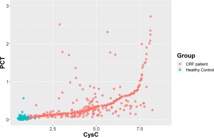
We further analyzed the correlation between PCT levels and other biochemistry parameters by employing linear regression. As seen in Table 3, BUN, CREA, CysC, Na, Cl, and CRP levels were significantly correlated with PCT levels, while no significant correlation was observed in the examination of K and Ca levels. Although we found a positive correlation among some biochemistry parameters, the R values for BUN (0.176), CREA (0.257), Na (−0.104), Cl (−0.220), and CRP (0.251) were not high enough to establish a linear correlation. However, we found that CysC, a biomarker for evaluating renal function, was significantly correlated with PCT level, with a moderate R value of 0.548. Figure 1 displays the scatter plot of PCT and CysC, along with the linear regression model.
Table 3.
Linear regression between PCT levels and other biochemistry parameters
| Variable | Standardized coefficient (R) | Standard error | P value |
|---|---|---|---|
| BUN (mmol/L) | 0.176 | 0.002 | <.001* |
| CREA (μmol/L) | 0.257 | 0.001 | <.001* |
| CysC (ng/mL) | 0.548 | 0.010 | <.001* |
| K (mmol/L) | 0.013 | 0.037 | .778 |
| Na (mmol/L) | ‐0.104 | 0.006 | .023* |
| Cl (mmol/L) | ‐0.220 | 0.005 | <.001* |
| Ca (mmol/L) | ‐0.034 | 0.110 | .460 |
| CRP (ng/mL) | 0.251 | 0.001 | <.001* |
P < .05.
Figure 1.

Scatter plot of PCT vs CysC
3.4. Impact of clinical stage on PCT level among CKD patients
To examine the impact of CKD stage on PCT level among CKD patients, we conducted a one‐way ANOVA to compare patients in different stages of CKD. The results are presented in Table 4, showing that the mean PCT level increased with advancing clinical stage. No significant difference was found in PCT levels between patients with stage 3 and 4 CKD. However, patients with stage 5 CKD maintained a significantly increased PCT level (0.50 ± 0.73 ng/mL), and a significant difference was found when comparing patients with both stage 3 and stage 4 CKD (P < .05).
Table 4.
One‐way ANOVA of the PCT level by stage among CKD patients
| Stage | Number | Mean ± SD | F value | P value |
|---|---|---|---|---|
| Stage 3 | 30 | 0.20 ± 0.31 | .991a | |
| Stage 4 | 42 | 0.21 ± 0.24 | <.001b , * | |
| Stage 5 | 289 | 0.50 ± 0.73 | 5.789 | <.001c , * |
Stage 3 vs Stage 4.
Stage 4 vs Stage 5.
Stage 3 vs Stage 5.
P < .05.
3.5. Impact of RRT on PCT and CRP levels among stage 5 CKD patients
In order to investigate whether RRT including hemodialysis and peritoneal dialysis can have an impact on PCT and CRP levels among stage 5 CKD patients or not, we conducted the one‐way ANOVA in these two parameters as well. The results showed that non‐dialysis patients have the highest PCT level (0.60 ± 1.38 ng/mL); both peritoneal dialysis (0.56 ± 0.51 ng/mL) and hemodialysis patients (0.48 ± 0.57 ng/mL) showed a slight reduction on PCT level, however, with no significance. Similarly, non‐dialysis patients also have the highest CRP level (21.33 ± 4.32 ng/mL), and dialysis can decrease the CRP level to some extent, but still no significance was found in the comparison (see Table 5).
Table 5.
One‐way ANOVA of PCT and CRP levels between different RRTs among stage 5 CKD patients
| Treatment (n) | PCT (ng/mL) | F value | P value | CRP (ng/mL) | F value | P value |
|---|---|---|---|---|---|---|
| None (39) | 0.60 ± 1.38 | .341 | 21.33 ± 4.32 | .394a | ||
| HD (230) | 0.48 ± 0.57 | .639 | 15.98 ± 2.58 | .303b | ||
| PD (20) | 0.56 ± 0.51 | 0.525 | .871 | 6.55 ± 4.94 | 0.987 | .165c |
None vs HD.
HD vs PD.
None vs PD.
3.6. ROC curve analysis of PCT and CRP levels
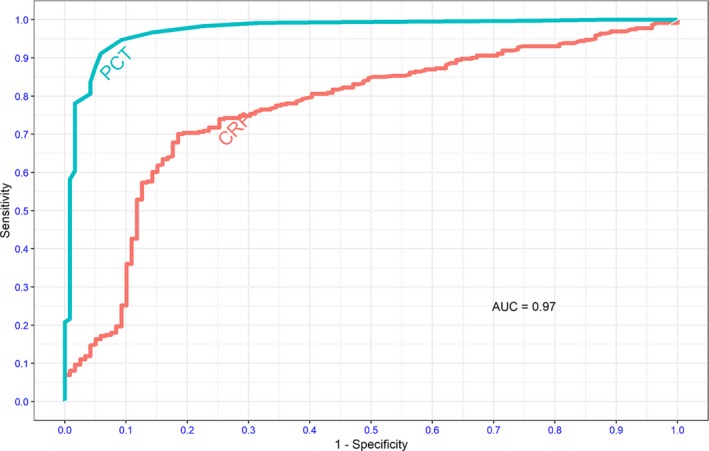
The ROC curve analysis was applied to evaluate the diagnostic performance of PCT in differentiating between CKD patients without infection and healthy controls. Moreover, we introduced CRP as a reference marker. Strikingly, Figure 2 demonstrates that PCT had an extremely high diagnostic performance with an AUC of 0.972, and the optimal cut value was 0.075. With this specific value, the PCT test yielded a sensitivity of 94.7% and specificity of 90.8%. In contrast, the AUC for CRP was 0.765, and the optimal cutoff value was 5.875. With this cutoff value, the CRP test yielded a sensitivity of 70.1% and specificity of 81.5%.
Figure 2.

Receiver operating characteristic curve analysis of PCT and CRP for differentiating between CKD patients without infection and healthy controls. Blue line: PCT; red line: CRP
4. DISCUSSION
The present study was designed to determine the PCT level in CKD patients without infection and to establish an optimal cutoff value of the PCT level in the diagnosis of infection among CKD patients, consequently avoiding the overuse of antibiotics and preserving residual renal function. Overall, our analysis results revealed a significant elevation in the PCT and CRP levels of CKD patients compared with the level of healthy controls. The most clinically relevant finding was that the PCT level increased approximately 10‐fold in CKD patients without infection compared with the level in control subjects, and the CRP level increased almost 3‐fold. Moreover, through one‐way ANOVA, we did observe that the PCT level was increased with advancing clinical stage in CRP patients. Although some biochemistry parameters, such as BUN, CREA, Cl, and CRP levels, were significantly correlated with PCT level, the Pearson's coefficients of the abovementioned parameters were too small to suggest a correlation. It is surprising that CysC, which is an important renal function parameter that has been correlated with PCT, had a coefficient of 0.548, suggesting a moderate correlation. We attempted to employ the ROC analysis to evaluate the diagnostic performance of PCT, which was the main objective of this study, and the PCT level demonstrated extremely high performance in differentiating between CKD patients and healthy controls; specifically, the optimal cutoff value was 0.075 ng/mL with a sensitivity of 94.7% and a specificity of 90.8%.
Procalcitonin is a 116‐amino acid peptide with an approximate molecular weight of 14.5 kDa, and PCT is a useful predictive marker of the inflammatory process with rapidly increased serum levels in inflammation or sepsis.11 The elevated serum PCT level we observed in CKD patients can be attributed to the following reasons. First, persistent and low‐intensity inflammation has been recognized as an important component of CKD pathology, and the intensity of inflammation, including IL‐1β, IL‐1 receptor antagonist, IL‐6, TNF‐α, and CRP levels, was inversely associated with residual renal function.12 With the accumulation of dysfunctional proinflammatory cytokines, which are produced by lymphocytes and various tissues,13 it is reasonable that PCT is elevated under such circumstances. The second probable cause of increased PCT levels is that impaired renal function could not provide sufficient clearance of circulating PCT. This finding contradicts the findings of previous studies conducted by Meisner et al,14 who observed the PCT half‐life among both patients with renal dysfunction and with normal renal function; the results showed no association between the PCT half‐life and creatinine clearance. The discrepancy between findings from the present study and the literature may be attributed to the different sample sizes. We included 361 CKD patients and 119 healthy controls, samples sizes that were both greater than those in previous studies. In contrast, a retrospective study also found that PCT level among CKD patients without infection was significantly higher when being compared with controls; in particular, stage 5 CKD patients without infection have a mean PCT level of 0.33 ng/mL, which also exceeded the reference range, and that partly supports our main finding.15 Moreover, we observed a moderate correlation between the CysC and PCT level, and this positive correlation corroborates the assumption that impaired renal function may lead to reduced clearance of PCT.
CysC is a protein with a low molecular weight of 13.3 kDa, which is very close to the molecular weight of PCT. CysC can serve as a more precise biomarker of renal function because it can only be removed from the system by glomerular filtration in the kidneys, and unlike BUN and CREA, it is more stable and free from the impacts caused by food and other factors.16 Therefore, we can assume that the PCT clearance was similar to the CysC clearance due to the similar molecular properties and positive correlations observed in the present study.
Our statistical analysis comparing the PCT level among CKD patients with different clinical stages further supported this assumption. As stated in the results section, patients with stage 5 CKD had higher PCT levels than patients with both stage 3 and stage 4 CKD. Although no significant difference was observed in the PCT levels between patients with stage 3 and stage 4 CKD, the difference observed in patients with stage 5 CKD could have been generated by the limited number of study participants in both groups. Another possible explanation is that in patients with stage 5 CKD, renal function degenerated significantly and caused massive accumulation of toxins and proinflammatory cytokines; consequently, an elevation in PCT emerged.
Using a cutoff value of 0.075 ng/mL yielded a sensitivity of 94.7% and a specificity of 90.8%, suggesting solid diagnostic performance in differentiating between CKD patients and healthy controls. This cutoff value is approximately three times higher than the reference value that is widely applied in clinical practice. It has been generally acknowledged that CKD patients are at high risk of various kinds of infections due to the invasive procedures of dialysis, and more importantly, dysfunctional immunity and infections are comment events in CKD. If patients are not properly sterilized, hemodialysis can be a vector for transmitting various infectious diseases, for instance, hepatitis C,17 and the infection can further worsen the patient's diagnosis. However, based on the current reference value, the application of PCT could lead to misunderstanding in terms of the diagnosis and treatment of CKD patients with suspected infection. Based on previous studies and the results of the present study, there is a great chance that the PCT test could misclassify CKD patients without infection if the current reference value still applies. Consequently, the PCT test could lead to the unnecessary administration of antibiotics, which would possibly further impair residual renal function. As can be seen, the present study recruited 289 patients in CKD stage 5, and the PCT level in those patients was significantly higher than patients in stages 3 and 4. Therefore, the PCT reference value used in end‐stage CKD patients should be further evaluated to avoid misunderstanding and the administration of unnecessary prescriptions. In addition to that, we failed to observe a significant reduction on PCT among stage 5 CKD patients between RRT and non‐dialysis, which are different with previous knowledge that dialysis can reduce the PCT level.18 Our results may be caused by insufficient number of study participants who did not receive RRT and should be interpreted cautiously. As suggested by the results of Sun et al15 and a meta‐analysis conducted by Lu et al,19 both PCT and CRP had poor sensitivity and specificity in diagnosing infection among CKD patients with current laboratory ranges, which is consistent with our finding that both of these two indicators would elevate even if without the presence of infection. That being said, CRP is more cost‐effective than PCT in terms of medical cost, as PCT measurement is widely conducted by using electrochemiluminescence immunoassay in clinical laboratories, an expensive method that costs three times higher than a CRP test. If the current laboratory ranges for PCT and CRP remain unchanged, CRP would be a more cost‐effective option in diagnosing infection among CKD patients.
The principal limitations of this study are in the number of healthy controls and that most of the CKD patients had advanced to stage 5 CKD upon enrollment. With regard to the insufficient number of CKD patients with stage 3 and stage 4 diseases, we were not able to observe a linear trend in the PCT levels and clinical stages. Moreover, the cutoff value of PCT we proposed in the present study could have higher accuracy and generalizability if the sample size of the healthy controls was further increased.
Despite the limitations described above, the present study offers a higher cutoff value of PCT with extremely high diagnostic performance that, if properly used, could contribute to the precise management of infection in end‐stage CKD patients and eventually avoid unnecessary damage to residual renal function.
CONFLICT OF INTEREST
The authors declare that they have no competing interests.
AUTHOR CONTRIBUTIONS
All authors contributed to the present study. WPH and SCW designed the research plan. The data were analyzed by CXL and YLZ. SCW wrote the article, and CXL, WPH, and YLZ were responsible for performing the experiments. All authors support the publication of the article.
ACKNOWLEDGMENTS
The authors also would like to express gratitude to all study participants for their consent and cooperation.
Wu S‐C, Liang C‐X, Zhang Y‐L, Hu W‐P. Elevated serum procalcitonin level in patients with chronic kidney disease without infection: A case‐control study. J Clin Lab Anal. 2020;34:e23065 10.1002/jcla.23065
Funding information
This study was financially supported by grants from Fujian Province Natural Science Foundation Program (No. 2018J01373).
REFERENCES
- 1. Chilelli NC, Cremasco D, Cosma C, et al. Effectiveness of a diet with low advanced glycation end products, in improving glycoxidation and lipid peroxidation: a long‐term investigation in patients with chronic renal failure. Endocrine. 2016;54:552‐555. [DOI] [PubMed] [Google Scholar]
- 2. GBD 2013 Mortality and Causes of Death Collaborators . Global, regional, and national age‐sex specific all‐cause and cause‐specific mortality for 240 causes of death, 1990‐2013: a systematic analysis for the Global Burden of Disease Study 2013. Lancet. 2015;385:117‐171. [DOI] [PMC free article] [PubMed] [Google Scholar]
- 3. Hill NR, Fatoba ST, Oke JL, et al. Global prevalence of chronic kidney disease – a systematic review and meta‐analysis. PLoS ONE. 2016;11:e0158765. [DOI] [PMC free article] [PubMed] [Google Scholar]
- 4. Malekmakan L, Malekmakan A, Daneshian A, Pakfetrat M, Roosbeh J. Hypertension and diabetes remain the main causes of chronic renal failure in Fars Province, Iran 2013. Saudi J Kidney Dis Transpl. 2016;27:423‐424. [DOI] [PubMed] [Google Scholar]
- 5. Stengel B, Couchoud C, Cénée S, Hémon D. Age, blood pressure and smoking effects on chronic renal failure in primary glomerular nephropathies. Kidney Int. 2000;57:2519‐2526. [DOI] [PubMed] [Google Scholar]
- 6. Stenvinkel P, Heimburger O, Paultre F, et al. Strong association between malnutrition, inflammation, and atherosclerosis in chronic renal failure. Kidney Int. 1999;55:1899‐1911. [DOI] [PubMed] [Google Scholar]
- 7. Kim HJ, Vaziri ND. Contribution of impaired Nrf2‐Keap1 pathway to oxidative stress and inflammation in chronic renal failure. Am J Physiol Renal Physiol. 2010;298:F662‐F671. [DOI] [PubMed] [Google Scholar]
- 8. Rosengren BI, Sagstad SJ, Karlsen TV, Wiig H. Isolation of interstitial fluid and demonstration of local proinflammatory cytokine production and increased absorptive gradient in chronic peritoneal dialysis. Am J Physiol Renal Physiol. 2013;304:F198‐F206. [DOI] [PubMed] [Google Scholar]
- 9. Hamad A, Ismail H, Elsayed M, et al. The epidemiology of acute peritonitis in end‐stage renal disease patients on peritoneal dialysis in Qatar: An 8‐year follow‐up study. Saudi J Kidney Dis Transpl. 2018;29:88‐94. [DOI] [PubMed] [Google Scholar]
- 10. Chan YL, Tseng CP, Tsay PK, Chang SS, Chiu TF, Chen JC. Procalcitonin as a marker of bacterial infection in the emergency department: an observational study. Crit Care. 2004;8:R12‐R20. [DOI] [PMC free article] [PubMed] [Google Scholar]
- 11. Ljungström L, Pernestig AK, Jacobsson G, Andersson R, Usener B, Tilevik D. Diagnostic accuracy of procalcitonin, neutrophil‐lymphocyte count ratio, C‐reactive protein, and lactate in patients with suspected bacterial sepsis. PLoS ONE. 2017;12:e0181704. [DOI] [PMC free article] [PubMed] [Google Scholar]
- 12. Gupta J, Mitra N, Kanetsky PA, et al. Association between albuminuria, kidney function, and inflammatory biomarker profile in CKD in CRIC. Clin J Am Soc Nephrol. 2012;7:1938‐1946. [DOI] [PMC free article] [PubMed] [Google Scholar]
- 13. Iglesias P, Díez JJ. Adipose tissue in renal disease: clinical significance and prognostic implications. Nephrol Dial Transplant. 2010;25:2066‐2077. [DOI] [PubMed] [Google Scholar]
- 14. Meisner M, Schmidt J, Hüttner H, Tschaikowsky K. The natural elimination rate of procalcitonin in patients with normal and impaired renal function. Intensive Care Med. 2000;26:S212‐216. [DOI] [PubMed] [Google Scholar]
- 15. Sun Y, Jiang L, Shao X. Predictive value of procalcitonin for diagnosis of infections in patients with chronic kidney disease: a comparison with traditional inflammatory markers C‐reactive protein, white blood cell count, and neutrophil percentage. Int Urol Nephrol. 2017;49(12):2205‐2216. [DOI] [PubMed] [Google Scholar]
- 16. Roos JF, Doust J, Tett SE, Kirkpatrick CM. Diagnostic accuracy of cystatin C compared to serum creatinine for the estimation of renal dysfunction in adults and children–a meta‐analysis. Clin Biochem. 2007;40:383‐391. [DOI] [PubMed] [Google Scholar]
- 17. Santos MA, Souto FJ. Infection by the hepatitis C virus in chronic renal failure patients undergoing hemodialysis in Mato Grosso state, central Brazil: a cohort study. BMC Public Health. 2007;7:32. [DOI] [PMC free article] [PubMed] [Google Scholar]
- 18. Grace E, Turner RM. Use of procalcitonin in patients with various degrees of chronic kidney disease including renal replacement therapy. Clin Infect Dis. 2014;59(12):1761‐1767. [DOI] [PubMed] [Google Scholar]
- 19. Lu XL, Xiao ZH, Yang MY, Zhu YM. Diagnostic value of serum procalcitonin in patients with chronic renal insufficiency: a systematic review and meta‐analysis. Nephrol Dial Transplant. 2013;28(1):122‐129. [DOI] [PubMed] [Google Scholar]


