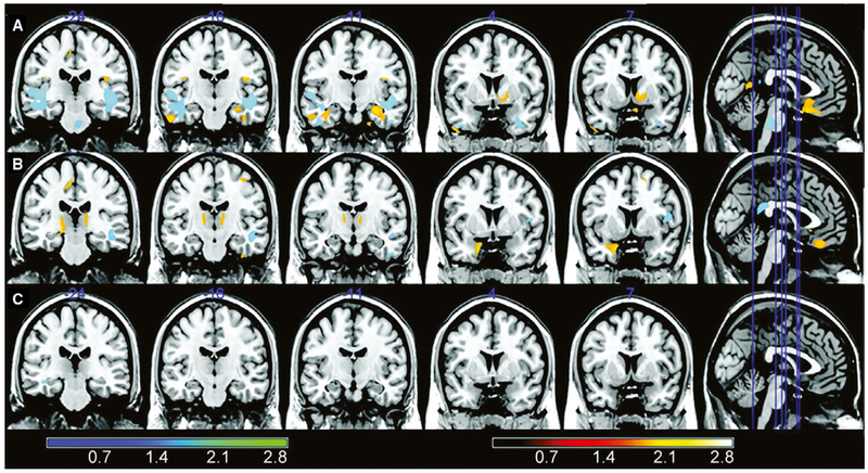
FIGURE 1.
Voxel-based morphometry (VBM) analyses comparing first and second magnetic resonance imaging (MRI) of left and right mesial temporal lobe epilepsy (mTLE) patients and controls. The VBM analysis (paired t test of MRI1 vs MRI2) of patients with left mTLE (row A) demonstrated significant gray matter reduction in the ipsilateral hippocampus, parahippocampal gyrus, and temporal lobes; bilateral frontal regions and cerebellum; and contralateral occipital region, fusiform gyrus, and cingulate. Patients with right mTLE (row B) had a significant reduction in gray matter volume in the ipsilateral uncus, fusiform gyrus, cerebellum, and occipital and frontal regions; bilateral thalamus and frontal region; and contralateral parietal region. A paired t test comparing the first and the second MRI of the control group (row C) showed only a small significant area of volume reduction in the right temporal lobe white matter. The minimum interval between the baseline and follow-up MRI was 7 months (range = 7-85 months, median = 39 months, SD = 27.5 months). Colored bars show z scores. Hot colors indicate gray matter; cold colors indicate white matter. Modified with permission from Coan et al15

