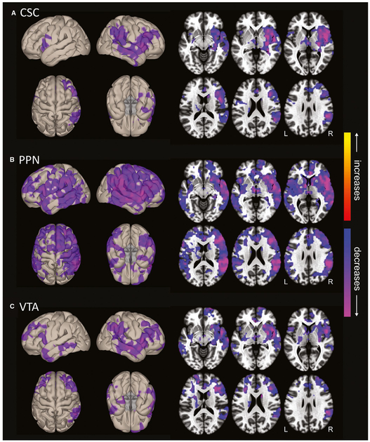
FIGURE 4.
Ascending reticular activating system (ARAS) functional connectivity decreases in mesial temporal lobe epilepsy patients. Surface (left) and axial (right) views are shown of voxelwise functional connectivity differences in patients compared to controls, seeded from cuneiform/subcuneiform nucleus (CSC; A), pedunculopontine nucleus (PPN; B), and ventral tegmental area (VTA; C). Seed-to-voxel functional connectivity (bivariate correlation) maps comparing patients and controls (t test) were generated for each ARAS region using the CONN toolbox 17 (https://www.nitrc.org/projects/conn/). In all three regions, connectivity decreases in patients are observed in insular, frontal, temporal, and parietal neocortical areas, with larger changes on the right side. Decreases appear most prominent in PPN-seeded connectivity maps, also involving subcortical structures such as thalamus and basal ganglia. No increases are seen in patients. Data represent t tests in 26 patients versus 26 matched controls (parametric cluster threshold level P < .01, with false discovery rate correction of multiple comparisons to reduce the false-positive rate). Modified with permission from Englot et al17

