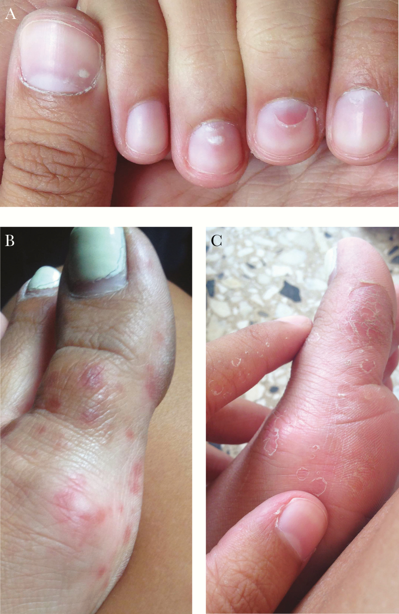
A 17-year-old female with no remarkable medical history completed a 7-week service project in the rural province of Bahoruco in the Dominican Republic. Three weeks after her return, she noticed white discolorations on several of her fingernails (Figure 1A). Several of these lesions began to flake, leading to regional loss of superficial layers of the nail. She also reported an illness that occurred 3 weeks after arrival in the Dominican Republic that consisted of body aches, a fever to 101.3ºF, and lightheadedness. This was followed 3 days later by a rash on the hands, elbows, feet, and ankles consisting of pink to violaceous papules (Figure 1B). The rash also involved the soles and palms. There was exquisite tenderness in the fingertips and soles of the feet lasting ~3 days. Previously present minor skin lesions were affected, becoming inflamed and erythematous. The rash faded gradually over a week, followed by desquamation (Figure 1C). Hyperpigmentation occurred in some areas affected by the rash.
Figure 1.
Nail and skin manifestations of Chikungunya infection. A, Scattered leukonychia and localized onychomadesis. B, Distal pink to violaceous thin papules on feet with occasional dusky center, healing with a collarette of scale. C, Desquamation after acute illness.
The patient had received malaria prophylaxis with atovaquone/proguanil, with which she was compliant. She also received oral typhoid vaccine before departure and was current with all standard immunizations. She denied any contact with animals. She used insect repellent and a mosquito net but did report many mosquito bites. She also bathed daily in the local canals.
Physical exam was unremarkable except for the nail findings and sequelae of the rash as shown. Complete blood count, differential, and chemistries were all within normal limits. Dengue IgG and IgM serologies were negative. Chikungunya IgM was positive at 2.33 (<0.9 negative), and IgG was negative.
The patient had a relatively mild, self-limited illness, possibly due to her youth and lack of other medical problems. The pain was confined to soft tissues rather than the joints, as commonly reported, and there was no subsequent arthritis.
Chikungunya has been associated with several distinctive cutaneous manifestations, including residual hyperpigmentation, particularly of the face, which eventually resolves [1]. Inflammation at sites of previous trauma as in this case has also been previously reported [1]. The nail findings of leukonychia and partial nail loss occurred as late complications in this case and may represent the nail equivalent of cutaneous desquamation. Leukonychia and onychomadesis have been described in 1 case each of Chikungunya infection [2]. Onychomadesis has also been described as a consequence of hand, foot, and mouth disease and varicella [3]. The distance of the leukonychia in this case from the nail bed was consistent with viral damage to the nail matrix occurring at the time of acute infection. This case demonstrates the diversity of presentation and severity of Chikungunya infection and a particularly unusual clinical manifestation involving the nails.
Acknowledgments
Potential conflicts of interest. Both authors: no reported conflicts of interest. Both authors have submitted the ICMJE Form for Disclosure of Potential Conflicts of Interest. Conflicts that the editors consider relevant to the content of the manuscript have been disclosed.
References
- 1. Inamadar AC, Palit A, Sampagavi VV, et al. . Cutaneous manifestations of chikungunya fever: observations made during a recent outbreak in south India. Int J Dermatol 2008; 47:154–9. [DOI] [PubMed] [Google Scholar]
- 2. Riyaz N, Riyaz A, Abdul Latheef EN, et al. ; Rahima Cutaneous manifestations of chikungunya during a recent epidemic in Calicut, North Kerala, South India. Indian J Dermatol Venereol Leprol 2010; 76:671–6. [DOI] [PubMed] [Google Scholar]
- 3. Salgado F, Handler MZ, Schwartz RA. Shedding light on onychomadesis. Cutis 2017; 99:33–6. [PubMed] [Google Scholar]


