Figure 4.
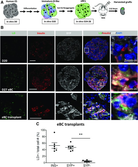
LDs detected in human ES cell–derived eBCs. A: A simplified schematic representing human ES cell differentiation into eBCs and the transplantation of eBCs into NSG mice. The eBCs are produced by FACS isolating the insulin+ synaptophysin+ cells from day 20 (D20), cell reaggregation, and then culturing for 4–8 days. eBCs improve their secretory properties upon transplantation (24). 3 & 9mon, 3 and 9 months. B: Representative images taken of day 20 spheres, day 27 eBCs, and a 9-month eBC graft immunostained with BODIPY (green), insulin (red), SYP (white), and DAPI (blue). LDs are enriched in insulin+ eBC cells but are also detectible in some insulin− cells. The white square illustrates the zoomed-in area. Scale bar = 50 μm. C: LD distribution within the eBC transplant insulin+, synaptophysin+, or synaptophysin− cell populations. n = 6. Error bars indicate SEM. **P < 0.01. INS, insulin; SYP, synaptophysin; Tx, transplant.

