Traumatic brain injury (TBI) is a leading cause of death and disability worldwide. Global assessments estimate that over 27 million cases of TBI occur annually, resulting in over 8 million years lived with disability (GBD 2016 Dementia Collaborators, 2019). Over 30 clinical trials have failed to show efficacy in TBI, and patients are currently left without any promising therapeutic options (Villapol et al., 2015). The pathophysiology of TBI is commonly divided into primary and secondary injuries. Primary injury refers to the parenchymal damage that occurs as an immediate consequence of acute kinetic energy transfer to the brain (i.e., membrane rupture, hemorrhage, axotomy, etc.). Secondary injury encompasses the deleterious molecular and cellular responses that occur in response to the primary injury in the minutes, hours or days following. The search for therapeutics that mitigate the effects of the secondary injury and/or assist endogenous repair processes remains a large focus of TBI research (Umschweif et al., 2014; Villapol et al., 2015; Janatpour et al., 2019).
Beyond its role in blood pressure modulation, the renin-angiotensin system (RAS) is implicated in many pathophysiological processes in the central nervous system (CNS), including those that occur after TBI. Following injury, activation of angiotensin II type I receptor (AT1R) in various cell types can promote inflammation, generate reactive oxygen species, increase glial proliferation, and reduce cerebral blood flow – physiological responses with known ability to damage brain parenchyma (Villapol et al., 2012, 2015). Thus, it stands to reason that blockade or countersignaling of AT1R would reduce damage in the traumatic penumbra. As anticipated, pre-clinical studies have demonstrated efficacy for angiotensin II receptor blockers (ARBs) in reducing pathological sequelae of TBI (Villapol et al., 2012, 2015).
In recent years, various biologically active cleavage products of angiotensinogen have been characterized. Those that are active within the brain include peptides such as angiotensin-(1–7) [Ang-(1–7)], angiotensin IV (Ang IV), and alamandine. Together, these constitute the neuroactive ligands of the extended RAS (eRAS). Importantly, many of these peptides have distinct effects, often in opposition to AT1R action, through signaling via separate receptors (Jackson et al., 2018). They therefore serve as promising targets for TBI pharmacotherapeutics. In this perspective, we will briefly highlight various RAS modulators and select eRAS ligands with potential as neurotherapeutic agents against TBI.
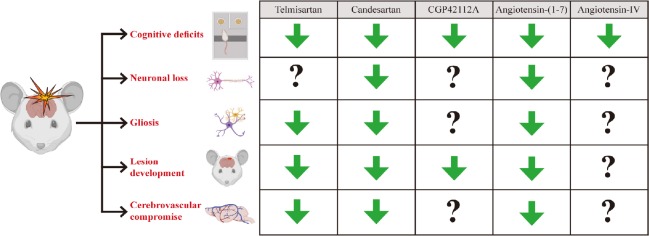
AT1R: The ARBs candesartan and telmisartan improve morphological and functional outcomes in various rodent models of TBI. When administered subcutaneously after injury, they decrease lesion volume, improve cerebral blood flow, decrease inflammation and reduce reactive gliosis at acute and chronic time points following moderate TBI (Figure 1) (Villapol et al., 2012, 2015). Different ARBs also have off-target beneficial effects that may assist in their neuroprotective abilities. In particular, the anti-inflammatory properties of telmisartan are preserved in the absence of AT1R and are likely mediated by AMPK- or PPARγ-related signaling (Xu et al., 2015). However, candesartan and losartan also mediate some actions through acting as PPARγ partial agonists (Villapol et al., 2012, 2015). Thus, in addition to their ability to block deleterious effects of AT1R signaling after insult, ARBs likely combat the molecular sequelae of TBI through multiple pathways. Since both telmisartan and candesartan are approved to treat hypertension in the USA (by the Food and Drug Administration) and are typically well tolerated, they merit consideration for use in TBI clinical trials. As these medications are widely prescribed, it would be interesting to compare TBI outcome in patients on telmisartan/candesartan versus appropriately matched controls in a retrospective study.
Figure 1.
Effects of RAS-modulating ligands on TBI.
Various ligands that act on receptors of the RAS and/or eRAS reduce sequelae of TBI in rodent models. Downward green arrows depict a ligand’s documented ability to reduce a particular sequela of TBI in rodents. Question marks depict unknown associations between a ligand and a particular sequela of TBI in rodents. eRAS: Extended renin-angiotensin system; RAS: renin-angiotensin system; TBI: traumatic brain injury.
Angiotensin II type 2 receptor (AT2R): Ang II signals through a second membrane bound G protein-coupled receptor, known as the AT2R, which is distinct from the AT1R. AT2R is also expressed throughout the brain and vasculature. Possibly due to activation of non-canonical G-protein and β-arrestin pathways, AT2R signaling can counter many downstream effects of AT1R activation (Zhang et al., 2017). Excitingly, both the peptide and small molecule AT2R agonists, respectively known as CGP42112A and Compound-21, can reduce neurological sequelae after ischemic brain insult in rodents (McCarthy et al., 2009; Joseph et al., 2014). However, only CGP42112A has demonstrated therapeutic efficacy in rodent models of TBI (Umschweif et al., 2014). Based on studies showing the neuroprotective potential of Compound-21, we predict that it too will reduce neurological sequalae following TBI in rodent models.
Ang IV: A far less discussed component of the extended RAS is Ang IV and its associated Ang IV receptor (AT4R). Ang IV is formed following enzymatic cleavage of Ang II by two peptidases: aminopeptidase A and aminopeptidase N, and signals through the AT4R, an insulin-regulated aminopeptidase. Though evidence suggests that Ang IV signaling increases transport of glucose transporter type 4 to the cell membrane in neurons, the mechanisms by which it signals remain unclear. Ang IV has many beneficial effects in the CNS, including reducing inflammation, enhancing cerebral blood flow, and increasing neuronal glucose uptake. Ang IV has consequently been recognized as having neuroprotective properties (Jackson et al., 2018). Ang IV’s enhancement of cognitive function is well established in in rodent models of CNS insult. Indeed, intracerebroventricular administration of Ang IV, or stabilized analogues, reverses the spatiotemporal memory deficits associated not only with scopolamine-induced amnesia or ischemic stroke, but with bilateral knife cuts to the brain. Even while utilizing systemic routes of administration, the cognitive benefits of Ang IV appear to be conserved (Wright et al., 1999; Jackson et al., 2018). As Ang IV treatment promotes various beneficial phenotypes within the CNS, it merits formal assessment in more established pre-clinical TBI models.
Ang-(1–7): Ang-(1–7) is produced from the octapeptide Ang II by angiotensin converting enzyme 2, which cleaves the terminal phenylalanine from Ang II’s C-terminus. Despite its similarity to Ang II, Ang-(1–7) mainly signals through a different G protein-coupled receptor known as the Mas receptor (MasR). Ang-(1–7)-induced MasR signaling acts in opposition to Ang II-induced AT1R signaling at various levels, and could therefore have significant beneficial effects in the CNS. Indeed, various pre-clinical studies have demonstrated the ability of Ang-(1–7) to mitigate non-traumatic neurologic sequelae after CNS insult, but most have utilized intracerebroventricular peptide administration to ensure direct access to the brain parenchyma (Jackson et al., 2018). Possibly due to the short half-life of the peptide or assumed poor blood-brain barrier permeability, few have assessed the ability of Ang-(1–7) to reduce neurologic damage when administered subcutaneously. We recently showed that Ang-(1–7), administered subcutaneously hours after insult, reduces both microgliosis and astrogliosis after TBI in rodents (Janatpour et al., 2019). However, the benefits of Ang-(1–7) in TBI span beyond its anti-inflammatory capabilities. Following severe TBI in rodents, subcutaneously-administered Ang-(1–7) also reduces capillary loss, neuronal loss, and brain lesion volume; and attenuates spatiotemporal memory deficits (Figure 1) (Janatpour et al., 2019).
Previous phase I and phase II clinical trials have demonstrated Ang-(1–7) is safe when administered subcutaneously in humans. There remain several ongoing clinical trials investigating both Ang-(1–7)/MasR and angiotensin converting enzyme 2 as therapeutic targets for different clinical indications including cognitive function. A recent trial of subcutaneous administration of Ang-(1–7) as a treatment for cognitive decline in heart failure patients (Sweitzer, 2019) has recently concluded but the results are not yet published. Ang-(1–7) continues to be tolerated with minimal side effects in clinical trials, and pre-clinical studies have demonstrated its neurotherapeutic potential. However, before Ang-(1–7) can be assessed clinically in the setting of acute TBI, we need to determine whether Ang-(1–7) has broad efficacy in different animals and different TBI models. It would also be advantageous to develop biomarkers of target engagement to enhance our understanding of events once in the clinical setting.
Bench to bedside: From what was once understood only as a modulator of blood pressure and fluid homeostasis, the RAS has implications in a wide spectrum of human conditions. Expression of RAS and eRAS components throughout the mammalian body provides many possibilities for targeted therapeutics and creates a promising system to explore for treating several different CNS insults particularly TBI. Manipulation of the RAS and eRAS share similar mechanisms of neuroprotection: direct neuroprotective action on neurons, reduction of inflammation and gliosis, increased cerebral blow flood, and preservation of both cerebrovascular and blood-brain barrier integrity. Each ligand and receptor perturbs the system uniquely, suggesting that each hold promise for TBI therapy. As many of the RAS and eRAS modulators are well-tolerated in humans, pre-clinical evidence in mammals of their therapeutic efficacy against acute neurologic damage narrows the bench to bedside gap in TBI pharmacotherapeutic research, and hopefully brings brain-injured patients one step closer towards better treatment options.
We are grateful to members of the Symes laboratory for their helpful comments and suggestions.
Additional file: Open peer review reports 1 (85.3KB, pdf) and 2 (85.1KB, pdf) .
Footnotes
Copyright license agreement: The Copyright License Agreement has been signed by both authors before publication.
Plagiarism check: Checked twice by iThenticate.
Peer review: Externally peer reviewed.
Open peer reviewers: He-Zuo Lü, Bangbu Medical College, China; Melanie G. Urbanchek, University of Michigan, USA.
P-Reviewers: Lü HZ, Urbanchek MG; C-Editors: Zhao M, Li JY; T-Editor: Jia Y
References
- 1.GBD 2016 Dementia Collaborators. Global, regional, and national burden of traumatic brain injury and spinal cord injury, 1990-2016: a systematic analysis for the Global Burden of Disease Study 2016. Lancet Neurol. 2019;18:88–106. doi: 10.1016/S1474-4422(18)30415-0. [DOI] [PMC free article] [PubMed] [Google Scholar]
- 2.Jackson L, Eldahshan W, Fagan SC, Ergul A. Int J Mol Sci. 2018. Within the brain: The renin angiotensin system. doi: 10.3390/ijms19030876. [DOI] [PMC free article] [PubMed] [Google Scholar]
- 3.Janatpour ZC, Korotcov A, Bosomtwi A, Dardzinski BJ, Symes AJ. Subcutaneous administration of angiotensin-(1-7) improves recovery after traumatic brain injury in mice. J Neurotrauma. 2019 doi: 10.1089/neu.2019.6376. doi: 101089/neu20196376. [DOI] [PubMed] [Google Scholar]
- 4.Joseph JP, Mecca AP, Regenhardt RW, Bennion DM, Rodríguez V, Desland F, Patel NA, Pioquinto DJ, Unger T, Katovich MJ, Steckelings UM, Sumners C. The angiotensin type 2 receptor agonist Compound 21 elicits cerebroprotection in endothelin-1 induced ischemic stroke. Neuropharmacology. 2014;81:134–141. doi: 10.1016/j.neuropharm.2014.01.044. [DOI] [PMC free article] [PubMed] [Google Scholar]
- 5.McCarthy CA, Vinh A, Callaway JK, Widdop RE. Angiotensin AT2 receptor stimulation causes neuroprotection in a conscious rat model of stroke. Stroke. 2009;40:1482–1489. doi: 10.1161/STROKEAHA.108.531509. [DOI] [PubMed] [Google Scholar]
- 6.Sweitzer N. Angiotensin (1-7) Treatment to improve cognitive functioning in heart failure patients. 2019. https://clinicaltrialsgov/ct2/show/NCT03159988 .
- 7.Umschweif G, Liraz-Zaltsman S, Shabashov D, Alexandrovich A, Trembovler V, Horowitz M, Shohami E. Angiotensin receptor type 2 activation induces neuroprotection and neurogenesis after traumatic brain injury. Neurotherapeutics. 2014;11:665–678. doi: 10.1007/s13311-014-0286-x. [DOI] [PMC free article] [PubMed] [Google Scholar]
- 8.Villapol S, Balarezo MG, Affram K, Saavedra JM, Symes AJ. Neurorestoration after traumatic brain injury through angiotensin II receptor blockage. Brain. 2015;138:3299–3315. doi: 10.1093/brain/awv172. [DOI] [PMC free article] [PubMed] [Google Scholar]
- 9.Villapol S, Yaszemski AK, Logan TT, Sánchez-Lemus E, Saavedra JM, Symes AJ. Candesartan, an angiotensin II AT(1)-receptor blocker and PPAR-gamma agonist, reduces lesion volume and improves motor and memory function after traumatic brain injury in mice. Neuropsychopharmacology. 2012;37:2817–2829. doi: 10.1038/npp.2012.152. [DOI] [PMC free article] [PubMed] [Google Scholar]
- 10.Wright JW, Stubley L, Pederson ES, Kramár EA, Hanesworth JM, Harding JW. Contributions of the brain angiotensin IV-AT4 receptor subtype system to spatial learning. J Neurosci. 1999;19:3952–3961. doi: 10.1523/JNEUROSCI.19-10-03952.1999. [DOI] [PMC free article] [PubMed] [Google Scholar]
- 11.Xu Y, Xu Y, Wang Y, Wang Y, He L, Jiang Z, Huang Z, Liao H, Li J, Saavedra JM, Zhang L, Pang T. Telmisartan prevention of LPS-induced microglia activation involves M2 microglia polarization via CaMKKbeta-dependent AMPK activation. Brain Behav Immun. 2015;50:298–313. doi: 10.1016/j.bbi.2015.07.015. [DOI] [PubMed] [Google Scholar]
- 12.Zhang H, Han GW, Batyuk A, Ishchenko A, White KL, Patel N, Sadybekov A, Zamlynny B, Rudd MT, Hollenstein K, Tolstikova A, White TA, Hunter MS, Weierstall U, Liu W, Babaoglu K, Moore EL, Katz RD, Shipman JM, Garcia-Calvo M, et al. Structural basis for selectivity and diversity in angiotensin II receptors. Nature. 2017;544:327–332. doi: 10.1038/nature22035. [DOI] [PMC free article] [PubMed] [Google Scholar]
Associated Data
This section collects any data citations, data availability statements, or supplementary materials included in this article.


