Figure 1.
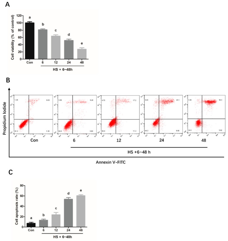
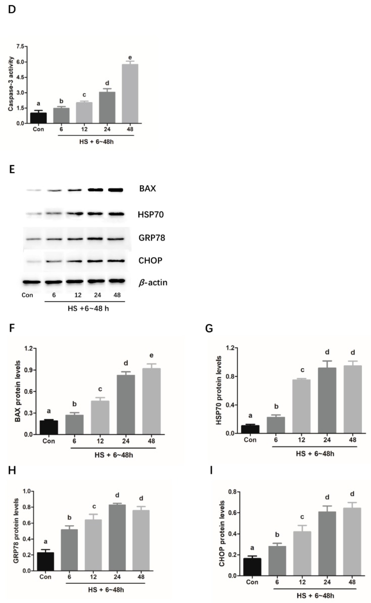
Chronic heat stress induces apoptosis and ER stress in mouse granulosa cells. Cells were cultured at 39 °C for heat stress for different periods of time (0, 6, 12, 24, and 48 h). Then, the treated cells were harvested for performing the CCK-8 assay (A) and flow cytometry-based analysis of apoptosis (B and C). Caspase-3 activity was measured by using a Caspase 3 Activity Assay Kit (D). Western blotting (E) was used to analyze the protein expression levels of cell apoptosis-related BAX (F), heat stress-related marker protein HSP70 (G) and ER stress activation markers, GRP78 (H) and CCAAT/enhancer binding protein homologous protein (CHOP) (I). β-actin was used for normalizing the level of protein expression. The results of data analysis are shown as the bar graphs. The data are presented as mean ± SEM of three independent experiments, and each independent experiment includes three technical replicates. Bars with different lowercase letters are significantly different (p < 0.05).


