Abstract
Background & objectives:
The burden of cardiovascular diseases is high in Kerala, India, and a considerable proportion of these occur in young people. The objective of this study was to estimate the severity of atherosclerosis in autopsies done for accidental and suicidal deaths in victims below 40 yr of age.
Methods:
Coronary arteries from 77 autopsies done for unnatural deaths in a population below 40 yr were graded, and the degree of stenosis, intimal thickness index (ITI) and the intima-media ratio (IMR) were measured.
Results:
There were 65 males and 12 females in the sample. The American Heart Association (AHA) type 3-6 (pathological intimal thickening) was seen in 55.4 per cent [95% confidence interval (CI): 42.5-67.7%] of males and 25 per cent (95% CI: 5.5-57.2%) of females and advanced lesions (type 4-6) in 44.6 per cent (95% CI: 32.3-57.5%) of males and 8.3 per cent (95% CI: 0.2-38.5%) of females. Types 5 or 6 lesions were seen in 32.2 per cent (95% CI: 21.2-45.1%) of males. The mean stenosis was 57.3 per cent in males and 40.6 per cent in females. More than 40 per cent stenosis was seen in 76.6 per cent cases, more than 50 per cent in 54.5 per cent cases and more than 75 per cent stenosis in 14.3 per of the sample. The mean ITI (MIT) was 1.85 and the mean IMR was 4.11. The degree of stenosis, MIT and IMR were significantly associated with male sex, overweight and smoking.
Interpretation & conclusions:
Morphometric data showed that the degree of atherosclerotic narrowing of coronary arteries in young non-diseased population was high. It portends a danger to the community unless preventive measures are taken up.
Keywords: Atherosclerosis, autopsy, coronary artery disease, IMR, ITI, morphometry, young adults
The overall age-adjusted prevalence of definite coronary artery disease (CAD) is 4.8 per cent in men and 2.6 per cent in women in Kerala, India. There was almost a three-fold increase since 19931. In the PROLIFE (Population Registry of Lifestyle Diseases) study conducted in Kerala, the standardized death rates for cardiovascular diseases (CVDs) in the one revenue block were 490 for men and 231 for women per 100,000 person-years2. An earlier hospital based study showed a good proportion of heart attacks in young individuals below 40 yr of age in Kerala3. We undertook this study to record the actual morphometric measurements of the artery in autopsy material of unnatural deaths occurred in a hospital in Kerala during a year.
Material & Methods
The study was conducted in the departments of Pathology and Forensic Medicine, Government TD Medical College, Alappuzha, Kerala, during July 2012 to June 2013. All cases autopsied for deaths due to accidents or suicides in those below 40 yr during the study period were included. Those having previously diagnosed ischemic heart diseases, were excluded. The study protocol was approved by the institutional ethics committee.
Relevant clinical data were obtained from the nearest relative of the deceased or the persons accompanying the body, using an open-ended questionnaire which included history regarding any previous illness in the deceased with special reference to heart disease, cerebrovascular disease, diabetes, hypertension and peripheral vascular disease. The personal habits of the deceased, especially related to smoking and alcohol consumption, were also recorded. General examination during autopsy including height and weight were recorded. Aorta was opened longitudinally, and gross examination of the ascending aorta, arch of the aorta and descending aorta was done. All the three coronaries were examined at 5 mm intervals and sections for histopathological examination were taken from area where there was greatest narrowing. The thickness of the left and right ventricles was measured.
Histology: Sections were stained by haematoxylin and eosin and Verhoeff-Van Gieson (VVG). The atheromatous lesions were graded on a six-point ordinal scale as per the American Heart Association (AHA) guidelines4. Medial changes such as thinning of media, calcification and inflammatory cell infiltration were recorded. The type and amount of inflammatory cells in the adventitia and periadventitial fibrosis were also noted.
Morphometric measurement: To take the morphometric measurements, microscopic images of the VVG-stained slides were captured on an Olympus BX43 microscope (Olympus Corporation, Tokyo, Japan) with camera and morphometric measurements made with cellSens imaging software (Olympus Corporation, Tokyo, Japan). In each section, the following variables were measured (Fig. 1), as detailed in Ruengsakulrach et al5. (i) luminal area (LA); (ii) internal elastic lamina area (IELA) which is the area encompassed by the internal elastic lamina; (iii) external elastic lamina area (EELA) which is the area encompassed by the external elastic lamina; (iv) width of the intima at maximal intimal thickness; and (v) width of the media at maximal intimal thickness. The intimal area was calculated by subtracting the LA from the IELA and medial area by subtracting the IELA from the EELA area.
Fig. 1.

Images from Verhoeff-Van Gieson-stained sections (×40) of coronary were used for morphometry using the cellSens software (Olympus Corporation, Tokyo, Japan). (1) Luminal border, (2) internal elastic lamina, (3) external elastic lamina, (4) intimal thickness, (5) medial thickness. The inner green line marks the luminal outline, the middle blue marks the internal elastic lamina and the outer red marks the external elastic lamina for measurement of areas.
Measures for assessing severity of atherosclerosis in the coronaries included (i) percentage of luminal narrowing=(intimal area/IEL area)×100, (ii) intimal thickness index (ITI)=intimal area/medial area, and (iii) intima-to-media ratio (IMR)=width of intima at maximal intimal thickness/width of media at maximal intima thickness.
Statistical analysis: Data were entered in Open Office spreadsheet and analyzed using Epi Info statistical software (Centers for Disease Control and Prevention, USA). Statistical tests employed were Chi-square test for proportions, t test for means of two independent variables and ANOVA for means of more than two variables. In case of t test and ANOVA, equality of variance assumption was tested by the Bartlett's test and Kruskal-Wallis test applied when the variances were not homogenous.
Results
A total of 77 autopsies were done (65 males, 12 females). The mean age of the sample was 30.3 [95% confidence interval (CI): 28.6-32.0] yr and the median 32 yr. The average body mass index (BMI) was 22.6 kg/m2 (95% CI: 21.8-23.4), with 27.7 per cent of males and 16.7 per cent of females being overweight (BMI more than 25 kg/m2). About 53.8 per cent of males were smokers. Traffic accidents (n=30, 39%), hanging (n=25, 32.5%), poisoning (n=9, 11.7%), drowning (n=6, 7.8%), homicide (n=4, 5.2%), electrocution (n=2, 2.6%) and burns (n=1) were the causes of death in the 77 cases studied.
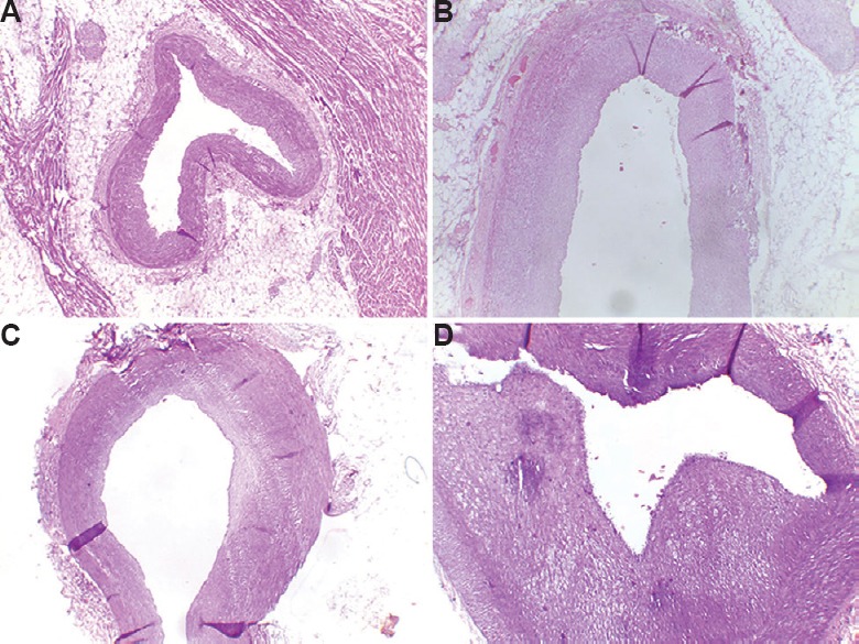
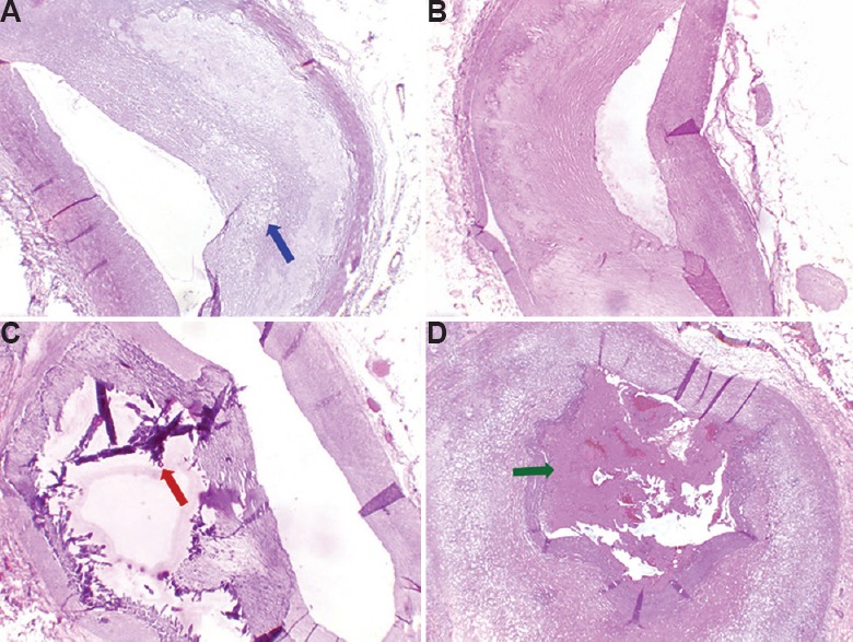
The arteries with the maximum narrowing sampled were left anterior descending in 49.4 per cent (n=38), right coronary in 28.6 per cent (n=22) and circumflex in 22.1 per cent (n=17) of the cases. The grading of the coronary arteries according to the AHA classification is shown in Table I, and the representative sections of some of the AHA types are shown in Figures 2 and 3. Among the 21 cases of type V lesions, nine were type Va and 12 type Vb. The sole type VI case showed a thrombus (type VIc) (Fig. 3).
Table I.
Grading of coronaries in the sample according to American Heart Association classification4 and sex
| Type | Male, n (%) | Female, n (%) | Total, n (%) |
|---|---|---|---|
| Normal | 5 (7.7) | 5 (41.7) | 10 (13.0) |
| I | 18 (27.7) | 3 (25.0) | 21 (27.3) |
| II | 6 (9.2) | 1 (8.3) | 7 (9.1) |
| III | 7 (10.8) | 2 (16.7) | 9 (11.7) |
| IV | 8 (12.3) | 0 (0.0) | 8 (10.4) |
| V | 20 (30.8) | 1 (8.3) | 21 (27.3) |
| VI | 1 (1.5) | 0 (0.0) | 1 (1.3) |
| Total | 65 (100.0) | 12 (100.0) | 77 (100.0) |
Fig. 2.

Representative sections of coronary arteries: Normal and American Heart Association (AHA) type I-III. (A) Normal coronary artery, (B) AHA type I lesion, (C) AHA type II lesion, (D) AHA type III lesion (H and E, ×40).
Fig. 3.

Sections of coronary arteries: American Heart Association (AHA) type IV-VI. (A) AHA type IV lesion, (B) AHA type Va lesion with lipid core (Blue arrow), (C) AHA type Vb lesion with calcification (red arrow), (D) AHA type VI lesion with thrombus (green arrow) (H and E, ×40).
The presence of AHA type III and upward [corresponding to pathological intimal thickening (PIT)] and its relation to different variables is shown in Table II. Almost half (n=39, 50.6%) of the samples had PIT. It was significantly high in those who were overweight. The proportion of those with advanced lesions (AHA 4, 5 and 6) was 39 per cent overall and 44.6 per cent in males. Table III shows mean coronary stenosis, ITI and IMR according to different variables. Table IV shows presence of more than 40 per cent stenosis according to different variables; 76.6 per cent sample showed more than 40 per cent stenosis. It was significantly higher in male sex, smokers, and overweight patients (P<0.05). Age groups showed no relation to PIT (AHA 3-6).
Table II.
Frequency of American Heart Association types III, IV, V, VI (corresponding to pathological intimal thickening) according to different variables
| Variable | n | Per cent of cases (95% CI) | P |
|---|---|---|---|
| Sex | |||
| Female | 3 | 25.0 (5.5-57.2) | 0.0530 |
| Male | 36 | 55.4 (42.5-67.7) | |
| Age group (yr) | |||
| 11-20 | 2 | 25.0 (3.2-65.1) | 0.2320 |
| 21-30 | 14 | 48.3 (29.5-67.5) | |
| 31-40 | 23 | 57.5 (40.9-73.0) | |
| Smoking | |||
| No | 18 | 42.9 (27.7-59.0) | 0.1340 |
| Yes | 21 | 60.0 (42.1-76.1) | |
| Overweight | |||
| No | 23 | 40.4 (27.6-54.2) | 0.0022 |
| Yes | 16 | 80.0 (56.3-94.3) | |
| Whole sample | 39 | 50.7 (39.0-62.2) | - |
CI, confidence interval
Table III.
Mean coronary stenosis, intimal thickness index (ITI) and intima-media ratio (IMR) according to different variables
| Variable | Stenosis Mean (95% CI) | P | ITI Mean (95% CI) | P | IMR Mean (95% CI) | P |
|---|---|---|---|---|---|---|
| Sex | ||||||
| Female | 40.6 (36.2-45.0) | 0.001 | 1.07 (0.75-1.39) | 0.027 | 2.05 (1.12-2.99) | 0.006 |
| Male | 57.3 (52.9-61.7) | 1.95 (1.60-2.31) | 4.48 (3.64-5.33) | |||
| Age group (yr) | ||||||
| 11-20 | 41.3 (31.2-51.2) | 0.085 | 0.95 (0.62-1.28) | 0.071 | 1.40 (0.89-1.92) | 0.004 |
| 21-30 | 56.1 (49.4-62.8) | 1.86 (1.36-2.36) | 4.0 (3.06-4.95) | |||
| 31-40 | 56.4 (50.9-61.9) | 1.96 (1.50-2.41) | 4.72 (3.52-5.92) | |||
| Smoking | ||||||
| No | 48.7 (43.3-54.1) | 0.001 | 1.46 (1.09-1.84) | 0.001 | 2.99 (2.23-3.74) | 0.001 |
| Yes | 61.9 (56.8-67.0) | 2.24 (1.75-2.41) | 5.45 (4.21-6.69) | |||
| Overweight | ||||||
| No | 51.4 (46.8-56.2) | 0.005 | 1.65 (1.28-2.02) | 0.0020 | 3.57 (2.75-4.38) | 0.0051 |
| Yes | 63.9 (57.4-70.4) | 2.28 (1.77-2.79) | 5.64 (4.11-5.92) | |||
| Whole sample | 54.7 (50.7-58.7) | - | 1.85 (1.61-2.09) | - | 4.11 (3.36-4.85) | - |
Table IV.
Presence of coronary artery stenosis ≥40% according to different variables
| Variable | Proportion of those with stenosis ≥40% with (95% CI) | P |
|---|---|---|
| Sex | ||
| Female | 50.0 (21.1-78.9) | 0.027 |
| Male | 81.5 (70.0-90.1) | |
| Age group (yr) | ||
| 11-20 | 50.0 (15.7-84.3) | 0.139 |
| 21-30 | 75.9 (56.5-89.7) | |
| 31-40 | 82.5 (67.2-92.7) | |
| Smoking | ||
| No | 64.3 (48.0-78.5) | 0.005 |
| Yes | 91.4 (76.9-98.2) | |
| Overweight | ||
| No | 70.2 (56.6-81.6) | 0.024 |
| Yes | 95.0 (75.1-99.9) | |
| Whole sample | 76.6 (65.6-85.5) | - |
CI, confidence interval
Discussion
More than 40 per cent stenosis was observed in about two third of sample. Studies done in the middle of the previous century showed that the coronary lesions were fewer and milder among Indians when compared to the developed countries6,7,8. In our sample, significant PIT and type 5+6 lesions were seen in 50.6 and 28.6 per cent samples, respectively. These were relatively high figures for a young population of India. Other autopsy studies from India showed a similar pattern, though the methodologies followed were not well defined, and in the absence of morphometric measurements, these were not helpful for comparisons9,10,11.
Coronary arteries have been studied in American combat casualties during the Korean and Vietnam wars in the 1950s and 1970s, respectively12,13. Other autopsy studies in young populations from the United States also reported the degree of stenosis14,15. A comparison of coronary stenosis from these studies with the current study is given in Table V.
Table V.
Proportion of cases having varying degrees of coronary artery stenosis in various studies (%)
| Study | Population | >40% stenosis (%) | >50% stenosis (%) | >75% stenosis (%) |
|---|---|---|---|---|
| Virmani et al16 | Korean war (US casualties) Mean age 20.5 yr | - | 19 | 6.4 |
| MacNamara et al13 | Vietnam war (US casualties) Mean age 22.1 yr | - | - | 5 |
| Joseph et al14 | Young trauma victims (US) <35 yr | - | 20.7 | 9 |
| McGill et al15 | Young trauma victims (US) 30-34 yr | 19 (men) 8 (women) |
||
| Current study | Accidents, suicide <40 yr | 76.6 | 54.5 | 14.3 |
A study from Spain in 200317 in the 12-35 yr age group dying of external causes found type IV lesions in 34 per cent of men and 0 per cent of women. The prevalence increased with age and was nearly 60 per cent in the 30-35 age group. However, there were no type V or VI lesions in their sample. In our study, 30.8 per cent of men and 8.3 per cent of women had AHA type V lesions. Severity of lesions was found to be related to BMI, male sex and smoking habits in our study. Lipid profiles could not be estimated because of non-availability of samples.
To conclude our study showed AHA types 3-6 PIT in about half of the autopsy samples of young individuals studied. More than 40 per cent stenosis was seen in >75 per cent cases. Urgent preventive measures need to be taken to stop these atherosclerotic changes in the coronaries of young adults.
Acknowledgment
Authors acknowledge the relatives of the deceased for their consent and co-operation.
Footnotes
Financial support & sponsorship: None.
Conflicts of Interest: None.
References
- 1.Krishnan MN, Zachariah G, Venugopal K, Mohanan PP, Harikrishnan S, Sanjay G, et al. Prevalence of coronary artery disease and its risk factors in Kerala, South India: A community-based cross-sectional study. BMC Cardiovasc Disord. 2016;16:12. doi: 10.1186/s12872-016-0189-3. [DOI] [PMC free article] [PubMed] [Google Scholar]
- 2.Soman CR, Kutty VR, Safraj S, Vijayakumar K, Rajamohanan K, Ajayan K. All-cause mortality and cardiovascular mortality in Kerala state of India: Results from a 5-year follow-up of 161,942 rural community dwelling adults. Asia Pac J Public Health. 2011;23:896–903. doi: 10.1177/1010539510365100. [DOI] [PubMed] [Google Scholar]
- 3.Mammi MV, Pavithran K, Abdu Rahiman P, Pisharody R, Sugathan K. Acute myocardial infarction in North Kerala – A 20 year hospital based study. Indian Heart J. 1991;43:93–6. [PubMed] [Google Scholar]
- 4.Stary HC, Chandler AB, Dinsmore RE, Fuster V, Glagov S, Insull W, Jr, et al. A definition of advanced types of atherosclerotic lesions and a histological classification of atherosclerosis. A report from the Committee on Vascular Lesions of the Council on Arteriosclerosis, American Heart Association. Circulation. 1995;92:1355–74. doi: 10.1161/01.cir.92.5.1355. [DOI] [PubMed] [Google Scholar]
- 5.Ruengsakulrach P, Sinclair R, Komeda M, Raman J, Gordon I, Buxton B. Comparative histopathology of radial artery versus internal thoracic artery and risk factors for development of intimal hyperplasia and atherosclerosis. Circulation. 1999;100:II139–44. doi: 10.1161/01.cir.100.suppl_2.ii-139. [DOI] [PubMed] [Google Scholar]
- 6.Gore I, Robertson WB, Hirst AE, Hadley GG, Koseki Y. Geographic Differences in the Severity of Aortic and Coronary Atherosclerosis: The United States, Jamaica, WI, South India, and Japan. Am J Pathol. 1960;36:559–74. [PMC free article] [PubMed] [Google Scholar]
- 7.Wig KL, Malhotra RP, Chitkara NL, Gupta SP. Prevalence of coronary atherosclerosis in northern India. Br Med J. 1962;1:510–3. doi: 10.1136/bmj.1.5277.510. [DOI] [PMC free article] [PubMed] [Google Scholar]
- 8.Mathur KS, Patney NL, Kumar V. Atherosclerosis in India. An autopsy study of the aorta and the coronary, cerebral, renal, and pulmonary arteries. Circulation. 1961;24:68–75. doi: 10.1161/01.cir.24.1.68. [DOI] [PubMed] [Google Scholar]
- 9.Puri N, Gupta PK, Sharma J, Puri D. Prevalence of atherosclerosis in coronary artery and internal thoracic artery and its correlation in North–West Indians. Indian J Thorac Cardiovasc Surg. 2010;26:243–6. [Google Scholar]
- 10.Singh V, Pai MR, Coimbatore RV, Naik R. Coronary atherosclerosis in Mangalore – A random post mortem study. Indian J Pathol Microbiol. 2001;44:265–9. [PubMed] [Google Scholar]
- 11.Singh H, Oberoi SS, Gorea RK, Bal MS. Atherosclerosis in coronaries in Malwa region. J Indian Assoc Forensic Med. 2005;27:236–9. [Google Scholar]
- 12.Virmani R, Robinowitz M, Geer JC, Breslin PP, Beyer JC, McAllister HA. Coronary artery atherosclerosis revisited in Korean war combat casualties. Arch Pathol Lab Med. 1987;111:972–6. [PubMed] [Google Scholar]
- 13.McNamara JJ, Molot MA, Stremple JF, Cutting RT. Coronary artery disease in combat casualties in Vietnam. JAMA. 1971;216:1185–7. [PubMed] [Google Scholar]
- 14.Joseph A, Ackerman D, Talley JD, Johnstone J, Kupersmith J. Manifestations of coronary atherosclerosis in young trauma victims – An autopsy study. J Am Coll Cardiol. 1993;22:459–67. doi: 10.1016/0735-1097(93)90050-b. [DOI] [PubMed] [Google Scholar]
- 15.McGill HC, Jr, McMahan CA, Zieske AW, Tracy RE, Malcom GT, Herderick EE, et al. Association of Coronary Heart Disease Risk Factors with microscopic qualities of coronary atherosclerosis in youth. Circulation. 2000;102:374–9. doi: 10.1161/01.cir.102.4.374. [DOI] [PubMed] [Google Scholar]
- 16.Virmani R, Robinowitz M, Geer JC, Breslin PP, Beyer JC, McAllister HA. Coronary artery atherosclerosis revisited in Korean war combat casualties. Arch Pathol Lab Med. 1987;111:972–6. [PubMed] [Google Scholar]
- 17.Bertomeu A, García-Vidal O, Farré X, Galobart A, Vázquez M, Laguna JC, et al. Preclinical coronary atherosclerosis in a population with low incidence of myocardial infarction: Cross sectional autopsy study. BMJ. 2003;327:591–2. doi: 10.1136/bmj.327.7415.591. [DOI] [PMC free article] [PubMed] [Google Scholar]


