Abstract
Background
Cigarette smoking is associated with an increased risk of developing respiratory diseases and various types of cancer. Early identification of such unfavorable outcomes in patients who smoke is critical for optimizing personalized medical care.
Methods
Here, we perform a comprehensive analysis using Systems Biology tools of publicly available data from a total of 6 transcriptomic studies, which examined different specimens of lung tissue and/or cells of smokers and nonsmokers to identify potential markers associated with lung cancer.
Results
Expression level of 22 genes was capable of classifying smokers from non-smokers. A machine learning algorithm revealed that AKR1B10 was the most informative gene among the 22 differentially expressed genes (DEGs) accounting for the classification of the clinical groups. AKR1B10 expression was higher in smokers compared to non-smokers in datasets examining small and large airway epithelia, but not in the data from a study of sorted alveolar macrophages. Moreover, AKR1B10 expression was relatively higher in lung cancer specimens compared to matched healthy tissue obtained from nonsmoking individuals. Although the overall accuracy of AKR1B10 expression level in distinction between cancer and healthy lung tissue was 76%, with a specificity of 98%, our results indicated that such marker exhibited low sensitivity, hampering its use for cancer screening such specific setting.
Conclusion
The systematic analysis of transcriptomic studies performed here revealed a potential critical link between AKR1B10 expression, smoking and occurrence of lung cancer.
Introduction
Worldwide, cigarette smoking is a life-style habit of approximately 1.1 billion individuals and is associated with more than 6 million deaths annually [1]. The immunological responses in persons chronically exposed to smoke from cigarettes are characterized by protracted secretion of inflammatory factors and by accumulation of several leukocytes in lung tissue and production of pro-fibrotic mediators such as transforming growth factor (TGF)-β [2, 3]. These inflammatory perturbations likely result in increased risk development of tobacco associated morbidity including several types of cancer [4], autoimmune disorders [5], chronic obstructive pulmonary diseases [6] and respiratory infections [7].
The role of tobacco smoking in the induction of disturbances in cell/tissue homeostasis and gene mutations, broadly or specifically associated with several types of tumors, have been investigated. Smoking-related malignancies have been reported to be associated with DNA methylation [8] and mutations in several proto-oncogenes, such as p53, KRAS, BRCA-1, BRCA-2, GPX2, GABP, TCF3, CRX, CYP2A13, CYP2A6, CYP2B6, among others [9–12]. In addition, it has also been reported that components of cigarette smoking modulate immune cell functions, which could lead to loss of T-cell proliferation and antibody responses [13]. Furthermore, chromosomal instability, epigenomic alterations and several mutations have been reportedly associated with lung cancer in particular [14]. Thus, in general, all of these events ultimately culminate with altered gene expression, even though the conversion of carcinogens to DNA adducts is more efficient in some individuals than in others [15]. Therefore, understanding the expression of these genes is important to fully understand the link between smoking exposure and risk of cancer development.
Identification of genetic markers predictive of cancer development is of utmost importance for promoting personalized medicine [16]. Such markers could be implemented as screening strategy for patients who exhibit strong risk factors for cancer, such as cigarette smoking. To identify such potential markers, we performed a systematic analysis of publicly available data from transcriptomic studies performed in lung tissue and/or cells and found that, among most of the studies investigated, increased expression of the gene AKR1B10 was associated with cigarette smoking as well as lung cancer. Development of a point-of-care assay to assess AKR1B10 expression in individuals exposed to cigarette smoking may serve as a relevant tool to identify those with high risk of cancer.
Methods
Ethics statement
There were no patients directly involved in the research. The present study used publicly available gene expression data from previous studies to perform a meta-transcriptome analysis. All information given to the research team were de-identified.
Description of discovery datasets
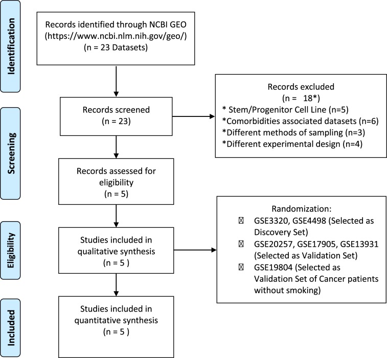
We searched for datasets using the Gene Expression Omnibus (GEO-NCBI -https://www.ncbi.nlm.nih.gov/geo/). The following terms were used: “Smoker”, “Smoking”, “Cigarrete” and “Homo sapiens” and found a total of 23 datasets. We next excluded 18 datasets for a number of reasons listed in Fig 1. Finally, 5 datasets were included. Those datasets were randomized in discovery and validation sets. Similar approach was used to find datasets on lung cancer in nonsmokers (with “nonsmoker”, “nonsmoking”and “cancer” serving as terms used for the GEO search (Fig 1). Thus, using this approach, two previously published microarray datasets were selected to be used as a discovery set (available from the GEO under accession no. GSE4498 [17] and GSE3320 [18]) and 3 have been used as validation set (GSE20257 [19], GSE17905 [20] and GSE13931 [21]). We found other three datasets using gene profiling by array. However, they could not be used for the following reasons: the dataset GSE57048 used mouse cells to measure expression, the GSE124265 used transformed lineage cells and the GSE92662 did not use cigarette-exposed patients. Moreover, there are other datasets by using RNA-seq, however in the present study we have focused on array data only. Due the data distribution differences from each methodology, the direct comparison is difficult. The Dataset GSE4498 [17] was designed with samples of human small airway bronchial epithelium of smokers (n = 10) compared to matched samples from non-smokers (n = 12). The dataset GSE3320 [18] was extracted from samples of human small airway bronchial epithelium to assess gene expression in phenotypically smokers (n = 6) compared to matched non-smokers (n = 5). These included datasets using the same method to collect the samples, by fiberoptic bronchoscopy and brushing. In addition, these studies used a similar transcriptional protocol using the platform Affymetrix Array, making possible to combine both datasets in a discovery set.
Fig 1. PRISMA flow chart of the microarray meta-analysis.
Selection of eligible GEO datasets for systems biology analysis according to PRISMA 2019 flow diagram.
In silico validation
We next performed validation of differentially expressed genes detected in the first phase of the investigation using 3 distinct datasets selected by examination of gene expression by smoking status: (i) GSE20257 was published by Shaykhievet al[19]. In this study, they used samples of small airway epithelium collected from individuals who were smokers (n = 51) and also from those who did not smoke (n = 42) and performed an analysis of microarray assays in theses samples. (ii) GSE17905 was published by Wang et al [20]. The authors used large airways samples collected by bronchoscopy of 31 smokers and 21 non-smoker individuals and also performed a microarray analysis. (iii) GSE13931 was published by Carolan et al [21]. The investigators used alveolar macrophages collected by bronchoalveolar lavage of 30 smokers and 19 non-smokers and performed a microarray analysis. (iv) Finally, GSE19804 was available in a publication from Lu et al [22]. This dataset had information of gene expression (assessed by microarray) of 60 pairs of lung cancer tissue and adjacent normal lung tissue from female patients who were not exposed to cigarette smoking.
The datasets were obtained using the GEOquerry[23] package and raw expression data of 22 samples present on GSE4498 and 11 samples on GSE3320 were normalized and log2 transformed by preprocesscoreR package [24]. Duplicated probes were collapsed by collapserows function in WGCNA package [25] and all common genes to both datasets were kept and used to merge the datasets. The expression data was submitted to a correction procedure of batch effect using an empirical Bayes framework implemented in the COMBAT function available in SVApackage[26].
Statistical analysis
Categorical data were presented as proportions whereas continuous data were plotted as mean and standard deviation (SD). Receiver operator characteristics (ROC) curves were employed to test the accuracy of 22 Genes signature values and AKR1B10 alone to distinguish smokers from those who not a smoker. The differentially expressed genes (DEGs) were identified by applying the absolute ≥1.0log2-fold-change threshold and p-value corrected with FDR adjustment for multiple testing (FDR = 5%), from limmapackage [27]. A volcano plot we used to identify changes in gene expression, the significance versus fold-change on the y and x axes, respectively. We use Venn diagrams to visualize all possible logical relations between all the DEGs between smokers and non-smokers in all datasets evaluated. The modular analysis was performed using the Cemitool package [28]. It is based on Weighted correlation network analysis (WGCNA) and default parameters was employed (Beta Parameter = 7). The module annotation was performed with the Kegg database v6.2 [29] and Gene Set Enrichment Analysis (GSEA) algorithm is available internally in the Cemitoolpackage and the Single sample Gene Set Enrichment analysis (ssGSEA) was performed with GSVA package [30]. The significant and annotated pathways were clustered using Euclidean distance as dissimilarity measure and average linkage for between-cluster separation (hclust function in the stats package in R 3.2.2). All The heatmap was generated in R via the heatmap.2 function from the gplots package, using the “scale = “row” switch to Z-score standardize the rows [31]. PCA was performed in order to compare and visualize the expression values of all genes to estimate the variance of the global gene expression with the function prcomp a native package in R. The decision trees were employed to validate and identify the minimal gene set that correctly classifies the smokers from nonsmokers from the 22-gene signature [32]. To estimate the decision tree models accuracy, we performed a 10-fold cross validation. The partition procedure was applied to avoid bias in the training/test sets sampling. Thus, the training set was used to tune the parameters, learning and building a model. The validation set was used to test the classifier performance. The sensibility and specificity were measured from the confusion matrix and visualized in the receiver–operating characteristic curve (ROC) [32]. Accuracy was evaluated by area under the curve of ROC plot.
Results
Meta-transcriptome signature of smoking
Two expression datasets for smoking were obtained with the accession number of GSE4498 [17] and GSE3320 [18]. Moreover, three datasets have been used as validation set (GSE20257 [19], GSE17905 [20] and GSE13931 [21]. The demographic characteristics of the study participants in each study are described in Table 1.
Table 1. Clinical and demographic characteristics of the study participants included in each dataset evaluated.
| Characteristics | Smoking datasets | p-value | Cancer dataset | ||||
|---|---|---|---|---|---|---|---|
| Discovery datasets | Validation datasets | ||||||
| GSE3320 | GSE4498 | GSE20257 | GSE17905 | GSE13931 | GSE19804 | ||
| Age, mean (SD) | 36.8 (5.6) | 43.0 (6.1) | 43.6 (9.9) | 42.4 (8.6) | 42.0 (7.0) | 0.1549 | 61.2 (10.2) |
| Gender, Male, n (%) | 7 (63.6%) | 17 (77.3%) | 95 (70.3%) | 107 (68.2%) | 73 (75.3%) | 0.6995 | 0 (0.0%) |
| Ethnic, n (%) | 0.9476 | ||||||
| Black | 4 (36.4%) | 11 (50.0%) | 67 (49.7%) | 86 (54.8%) | 56 (57.7%) | 0 (0.0%) | |
| White | 5 (45.5%) | 9 (40.9%) | 44 (32.6% | 46 (29.3%) | 32 (33.0%) | 0 (0.0%) | |
| Hispanic/Latino | 2 (18.2%) | 2 (9.1%) | 21 (15.5%) | 21 (13.4%) | 10 (10.3%) | 0 (0.0%) | |
| Afro-Hispanic | 0 (0.0%) | 0 (0.0%) | 1 (0.7%) | 2 (1.3%) | 0 (0.0%) | 0 (0.0%) | |
| Asian | 0 (0.0%) | 0 (0.0%) | 2 (1.5%) | 2 (1.3%) | 0 (0.0%) | 60 (100.0%) | |
| Smoke status, n (%) | 0.6102 | ||||||
| non-smoker | 5 (45.5%) | 12 (45.5%) | 53 (39.3%) | 67 (42.7%) | 38 (39.2%) | 0 (0.0%) | |
| smoker | 6 (54.5%) | 10 (54.5%) | 59 (43.7%) | 90 (57.3%) | 60 (61.9%) | 0 (0.0%) | |
| COPD, n (%) | 0 (0.0%) | 0 (0.0%) | 23 (17.8%) | 0 (0.0%) | 0 (0.0%) | 0 (0.0%) | |
| Lung Cancer, n (%) | 0 (0.0%) | 0 (0.0%) | 0 (0.0%) | 0 (0.0%) | 0 (0.0%) | 60 (100.0%) | |
COPD: Chronic obstructive pulmonary disease.
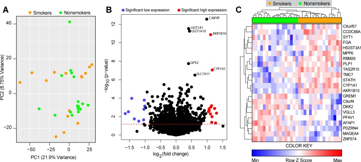
After preprocessing and merging the datasets, we applied a Principal Component Analysis (PCA) algorithm using the expression values of all genes to estimate the variance of the global gene expression. This analysis revealed that the subgroups of smokers and non-smokers could not be separated, and 2 main groups containing both smoker and non-smoker individuals were observed (Fig 2A). To visualize the overall profile of individual gene expression, we used a volcano plot (Fig 2B). This approach indicated presence of a total of 800 statistically significant genes (p<0.05, corrected by Benjamini–Hochberg false discovery rate [FDR]), of which 375 genes were upregulated and 425 genes were downregulated (Fig 2B). Additional analyses identified 22 the differentially expressed genes (DEGs), defined here and genes which exhibited more than ± 1-fold-difference variation (smokers vs. non-smokers) and a significant p-value after FDR adjustment (p<0.05). Such DEGs were inputted in an unsupervised two-way hierarchical clustering analysis. The results demonstrated that when considered together, the 22-gene signature was capable of classifying smokers from non-smokers into completely separate clusters (Fig 2C).Moreover, using canonical discriminant models to further characterize the association of all 22 genes signatures used the validation set GSE20257 [19], GSE17905 [20] and GSE13931 [21]. The area under the ROC curve (AUC) for GSE17905 [20] was 0.86 (P<0.0001), for GSE20257 [19] AUC was 0.86 (P<0.0001) and GSE13931 [21] was 0.60 (P = 0.4236). The ROC curve analyses are summarized in Table 2. This table presents the overall accuracy, sensitivity and specificity of the DEGs identified in human small airway bronchial epithelium (GSE20257 and GSE17905) and in alveolar macrophages (GSE13931).To answer whether sex, age and ethnicity had any influence in the overall gene expression profiles, we performed a Principal Component Analysis (S1 Fig) using both the discovery datasets and the three independent validation sets. Using this approach, we found that such demographic characteristics were not associated with unique expression profiles.
Fig 2. Differentially expressed genes associated with cigarette smoking.
We analyzed publicly available data of 2 datasets of small airways transcriptome (RNAseq). (A) A principal component analysis (PCA) model of 13,516 genes was used to distinguish smokers from nonsmokers. (B) Volcano plot of all genes (smoker vs. nonsmokers). (C) 22 differentially expressed genes (DEGs), defined as p<0.05 after 1%FDR and 1.0-fold change expression, were found and together were able to discriminate the clinical conditions.
Table 2. Detailed information obtained from the ROC curve analysis used in the study.
| Dataset | Tissue | Genes/signature | AUC | 95% CI | p-value | Sensibility | 95% CI | Specificity | 95% CI |
|---|---|---|---|---|---|---|---|---|---|
| GSE17905* | Small and large airway bronchial epithelium | 22-gene | 0.864 | 0.808–0.989 | <0.0001 | 83.87 | 66.2%-94.5% | 95.24 | 76.1%-99.8% |
| GSE20257* | Small airway bronchial epithelium | 22-gene | 0.862 | 0.845–0.973 | <0.0001 | 69.05 | 52.9%-82.3% | 98.04 | 89.5%-99.9% |
| GSE13931* | Alveolar Macrophages | 22-gene | 0.607 | 0.396–0.740 | 0.4236 | 80.00 | 61.4%-92.2% | 42.11 | 20.2%-66.5% |
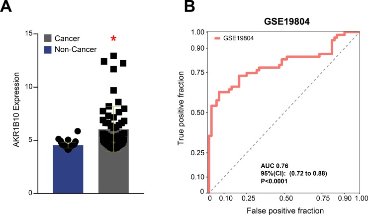
| GSE19804** | Lung tissue | AKR1B10 | 0.760 | 0.720–0.880 | <0.0001 | 35.00 | 23.1%-48.4% | 98.31 | 90.9%-99.9% |
*Smokers versus nonsmokers comparison
**Cancer versus non cancer comparison
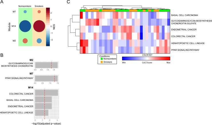
To delineate the gene pathways from which the overall transcription profile in smokers vs. non-smokers were involved, we used the CemiTool package [28]. We detected 3 distinct co-expressed gene modules, annotated in Kyoto Encyclopedia of Genes and Genomes (Kegg) database (Module [M] 2, M7, M14) (Fig 3A). Two modules were enriched in the non-smoking samples compared to smokers, based on the normalized enrichment scores (NES). The first module (M2), found to be enriched in non-smokers was Glycosaminoglycan biosynthesis chondroitin (log10 p = 1.57). A second module (M7) was overrepresented in smokers compared to non-smokers and showed to be enriched in the Peroxisome proliferator-activated receptor (PPAR) signaling pathway (log10 p-value = 1.5). A third module, also more representative in smokers encompassed colorectal cancer (log10 p-value = 3.2) and basal cell carcinoma (log10 p-value = 2.4) (Fig 3B). We next calculated the NES for each top ranked pathway identified per individual study subject and found that, when considered together, such pathways were not able to cluster smokers and non-smokers separately (Fig 3C).
Fig 3. Gene pathway analysis in smokers and nonsmokers.
(A) Co-expressed modules of all genes. Circle sizes are proportional to the normalized enrichment scores (NES). (B) The modules were annotated using Keg package for R. Dashed lines represent significance threshold. (C) Hierarchical cluster analysis (Ward’s method) using the NES scores for each annotated module and calculated for each person was employed test discrimination between smokers and nonsmokers.
A 22-gene signature in lung tissue, but not in alveolar macrophages, including AKR1B10 as the most informative marker, discriminates smoking from nonsmoking individuals
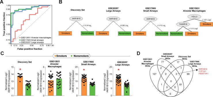
To validate our discoveries, we tested the 22 DEGs identified in our analyses in 3 distinct datasets that compared smokers and non-smokers: (i) GSE20257, that was composed by data from small airways samples, (ii) GSE17905, which compared gene expression from large airways samples and (iii) GSE13931, which used data from alveolar macrophages. Discriminant analyses using Receiver Operating Characteristic (ROC) curves were able to reveal high accuracy of such gene signature to distinguish smokers from nonsmokers in the 2 datasets that large and small airway samples (GSE20257 Area under the curve [AUC]: 0.862, p<0.0001; GSE17905 AUC: 0.864, p<0.0001). The same approach indicated that when a dataset from alveolar macrophages was considered, the 22-gene signature was not able to distinguish the study groups (GSE13931AUC: 0.607, p = 0.423) (Fig 4A). We next employed a machine-learning approach using decision trees to identify which markers from the 22-gene signature would exhibit more robust discrimination power in each dataset evaluated. Of note, the gene AKR1B10 was the most informative gene in the discovery set and also in the 2 distinct datasets that used large or small airway tissue (Fig 3B). In the dataset that used gene expression values form alveolar macrophages, AKR1B10 was not shown to be relevant in discrimination, and a combination of 2 other genes (VGLL3 and TAS2R10) accounted for the differences between smokers and non-smokers (Fig 4B). AKR1B10 expression was higher in smokers compared to non-smokers in all datasets evaluated, except again in the GSE13931, which used data on alveolar macrophages (Fig 4C). Furthermore, we plotted Venn diagrams of all the DEGs between smokers and non-smokers in each dataset to verify overlaps. We confirmed that AKR1B10 was a DEG commonly shown in the discovery set as well as in the databanks which used airway tissue samples, but not in the alveolar macrophage dataset (Fig 4D). The 2 other DEGs found in smokers were CYP1A1 and HS3ST3A1 (Fig 4D). CYP1A1 encodes a protein that localizes at the endoplasmic reticulum and its expression is induced by polycyclic aromatic hydrocarbons, some of which are found in cigarette smoke [33]. HS3ST3A1is a member of the heparan sulfate biosynthetic enzyme family [34].
Fig 4. Defining the molecular signatures of smoking.
(A) Data on the 22 DEGs found in our discovery analyses were used to validate discrimination between smokers and nonsmokers in 3 different previously published datasets. (B) Machine-learning decision trees were built for each dataset to describe the most relevant genes driving discrimination. Of note, the gene AKR1B10 was found to be the main discriminator in 3 out of the 4 datasets examined. (C) Scatter plots of the AKR1B10 gene expression in the 4 datasets. (D) Venn diagram of the DEGs in each dataset shows AKR1B10 in the intersection of 3 datasets extracted from lung tissue specimens but not included among DEGs from alveolar macrophages. *p<0.05 (Student’s t-test).
Testing AKR1B10 as a potential biomarker of lung cancer in patients who do not smoke
The results described above demonstrate that higher AKR1B10 expression hallmarks tissue airways from smokers. Smoking is a well-established risk factor for lung cancer [9]. We next tested whether AKR1B10 gene expression could also be useful to inform presence of cancer in the absence of exposure to smoking. We downloaded the dataset GSE19804, which included tissue samples from non-small cell lung cancer as well as ipsilateral healthy lung tissue obtained from patients who did not present history of cigarette smoking. The AKR1B10 gene expression was substantially higher in the specimen collected from the tumor compared to the healthy lung tissue in the same patients (Fig 5A). ROC curve analysis indicated that AKR1B10 gene expression value was able to correctly identify non-cancer tissue (AUC 0.76, P<0.0001), with relatively high specificity (98.31%). Nevertheless, the results demonstrated low sensitivity (35%), which limits the use of such biomarker for screening in the clinical setting (Fig 5B and Table 2).
Fig 5. In nonsmokers, higher AKR1B10 expression is detected in lung cancer.
(A) We analyzed AKR1B10 gene expression values in a published dataset of neoplastic lung tissue microarray in nonsmoking individuals who were diagnosed with lung cancer and compared to ipsilateral healthy lung tissue specimens (controls.) Scatter plots of AKR1B10 gene expression in the groups. *p<0.05 (Student’s t-test). (B) Receiver Operator Characteristics (ROC) indicated a high accuracy to discriminate cancer tissue from controls.
Discussion
In the present study, we examined a number of publicly available transcriptome data to identify a 22-gene signature that could distinguish lung tissue specimens from smokers vs. non-smoking individuals. The most relevant finding of the initial part of analyses using the 22-gene signature was the AKR1B10 expression level was the most informative in such discrimination among 3 different datasets obtained from lung tissue, but not in the transcriptome data originated from alveolar macrophages. Moreover, ROC curve analysis indicated that AKR1B10 gene expression level exhibited high specificity to but low sensitivity to identify neoplastic from healthy lung tissue in persons not exposed to cigarette smoking. Such analysis however revealed that the overall accuracy is below 80%, and thus not an ideal biomarker for diagnostic purposes. Nevertheless, these findings are important because they have identified AKR1B10 as a biomarker which expression is triggered by cigarette smoking and can be simultaneously observed in lung cancer specimens. It is possible that such gene may be involved in carcinogenesis associated with cigarette smoking. In fact, among the multiple carcinogens from cigarette smoke, the nitrosamine 4-(methylnitrosamino)-1-(3-pyridyl)-1-butanone (NNK) is described to play a critical role in lung carcinogenesis [35]. Carbonyl reduction takes place in both microsomal and cytosolic fractions from different human tissues such as lung and liver [36]. Within these subcellular fractions, several enzymes have been described to mediate NNK reduction, including the protein encoded by AKR1B10, which is from the aldo-keto reductase superfamily (AKR) [37]. Our findings suggest an association between AKR1B10 and smoking, however, the direct relationship with occurrence of lung cancer was not completely validated here. Moreover, if validated in other settings, this gene could be suitable to be used as rule-out test in which non-smoking individuals presenting low AKR1B10 expression would have low risk of having lung cancer. Additional studies are warranted to directly test this hypothesis.
The gene AKR1B10 found differentially expressed in lung tissue from smokers vs. non-smokers has been previously described in experimental studies to play an important role in the pathophysiology of lung cancer [38]. AKR1B10 is a regulator of the synthesis of fatty acid and participates in the metabolic pathway of lipids and isoprenoids [39]. In addition, the protein encoded by AKR1B10 exhibits a high retinaldehyde reductase activity [40]. Importantly, AKR1B10 can metabolize specific substrates, such as aldo-ketoreductases; farnesal, geranylgeranil, retinal and carbonyls [41]. Such activity is associated with promotion of carcinogenesis [42]. AKR1B10 has also been shown to promote cancer cell survival by 2 distinct studies [43, 44]. These previous investigations revealed that knocking down AKR1B10 expression induces cancer cell apoptosis and inhibited cancer cell proliferation, suggesting AKR1B10 could serve as a potential therapeutic target.
Aside from being associated with lung carcinogenesis, AKR1B10 expression has also been linked to the development of several additional types of cancers. In hepatocellular carcinoma (HCC), AKR1B10 expression is found upregulated, and experimental deletion of such gene inhibited the proliferation of HCC cells tumor growth in a xenograft mice model [45]. In HCT-8, a human colon adenocarcinoma cell line, and NCI-H460, a human lung carcinoma cell line, AKR1B10 gene deletion has been shown to induce cell apoptosis and mitochondrial degeneration, leading to oxidative stress [43]. Furthermore, higher AKR1B10 expression has been observed in squamous cell lung carcinoma (SCC) associated with smoking [46]. Finally, our findings indicate that AKR1B10 is overexpressed in lungs of healthy people who smoke but had no cancer as well as in lung carcinoma from non-smokers. These observations argue that cigarette smoking already modifies the microenvironment of the lung epithelium probably creating a favorable scenario for carcinogenesis. This idea corroborates with previously published studies which demonstrated that smoking per se mediates upregulation of AKR1B10 expression in the airway epithelia of healthy smokers with no evidence of lung cancer [47]. Thus, there is strong evidence to suggest that cigarette smoking-induced upregulation of AKR1B10 may represent an initial critical step in the cascade of events leading to lung cancer.
In addition to AKR1B10, our analysis revealed that 2 additional genes, CYP1A1 and HS3ST3A1, overlapped in the datasets as DEGs capable of discriminating smokers from nonsmokers. Of note, CYP1A1 has also been described to induce carcinogenesis, by promoting CYP‐catalyzed epoxidation reactions, resulting in the formation of reactive metabolites that can cause DNA [48, 49]. Moreover, CYP1A1 polymorphisms in smokers increase susceptibility to stomach cancer [50]. Furthermore, HS3ST3A1gene encodes the enzyme 3-O-sulfotransferase, which catalyzes the biosynthesis of a specific subtype of heparan sulfate (HS), 3-O-sulfated heparan sulfate, which is found to be upregulated in human lung cancer specimens and to contribute to its elevated metastatic potential [34]. Thus, the 3 genes found commonly differentiate regulated in individuals exposed to cigarette smoking are all known to favor development of cancer and could be used as an early biomarker of disease progression in high risk populations, but future studies specifically designed to test this hypothesis are necessary.
Our study has several strengths such as the large number of samples evaluated, the use of discovery and validation datasets using different lung tissue/cellular types and different clinical conditions. An important limitation was the low number of studies included, which was dependent on publicly available datasets. In addition, we have not performed validation in experimental systems. Regardless, by performing a systematic analysis of publicly available data from transcriptomic studies of lung tissue and cells, our study provides strong evidence to support a potential role of AKR1B10 in smoking-associated lung cancer.
Supporting information
A principal component analysis (PCA) was employed to test whether the sex, ethnicity and age could cluster patients in the two discovery datasets (GSE4498 [17] and GSE3320 [18]) and in the three validation sets separately (GSE20257 [19], GSE17905 [20] and GSE13931 [21]).
(TIF)
Acknowledgments
We thank Fundação Oswaldo Cruz for the support. Mr. Olival Rocha, Mr. Jose Lima, Mr. Luiz Matos, Mr. Getúlio Pacheco, Mr. Humberto and Mr. Edvan Santana (Universidade Salvador) for the technical support. KFF thanks Alana Alves Farias, Fernanda Freitas Lemos Lopes and Maísa Almeida Silva for the inspiration.
Data Availability
We accessed the Gene Expression Omnibus (GEO-NCBI -https://www.ncbi.nlm.nih.gov/geo/) and have looked for datasets with tobacco smoking information in human biopsies or tissues, without diagnosis of other comorbidities. Five datasets have been found in GEO: GSE4498, GSE3320, GSE20257, GSE17905, GSE13931.
Funding Statement
This study was supported by Universidade Salvador, the Intramural Program of Fundação Oswaldo Cruz (FIOCRUZ), Fundação José Silveira and by the Brazilian National Council for Scientific and Technological Development (CNPq). K.F.F. received a fellowship from the Programa Nacional de Pós-Doutorado, Coordenação de Aperfeiçoamento de Pessoal de Nível Superior (CAPES) (Finance Code 001). The work of B.B.A. was supported by grants from the NIH (U01AI115940, R01AI069923-08, R01AI20790-02). B.B.A. and A.T.L.Q. are senior investigators from CNPq. J. M. C.-A. was supported by the Organization of American States - Partnerships Program for Education and Training (OAS-PAEC) and his study was financed in part by the Coordenação de Aperfeiçoamento de Pessoal de Nível Superior - Brasil (CAPES) - Finance Code 001. M.B.A. received PhD fellowship from Fundação de Amparo à Pesquisa da Bahia (FAPESB) and FIOCRUZ. L.A.B.C. was supported by a research fellowship from CNPq. The funders had no role in study design, data collection and analysis, decision to publish, or preparation of the manuscript.
References
- 1.WHO. World Health Organization global report on trends in prevalence of tobacco smoking. Publications of the World Health Organization; 2015. [Google Scholar]
- 2.Barnes PJ. Inflammatory mechanisms in patients with chronic obstructive pulmonary disease. J Allergy Clin Immunol. 2016;138(1):16–27. Epub 2016/07/05. 10.1016/j.jaci.2016.05.011 . [DOI] [PubMed] [Google Scholar]
- 3.Sohal SS, Ward C, Danial W, Wood-Baker R, Walters EH. Recent advances in understanding inflammation and remodeling in the airways in chronic obstructive pulmonary disease. Expert Rev Respir Med. 2013;7(3):275–88. Epub 2013/06/06. 10.1586/ers.13.26 . [DOI] [PubMed] [Google Scholar]
- 4.Ranjit S, Kumar S. Recent advances in cancer outcomes in HIV-positive smokers. F1000Res. 2018;7 Epub 2018/06/28. 10.12688/f1000research.12068.1 [DOI] [PMC free article] [PubMed] [Google Scholar]
- 5.Perricone C, Versini M, Ben-Ami D, Gertel S, Watad A, Segel MJ, et al. Smoke and autoimmunity: The fire behind the disease. Autoimmun Rev. 2016;15(4):354–74. Epub 2016/01/17. 10.1016/j.autrev.2016.01.001 . [DOI] [PubMed] [Google Scholar]
- 6.Cockcroft DW. Environmental Causes of Asthma. Semin Respir Crit Care Med. 2018;39(1):12–8. Epub 2018/02/11. 10.1055/s-0037-1606219 . [DOI] [PubMed] [Google Scholar]
- 7.Feldman C, Anderson R. Cigarette smoking and mechanisms of susceptibility to infections of the respiratory tract and other organ systems. J Infect. 2013;67(3):169–84. Epub 2013/05/28. 10.1016/j.jinf.2013.05.004 . [DOI] [PubMed] [Google Scholar]
- 8.Lee KW, Pausova Z. Cigarette smoking and DNA methylation. Front Genet. 2013;4:132 Epub 2013/07/25. 10.3389/fgene.2013.00132 [DOI] [PMC free article] [PubMed] [Google Scholar]
- 9.Gibbons DL, Byers LA, Kurie JM. Smoking, p53 mutation, and lung cancer. Mol Cancer Res. 2014;12(1):3–13. Epub 2014/01/21. 10.1158/1541-7786.MCR-13-0539 [DOI] [PMC free article] [PubMed] [Google Scholar]
- 10.Blackford A, Parmigiani G, Kensler TW, Wolfgang C, Jones S, Zhang X, et al. Genetic mutations associated with cigarette smoking in pancreatic cancer. Cancer Res. 2009;69(8):3681–8. Epub 2009/04/09. 10.1158/0008-5472.CAN-09-0015 [DOI] [PMC free article] [PubMed] [Google Scholar]
- 11.Hecht SS. Progress and challenges in selected areas of tobacco carcinogenesis. Chem Res Toxicol. 2008;21(1):160–71. Epub 2007/12/07. 10.1021/tx7002068 [DOI] [PMC free article] [PubMed] [Google Scholar]
- 12.Jin Y, Xu P, Liu X, Zhang C, Tan C, Chen C, et al. Cigarette Smoking, BPDE-DNA Adducts, and Aberrant Promoter Methylations of Tumor Suppressor Genes (TSGs) in NSCLC from Chinese Population. Cancer Invest. 2016;34(4):173–80. Epub 2016/04/05. 10.3109/07357907.2016.1156689 . [DOI] [PubMed] [Google Scholar]
- 13.Sopori M. Effects of cigarette smoke on the immune system. Nat Rev Immunol. 2002;2(5):372–7. Epub 2002/05/30. 10.1038/nri803 . [DOI] [PubMed] [Google Scholar]
- 14.Tan Q, Wang G, Huang J, Ding Z, Luo Q, Mok T, et al. Epigenomic analysis of lung adenocarcinoma reveals novel DNA methylation patterns associated with smoking. Onco Targets Ther. 2013;6:1471–9. Epub 2013/11/10. 10.2147/OTT.S51041 [DOI] [PMC free article] [PubMed] [Google Scholar]
- 15.Peluso M, Munnia A, Piro S, Armillis A, Ceppi M, Matullo G, et al. Smoking, DNA adducts and number of risk DNA repair alleles in lung cancer cases, in subjects with benign lung diseases and in controls. J Nucleic Acids. 2010;2010:386798 Epub 2010/10/27. 10.4061/2010/386798 [DOI] [PMC free article] [PubMed] [Google Scholar]
- 16.Hamburg MA, Collins FS. The path to personalized medicine. N Engl J Med. 2010;363(4):301–4. Epub 2010/06/17. 10.1056/NEJMp1006304 . [DOI] [PubMed] [Google Scholar]
- 17.Tilley AE, Harvey BG, Heguy A, Hackett NR, Wang R, O'Connor TP, et al. Down-regulation of the notch pathway in human airway epithelium in association with smoking and chronic obstructive pulmonary disease. Am J Respir Crit Care Med. 2009;179(6):457–66. Epub 2008/12/25. 10.1164/rccm.200705-795OC [DOI] [PMC free article] [PubMed] [Google Scholar]
- 18.Harvey BG, Heguy A, Leopold PL, Carolan BJ, Ferris B, Crystal RG. Modification of gene expression of the small airway epithelium in response to cigarette smoking. J Mol Med (Berl). 2007;85(1):39–53. Epub 2006/11/23. 10.1007/s00109-006-0103-z . [DOI] [PubMed] [Google Scholar]
- 19.Shaykhiev R, Otaki F, Bonsu P, Dang DT, Teater M, Strulovici-Barel Y, et al. Cigarette smoking reprograms apical junctional complex molecular architecture in the human airway epithelium in vivo. Cell Mol Life Sci. 2011;68(5):877–92. Epub 2010/09/08. 10.1007/s00018-010-0500-x [DOI] [PMC free article] [PubMed] [Google Scholar]
- 20.Wang G, Wang R, Ferris B, Salit J, Strulovici-Barel Y, Hackett NR, et al. Smoking-mediated up-regulation of GAD67 expression in the human airway epithelium. Respir Res. 2010;11:150 Epub 2010/11/03. 10.1186/1465-9921-11-150 ; PubMed Central PMCID: PMC2988726. [DOI] [PMC free article] [PubMed] [Google Scholar]
- 21.Carolan BJ, Harvey BG, Hackett NR, O'Connor TP, Cassano PA, Crystal RG. Disparate oxidant gene expression of airway epithelium compared to alveolar macrophages in smokers. Respir Res. 2009;10:111 Epub 2009/11/19. 10.1186/1465-9921-10-111 [DOI] [PMC free article] [PubMed] [Google Scholar]
- 22.Lu TP, Tsai MH, Lee JM, Hsu CP, Chen PC, Lin CW, et al. Identification of a novel biomarker, SEMA5A, for non-small cell lung carcinoma in nonsmoking women. Cancer Epidemiol Biomarkers Prev. 2010;19(10):2590–7. Epub 2010/08/31. 10.1158/1055-9965.EPI-10-0332 . [DOI] [PubMed] [Google Scholar]
- 23.Davis S, Meltzer PS. GEOquery: a bridge between the Gene Expression Omnibus (GEO) and BioConductor. Bioinformatics. 2007;23(14):1846–7. Epub 2007/05/15. 10.1093/bioinformatics/btm254 . [DOI] [PubMed] [Google Scholar]
- 24.Bolstad B. preprocessCore: A Collection of Pre-Processing Functions. R package. 2018.
- 25.Langfelder P, Horvath S. WGCNA: an R package for weighted correlation network analysis. BMC Bioinformatics. 2008;9:559 Epub 2008/12/31. 10.1186/1471-2105-9-559 [DOI] [PMC free article] [PubMed] [Google Scholar]
- 26.Johnson WE, Li C, Rabinovic A. Adjusting batch effects in microarray expression data using empirical Bayes methods. Biostatistics. 2007;8(1):118–27. Epub 2006/04/25. 10.1093/biostatistics/kxj037 . [DOI] [PubMed] [Google Scholar]
- 27.Ritchie ME, Phipson B, Wu D, Hu Y, Law CW, Shi W, et al. limma powers differential expression analyses for RNA-sequencing and microarray studies. Nucleic Acids Res. 2015;43(7):e47 Epub 2015/01/22. 10.1093/nar/gkv007 [DOI] [PMC free article] [PubMed] [Google Scholar]
- 28.Russo PST, Ferreira GR, Cardozo LE, Burger MC, Arias-Carrasco R, Maruyama SR, et al. CEMiTool: a Bioconductor package for performing comprehensive modular co-expression analyses. BMC Bioinformatics. 2018;19(1):56 Epub 2018/02/21. 10.1186/s12859-018-2053-1 [DOI] [PMC free article] [PubMed] [Google Scholar]
- 29.Kanehisa M, Goto S. KEGG: kyoto encyclopedia of genes and genomes. Nucleic Acids Res. 2000;28(1):27–30. Epub 1999/12/11. 10.1093/nar/28.1.27 [DOI] [PMC free article] [PubMed] [Google Scholar]
- 30.Hanzelmann S, Castelo R, Guinney J. GSVA: gene set variation analysis for microarray and RNA-seq data. BMC Bioinformatics. 2013;14:7 Epub 2013/01/18. 10.1186/1471-2105-14-7 [DOI] [PMC free article] [PubMed] [Google Scholar]
- 31.Wickham H. ggplot2 Elegant Graphics for Data Analysis: Springer-Verlag; 2016. 260 p. [Google Scholar]
- 32.Fukutani KF, Kasprzykowski JI, Paschoal AR, Gomes MS, Barral A, de Oliveira CI, et al. Meta-Analysis of Aedes aegypti Expression Datasets: Comparing Virus Infection and Blood-Fed Transcriptomes to Identify Markers of Virus Presence. Front BioengBiotechnol. 2017;5:84 Epub 2018/01/30. 10.3389/fbioe.2017.00084 [DOI] [PMC free article] [PubMed] [Google Scholar]
- 33.Pavanello S, Clonfero E. Biological indicators of genotoxic risk and metabolic polymorphisms. Mutat Res. 2000;463(3):285–308. Epub 2000/10/06. 10.1016/s1383-5742(00)00051-x . [DOI] [PubMed] [Google Scholar]
- 34.Nakano T, Shimizu K, Kawashima O, Kamiyoshihara M, Kakegawa S, Sugano M, et al. Establishment of a human lung cancer cell line with high metastatic potential to multiple organs: gene expression associated with metastatic potential in human lung cancer. Oncol Rep. 2012;28(5):1727–35. Epub 2012/08/28. 10.3892/or.2012.1972 . [DOI] [PubMed] [Google Scholar]
- 35.Akopyan G, Bonavida B. Understanding tobacco smoke carcinogen NNK and lung tumorigenesis. Int J Oncol. 2006;29(4):745–52. Epub 2006/09/12. . [PubMed] [Google Scholar]
- 36.Maser E, Stinner B, Atalla A. Carbonyl reduction of 4-(methylnitrosamino)-1-(3-pyridyl)-1-butanone (NNK) by cytosolic enzymes in human liver and lung. Cancer Lett. 2000;148(2):135–44. Epub 2000/03/01. 10.1016/s0304-3835(99)00323-7 . [DOI] [PubMed] [Google Scholar]
- 37.Stapelfeld C, Neumann KT, Maser E. Different inhibitory potential of sex hormones on NNK detoxification in vitro: A possible explanation for gender-specific lung cancer risk. Cancer Lett. 2017;405:120–6. Epub 2017/07/27. 10.1016/j.canlet.2017.07.016 . [DOI] [PubMed] [Google Scholar]
- 38.Kang MW, Lee ES, Yoon SY, Jo J, Lee J, Kim HK, et al. AKR1B10 is associated with smoking and smoking-related non-small-cell lung cancer. J Int Med Res. 2011;39(1):78–85. Epub 2011/06/16. 10.1177/147323001103900110 . [DOI] [PubMed] [Google Scholar]
- 39.Ma J, Yan R, Zu X, Cheng JM, Rao K, Liao DF, et al. Aldo-keto reductase family 1 B10 affects fatty acid synthesis by regulating the stability of acetyl-CoA carboxylase-alpha in breast cancer cells. J BiolChem. 2008;283(6):3418–23. Epub 2007/12/07. 10.1074/jbc.M707650200 . [DOI] [PubMed] [Google Scholar]
- 40.Gallego O, Ruiz FX, Ardevol A, Dominguez M, Alvarez R, de Lera AR, et al. Structural basis for the high all-trans-retinaldehyde reductase activity of the tumor marker AKR1B10. Proc Natl Acad Sci U S A. 2007;104(52):20764–9. Epub 2007/12/19. 10.1073/pnas.0705659105 [DOI] [PMC free article] [PubMed] [Google Scholar]
- 41.Martin HJ, Maser E. Role of human aldo-keto-reductase AKR1B10 in the protection against toxic aldehydes. Chem Biol Interact. 2009;178(1–3):145–50. Epub 2008/11/18. 10.1016/j.cbi.2008.10.021 . [DOI] [PubMed] [Google Scholar]
- 42.Berndt N, Hamilton AD, Sebti SM. Targeting protein prenylation for cancer therapy. Nat Rev Cancer. 2011;11(11):775–91. Epub 2011/10/25. 10.1038/nrc3151 [DOI] [PMC free article] [PubMed] [Google Scholar]
- 43.Wang C, Yan R, Luo D, Watabe K, Liao DF, Cao D. Aldo-keto reductase family 1 member B10 promotes cell survival by regulating lipid synthesis and eliminating carbonyls. J Biol Chem. 2009;284(39):26742–8. Epub 2009/08/01. 10.1074/jbc.M109.022897 [DOI] [PMC free article] [PubMed] [Google Scholar]
- 44.Chung YT, Matkowskyj KA, Li H, Bai H, Zhang W, Tsao MS, et al. Overexpression and oncogenic function of aldo-keto reductase family 1B10 (AKR1B10) in pancreatic carcinoma. Mod Pathol. 2012;25(5):758–66. Epub 2012/01/10. 10.1038/modpathol.2011.191 [DOI] [PMC free article] [PubMed] [Google Scholar]
- 45.Satow R, Shitashige M, Kanai Y, Takeshita F, Ojima H, Jigami T, et al. Combined functional genome survey of therapeutic targets for hepatocellular carcinoma. Clin Cancer Res. 2010;16(9):2518–28. Epub 2010/04/15. 10.1158/1078-0432.CCR-09-2214 . [DOI] [PubMed] [Google Scholar]
- 46.Fukumoto S, Yamauchi N, Moriguchi H, Hippo Y, Watanabe A, Shibahara J, et al. Overexpression of the aldo-keto reductase family protein AKR1B10 is highly correlated with smokers' non-small cell lung carcinomas. Clin Cancer Res. 2005;11(5):1776–85. Epub 2005/03/10. 10.1158/1078-0432.CCR-04-1238 . [DOI] [PubMed] [Google Scholar]
- 47.Wang R, Wang G, Ricard MJ, Ferris B, Strulovici-Barel Y, Salit J, et al. Smoking-induced upregulation of AKR1B10 expression in the airway epithelium of healthy individuals. Chest. 2010;138(6):1402–10. Epub 2010/08/14. 10.1378/chest.09-2634 [DOI] [PMC free article] [PubMed] [Google Scholar]
- 48.Badal S, Delgoda R. Role of the modulation of CYP1A1 expression and activity in chemoprevention. J Appl Toxicol. 2014;34(7):743–53. Epub 2014/02/18. 10.1002/jat.2968 . [DOI] [PubMed] [Google Scholar]
- 49.Buterin T, Hess MT, Luneva N, Geacintov NE, Amin S, Kroth H, et al. Unrepaired fjord region polycyclic aromatic hydrocarbon-DNA adducts in ras codon 61 mutational hot spots. Cancer Res. 2000;60(7):1849–56. Epub 2000/04/15. . [PubMed] [Google Scholar]
- 50.Li H, Chen XL, Li HQ. Polymorphism of CYPIA1 and GSTM1 genes associated with susceptibility of gastric cancer in Shandong Province of China. World J Gastroenterol. 2005;11(37):5757–62. Epub 2005/11/05. 10.3748/wjg.v11.i37.5757 [DOI] [PMC free article] [PubMed] [Google Scholar]
Associated Data
This section collects any data citations, data availability statements, or supplementary materials included in this article.
Supplementary Materials
A principal component analysis (PCA) was employed to test whether the sex, ethnicity and age could cluster patients in the two discovery datasets (GSE4498 [17] and GSE3320 [18]) and in the three validation sets separately (GSE20257 [19], GSE17905 [20] and GSE13931 [21]).
(TIF)
Data Availability Statement
We accessed the Gene Expression Omnibus (GEO-NCBI -https://www.ncbi.nlm.nih.gov/geo/) and have looked for datasets with tobacco smoking information in human biopsies or tissues, without diagnosis of other comorbidities. Five datasets have been found in GEO: GSE4498, GSE3320, GSE20257, GSE17905, GSE13931.


