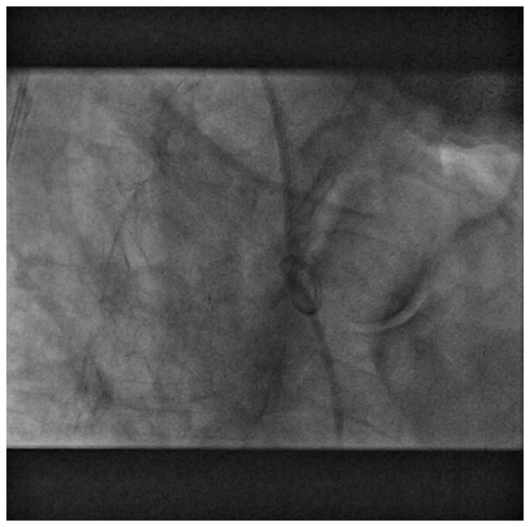
A 77-year-old male patient was transferred to our hospital after a complicated bedside right heart catheterization by a Swan-Ganz Catheter (SGC) inserted through the subclavian vein which was indicated to investigate the aetiology of new-onset severe pulmonary arterial hypertension in the context of respiratory insufficiency. During the procedure, while placing the SGC through the right ventricle by the operating physician it got tangled, and knotted then entrapped, thereby the physician encountered a resistance with impossibility to remove the SGC. At admission in intensive care unit (ICU), patient was haemodynamically stable. A transthoracic echocardiography (TTE) shows the presence of guiding catheter at the base of the pulmonary artery with a dilated right ventricle. In the catheterization lab, fluoroscopy showed the SGC forming a kinked knot hanging in the right ventricle (Figure 1). Avoiding to perform a stressful surgical approach, we choose to start an interventional radiologic procedure by passing a size 0.25 Fr guide-wire through the common femoral vein. The guide-wire was introduced into a hallow in the centre of the knot, and subsequently a balloon was successively inflated by 8, 15, and 23 mm in order to expand progressively the diameter of the knot reversing thereafter the preformed knot (Supplementary material online, Video S1). We pursue a right heart catheterization that revealed a pre-capillary pulmonary arterial hypertension of 43 mmHg, a pulmonary capillary wedge pressure of 13 mmHg, and a cardiac output of 3.7 mL/min/m2. The SGC was successfully removed, and the patient returned to the ICU where he was monitored.
Figure 1.
Fluoroscopy showing the Swan Ganz catheter entrapped in the right ventricle.
Multiple removal strategies were noted in the literature such as using a dormier basket,1 an endomyocardial biopsy forceps,2 and in some critical situations, clinicians prefer to keep the knotted catheter regardless of the potential associated complications.3 In summary, we report an interventional technique to proceed with this serious complication for right heart catheterization which is an invasive procedure that must be performed under fluoroscopic guidance. Physicians should be aware of this rare complication when a guide-wire is advanced if any resistance is encountered, and of several available strategies to manage this unusual situation.
Supplementary material
Supplementary material is available at European Heart Journal - Case Reports online.
Consent: The author/s confirm that written consent for submission and publication of this case report including image(s) and associated text has been obtained from the patient in line with COPE guidance.
Conflict of interest: none declared.
Supplementary Material
References
- 1. Hood S, McAlpine HM, Davidson SA.. Successful retrieval of a knotted pulmonary artery catheter trapped in the right ventricle using a dormier basket. Scott Med J 1997;147:184.. [DOI] [PubMed] [Google Scholar]
- 2. Mehta N, Lochab SS, Tempe DK, Trehan V, Nigam M.. Successful non surgical removal of a knotted and entrapped pulmonary artery catheter. Catheter Cardiovasc Diagn 1998;43:87–89. [DOI] [PubMed] [Google Scholar]
- 3. Karanikas ID, Polychronidis A, Vrachatis A, Arvanitis DP, Simopoulos CE, Lazarides MK.. Removal of knotted intravascular devices. Case report and review of the literature. Eur J Vasc Endovasc Surg 2002;14:518–520. [DOI] [PubMed] [Google Scholar]
Associated Data
This section collects any data citations, data availability statements, or supplementary materials included in this article.


