Abstract
Tolerance to innocuous antigens from the diet and the commensal microbiota is a fundamental process essential to health. Why tolerance is efficiently induced to substances arising from the hostile environment of the gut lumen is incompletely understood but may be related to how these antigens are encountered by the immune system. We observed that goblet cell associated antigen passages (GAPs), but not other pathways of luminal antigen capture, correlated with the acquisition of luminal substances by lamina propria (LP) antigen presenting cells (APCs) and with the sites of tolerance induction to luminal antigens. Strikingly this role extended beyond antigen delivery. The GAP function of goblet cells facilitated maintenance of pre-existing LP T regulatory cells (Tregs), imprinting LP-dendritic cells with tolerogenic properties, and facilitating LP macrophages to produce the immunomodulatory cytokine IL-10. Moreover, tolerance to dietary antigen was impaired in the absence of GAPs. Thus, by delivering luminal antigens, maintaining pre-existing LP Tregs, and imprinting tolerogenic properties on LP-APCs GAPs support tolerance to substances encountered in the hostile environment of the gut lumen.
Keywords: Oral Tolerance, Intestine, Goblet Cell, T Regulatory Cell, Dietary Antigens
Introduction
The single layer epithelium lining the gastrointestinal (GI) tract is the interface between the host and the luminal environment containing trillions of microbes. At this site, the immune system encounters an array of foreign substances, ranging from innocuous dietary antigens and commensal microbes to pathogens. Responding appropriately to each of these is critical to maintaining immune homeostasis in this potentially hostile environment. Paradoxical to the hostile environment of the gut lumen, steady state encounters with non-pathogenic antigens originating from this site result in the induction of antigen specific tolerance, which is largely mediated by CD4+ Foxp3+ T regulatory cells (Tregs)1. Despite advances in our understanding of mechanisms inducing antigen specific Tregs, why tolerance is so efficiently induced to antigens originating from this particularly hostile environment of the gut lumen remains incompletely understood.
The intestinal lamina propria (LP) contains an array of antigen presenting cells (APCs), including classical CD103+ CD11b− IRF8 dependent dendritic cells (DCs), IFR4 dependent CD103+ CD11b+ DCs, and CD103− CD11b+ cells that can express IRF4 and can include resident macrophages, with the CD103+ CD11b+ and CD103− CD11b+ APCs making of the majority of the population in the LP 2–7. Collectively these cellular populations, excluding B lymphocytes, will be referred to as LP-APCs. While each subset preferentially supports various phenotypes of antigen specific T cell responses, there is an evolving understanding that they may play redundant roles in the induction of oral tolerance 8. While tolerogenic responses can be induced in Peyer’s Patches and potentially in other mucosal lymphoid tissues, it has become appreciated that the gut draining lymph nodes are critical sites for the induction of oral tolerance 9,10. Current understanding is that this process requires the acquisition of antigens by LP-APCs underlying the villous epithelium, their trafficking to the draining lymph nodes to induce naive CD4+ T cells to differentiate into peripherally induced Tregs (pTregs), and homing of these pTregs to the LP where they are maintained by continued stimulation by LP-APCs acquiring the cognate antigen for these pTregs from the lumen 11,12. Tolerance to luminal antigens occurs in the small intestine (SI) 13 and in the distal colon 14, indicating these are the sites where luminal antigens cross the epithelium and are acquired by LP-APCs. How antigens are captured by LP-APCs at these sites may be the basis for why tolerance is effectively induced in this hostile setting.
Several routes by which luminal substances cross the epithelium have been identified including paracellular leak, the direct capture by LP-APCs via extension of trans-epithelial dendrites (TEDs) into the gut lumen, passage from the lumen via villous M cells, and passage from the lumen via goblet cell associated antigen passages (GAPs) 15–22. Of these, LP-APC extension of TEDs is the currently favored route to support the induction and maintenance of tolerance to luminal substances in the steady state, as the extension of TEDs does not compromise the epithelial barrier and would allow direct acquisition of luminal antigens by LP-APCs16. However, this process directly exposes the LP-APCs to luminal contents, which in vitro studies indicate induces mixed Th1 and Th2 responses23. In addition, TEDs are absent in some mouse strains, which do not display defects in oral tolerance 24 and are lacking in regions of the gut where gavaged antigen is captured by LP-APCs 25,26 suggesting that other luminal antigen acquisition pathways could support oral tolerance. Thus, how luminal antigens are acquired by LP-APCs for the induction of tolerance and if this process is integral to efficiently inducing tolerance in the hostile gut luminal environment remain unclear. Here we evaluated steady state routes of luminal antigen capture by LP-APCs. We found that LP-APC extension of TEDs, villous M cells and paracellular leak did not correlate with effective antigen capture by LP-APCs. In contrast the density of GAPs directly correlated with LP-APC luminal antigen capture and with the regions within the gut where tolerance is induced to luminal substances. Moreover, beyond the role of antigen delivery, we find that the GAP function of goblet cells imprints and maintains LP-DCs and macrophages with tolerogenic properties, maintains pre-existing Tregs in the SI LP, and in the absence of GAP tolerance to dietary antigens is impaired. Thus, the GAP function of goblet cells acts as both a pathway to deliver luminal substances to LP-APCs and as a mechanism imprinting LP-APCs with tolerogenic properties to maintain and induce tolerance to antigens encountered in the hostile environment of the gut lumen.
Results
The presence of goblet cell associated antigen passages (GAPs), but not LP-APC extension of TEDs or villous M cells, correlates with the sites of luminal antigen capture for the induction of tolerance in the steady state
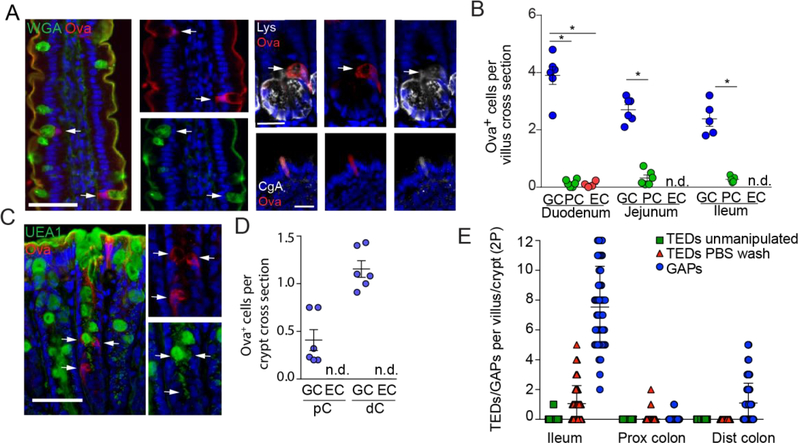
In the steady state, tolerance to luminal substances is induced in the SI and distal colon 13,14. How luminal substances cross the epithelium to be encountered by the immune system is a fundamental process that may underlie why tolerance is so efficiently induced to substances arising from an unfavorable environment with abundant microbes and microbial products. To evaluate how dietary antigen traverses the intestinal epithelium we performed intraluminal injections of fluorescently labeled ovalbumin (Ova) and evaluated fixed intestinal sections by fluorescent microscopy. Immunofluorescent staining of fixed tissue sections demonstrated that goblet cells containing the luminally administered fluorescent Ova could be identified throughout the SI and in the distal descending colon and sigmoid colon, referred to as the distal colon, but were less common in the cecum, ascending colon, transverse colon, and proximal descending colon, referred to as the proximal colon (Figure 1A–D). The presence of GAPs in the distal colon was not appreciated in the work initially identifying GAPs using the in vivo imaging approach due to the difficulty of imaging the distal colon with this approach. This regional distribution of GAPs correlates with the previously identified lymph nodes draining the regions of the gut supporting tolerance 13,14. Secretory intestinal epithelial cell lineages other than goblet cells have been observed to take up luminal antigens 27,28. We observed that Paneth cells containing luminally administered Ova were present throughout the length of the SI but significantly less common when compared to goblet cells containing fluorescent Ova (Figure 1A–B). We identified a small number of enteroendocrine cells containing luminally administered Ova in the steady state that were restricted to the duodenum; these were also significantly less common than goblet cells containing fluorescent Ova (Figure 1A–B). In addition, we did not observe M cells in the non-follicle bearing epithelium in the SI or colon in the steady state (Figure S1).
Figure 1: Goblet cell associated antigen passages (GAPs) are present at the sites of antigen acquisition where tolerance to luminal substances is induced in the steady state.
A) representative images and B) quantification of goblet cells (GC; wheat germ agglutinin (WGA)+ in SI), Paneth cells (PC; lysozyme (Lys) +), and enteroendocrine cells (EC; chromogranin A (CgA) +) taking up luminal fluorescent ovalbumin (Ova) in regions of the SI determined by immunofluorescent staining on fixed tissue sections. C) Representative images and D) quantification and of goblet cells (Ulex Europaeus Agglutinin I (UEAI) + in colon) and ECs taking up luminal fluorescent Ova in the proximal colon (pC) and distal colon (dC) as determined by immunofluorescent staining of fixed tissue sections. E) Quantification of TEDs and GAPs per SI villus or colon crypt obtained via in vivo two photon imaging. Scale bar = 50μm in large panels A and C and 20μm in small panels A, Each data point represents an individual mouse with 30 or more villi and 40 or more crypts evaluated per mouse in panels B and D. Each data point in panel E represents an individual crypt or villus. Data is presented as the mean +/− SEM. * = P < 0.05, ns = not significant, n.d. = not detected
The currently favored route of luminal antigen acquisition by LP-APCs for tolerance induction is direct capture through the extension of TEDs. We evaluated the frequency and regional distribution of TED extension by LP-APCs by in vivo two photon imaging of CD11cYFP and CX3CR1GFP reporter mice. Mice were imaged at various times throughout the day and were not deprived of food or water prior to imaging. At steady state conditions, we observed LP-APC extension of TEDs to be very rare in the distal SI and absent in the proximal SI (Figure S1 B–E). We observed two TEDs that were located in the distal SI out of greater than 500 villi imaged from tip to base from multiple CD11cYFP reporter mice (Figure S1D left side) and four TEDs forming in the distal SI out of greater than 350 villi imaged from tip to base throughout the SI from CX3CR1GFP/WT reporter mice (Figure S1E left side). We did not observe any TED extension in either the proximal or distal colon after analyzing 260 colonic crypts in the CD11cYFP reporter mice and 263 crypts in the CX3CR1GFP reporter mice (Figure S1D and E, left side).
Previous studies removing the luminal contents mucus by washing, identified TED extension by APCs occurred at a rate of ~1.5–2.0 TEDs/villus 15,24,25,29,30. Approximately ten minutes following the removal of the luminal contents and mucus by rinsing with PBS, LP-APCs became less compact and extended multiple dendrites within the LP, into the epithelium, and into the lumen, with some LP-APCs traversing the epithelium (Supplemental Movie S1). However, consistent with prior observations 15,25, CX3CR1GFP+ LP-APC TED formation did not occur in the duodenum, the site where gavage antigen is acquired by CX3CR1GFP+ LP-APCs (Figure S1E right side), and TED formation was not observed in the distal colon in any condition (Figure S1B–E), consistent with observations by others that TEDs are rare or absent in the colon 31,32.
Using the in vivo two photon imaging approach we used to evaluate the frequency of TEDs, we evaluated the frequency of GAPs in villi and colonic crypts. We did not observe an effect of removal of the mucus layer on the frequency of GAPs and the regional distribution of GAPs remained similar to our findings of GAPs using fluorescent microscopy on fixed tissue sections (data not shown). In the SI GAPs were ~1000 fold more common than TEDs when the mucus layer was left intact and ~10 fold more common when the mucus layer was removed (Figure 1E). Thus, the frequency and regional distribution of GAPs, but not luminal antigen acquisition by Paneth cells, or enteroendocrine cells, the presence M cells, or LP-APC extension of TEDs correlated with regions of the gut where tolerance to luminal substances can be induced 13,14.
GAPs support LP-APC capture of, and CD4+ T cell responses to, luminal antigen
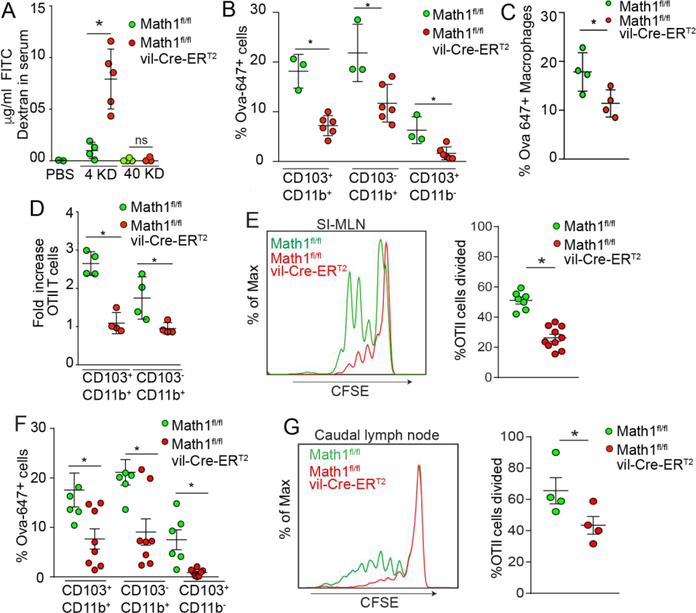
Mouse atonal homologue 1 (Math1) is a transcription factor required for the development of neurons and intestinal secretory intestinal epithelial lineages, which includes goblet cells, enteroendocrine cells, and Paneth cells 33–36. Paneth cells have a significantly longer half-life than goblet cells 37,38, and accordingly, ten days after treatment with tamoxifen, mice with an inducible deletion of Math1 in intestinal epithelial cell lineages (Math1fl/flvil-Cre-ERT2 mice) lose goblet cells (Figure S2A), but retain Paneth cells, albeit at a somewhat reduced number when compared to their littermate controls (Figure S2B). Goblet cells acquiring luminal fluorescent dextran in the SI and distal colon decreased significantly ten days following the deletion of Math1 in intestinal epithelial cells (Figure S2C and D). In contrast to the decrease in GAPs, intestinal permeability increased, as evidenced by serum levels of 4kD FITC dextran following gavage (Figure 2A), and as evidence by the presence of 3kD fixable FITC dextran between epithelial cells and within the lamina propria of goblet cell deficient mice following gavage (Figure S2E). The increased permeability might be attributed to the loss of the mucus barrier following goblet cell deletion. Despite the increase in intestinal permeability, SI LP-APCs, identified by flow cytometry (Figure S3), acquired less luminally administered fluorescent Ova (Figure 2B and C). In addition to the effects rising from the loss of GAPs, this may in part be related to the size of intact Ova (~43kD), as gavage of 40kD FITC dextran did not result in increased serum levels in goblet cell deficient mice (Figure 2A) and gavaged fluorescent Ova was not found leaking between SI epithelial cells or in the lamina propria of goblet cell deficient mice (Figure S2E). Isolation of the CD103+ CD11b+ APC population and the CD103− CD11b+ APC population, which may contain DCs and macrophages, following Ova gavage revealed that the APCs were no longer able to acquire gavaged Ova in a manner capable of inducing CD4+ T cell responses in ex vivo co-cultures with Ova specific T cells from OTII T cell receptor (TCR) transgenic mice when goblet cells and GAPs were absent (Figure 2D). We were unable to isolate sufficient numbers of CD103+ CD11b− SI LP-DCs for this ex vivo assay. The impaired ability of LP-APCs to induce T cell proliferation to luminal antigen was not due to an intrinsic defect in antigen acquisition or presentation, as LP-APCs isolated from mice with Math1 deleted in intestinal epithelial cells displayed no defects in capture of fluorescent antigen in culture (Figure S4A) and no defect in induction of T cell proliferation when exogenous Ova was added to ex vivo co-cultures (Figure S4B). We attributed the decrease in LP-APC antigen acquisition to the loss of goblet cells and GAPs, as we saw very few intestinal enteroendocrine cells and few Paneth cells acquiring luminal Ova in wildtype mice (Figure 1A and B), and Paneth cells were still present at this time following deletion of Math1 (Figure S2B). Moreover, mice lacking goblet cells/GAPs were significantly impaired at inducing antigen specific CD4+ T cell responses to gavaged antigen in the SI draining MLN (Figure 2E), the site of tolerance induction to dietary antigens. The impaired responses to luminal antigen were not attributable to defects in the ability of MLN T cells to respond to Ova, as responses to systemically administered Ova were not impaired (Figure S4C). The CSFE dilution seen in the mice lacking goblet cells may be due to antigen acquired at other sites, such as the Peyer’s Patches and migration of DCs to the MLN, as we saw no proliferation of OTII Rag−/− T cells, which have TCR specificity only for Ova, in the MLN in the absence of Ova gavage, and reduced but detectable proliferation of OTII Rag−/− T cells in the MLN in response to Ova in mice lacking goblet cells and GAPs when compared with their Cre-littermates (Figure S4D). We also observed that GAPs were decreased in the distal colon of mice lacking goblet cells (Figure S2D) and that LP-APC acquisition of intra-colonic fluorescent Ova was impaired in the distal colon in the absence of goblet cells and GAPs (Figure 2F). Moreover, deletion of goblet cells impaired the induction of CD4+ T cell responses to Ova via enema in the distal colon draining LN in vivo (Figure 2G). Thus, loss of goblet cells and GAPs impairs the ability of LP-APCs to acquire luminal antigen and impairs immune responses to luminal antigen in vivo despite the presence of increased intestinal leak.
Figure 2: Goblet cells support antigen presenting cell acquisition of luminal antigen and CD4+ T cell responses to luminal antigen in the gut draining lymph nodes.
A) 4kD and 40kD FITC-dextran in serum after oral gavage in Math1f/fvil-Cre-ERT2 mice and Math1f/f littermate controls. B and C) Luminal Ova acquisition by SI LP-APCs assessed by flow-cytometric analysis two hours post oral gavage. D) Antigen presentation capacity of SI LP-APCs isolated from mice Math1f/f and Math1f/fvil-Cre-ERT2 mice given luminal Ova as assessed by expansion of Ova specific OTII T cells in ex vivo cultures. E) Histograms and quantification of in vivo proliferation of CFSE labeled OTII T cells in SI draining MLN of Math1f/f and Math1f/fvil-Cre-ERT2 mice 2 days after oral Ova gavage. F) Antigen acquisition by distal colon LP-APCs assessed by flow cytometry 2 hours following intra-colonic administration of fluorescent Ova. G) Histograms and quantification of in vivo proliferation of CFSE labeled OTII T cells in distal colon draining caudal LN of Math1f/f and Math1f/fvil-Cre-ERT2 mice 2 days after Ova enema. Data are representative of two or more replicates with ≥ 3 mice per group, each data point represents an individual mouse. Data is presented as mean +/− SEM, * = P < 0.05, ns = not significant.
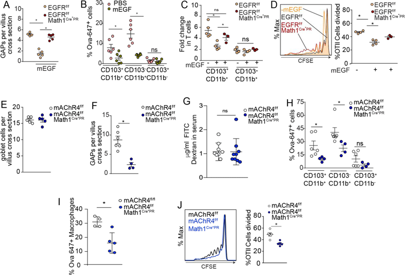
Goblet cells play an important role in maintaining the intestinal barrier through mucus production and release of anti-microbial products, and accordingly deletion of goblet cells may have effects unrelated to the loss of GAPs. Therefore, to examine the role of the GAP function of goblet cells in luminal antigen delivery, we evaluated the effect of GAP inhibition on luminal antigen capture by LP-APCs and immune responses independent of deletion of goblet cells. GAPs form in response to acetylcholine (ACh) acting on the muscarinic ACh receptor 4 (mAChR4) on goblet cells, and conversely GAPs are inhibited by activation of the epidermal growth factor receptor (EGFR) in goblet cells21. Inhibition of GAPs by luminal recombinant murine epidermal growth factor (mEGF) significantly impaired LP-APC capture of luminally administered fluorescent Ova (Figure 3A and B), as well as the ability of LP-APCs to acquire gavaged Ova in a manner capable of inducing antigen specific CD4+ T cell proliferation in ex vivo cultures (Figure 3C). Moreover, mEGF significantly impaired antigen specific CD4+ T cell responses to oral Ova in vivo in the MLN (Figure 3D). Importantly, deletion of the EGFR in goblet cells using an inducible Math1 driven Cre recombinase, EGFRf/fMath1Cre*PR mice, reversed the effects of mEGF on GAP inhibition, and T cell responses to luminal Ova in ex vivo cultures and in vivo (Figure 3A, C, and D), demonstrating that the defect in antigen capture could not be attributed to effects of EGF on LP-APCs or T cells. This is consistent with the effect of EGF being mediated by effecting goblet cells and GAPs. Likewise, we observed that inducible deletion of mAChR4 on goblet cells, (mAChR4f/fMath1Cre*PR mice) did not affect goblet cell numbers (Figure 3E), but impaired GAP formation (Figure 3F). Unlike the deletion of goblet cells, we did not see an increase in leak when GAPs were inhibited (Figure 3G) and accordingly we did not see a reduction in the mucus barrier when GAPs were inhibited and goblet cells remained intact (Figure S5A and B). Inhibition of GAPs by deletion of the mAChR4 on goblet cells impaired luminal fluorescent Ova acquisition by LP-APCs (Figure 3H and I), and impaired antigen specific CD4+ T cell responses to gavaged Ova in the SI draining MLN (Figure 3J). We found that GAPs in the distal colon were inhibited by the pan-muscarinic acetylcholine receptor antagonist atropine and were induced by the ACh analogue carbamylcholine (Figure S6A), but were not inhibited by deletion of mAChR4 in goblet cells (Figure S6B), indicating that GAPs in the distal colon are induced by ACh acting on receptors other than mAChR4 and that there are yet to be identified pathways inducing GAP formation in the distal colon. While this prevented us from performing analogous studies in the distal colon to inhibit GAPs, these data support that the GAP function of goblet cells plays a role in delivering luminal antigens to LP-APCs for the induction of immune responses in the steady state.
Figure 3: The GAP function of goblet cells supports the acquisition of, and CD4+ T cell responses to, luminal antigen.
A) GAPs per villus as assessed by immunofluorescent staining B) luminal fluorescent Ova capture by LP-APCs as assessed by flow cytometry C) ability of LP-APCs to stimulate Ova specific T cells ex vivo in response to luminal Ova and D) ability of Ova specific T cells to expand in vivo in response to luminal Ova in wildtype mice (panel B) and in mice lacking EGFR in goblet cells (EGFRf/f Math1Cre*PR mice) and littermate controls given EGF to inhibit GAPs. E) Goblet cells per villus as assessed by WGA staining, F) GAPs per villus as assessed by luminal fluorescent Ova uptake, G) FITC-dextran (4kD) in serum after oral gavage, H and I) luminal fluorescent Ova capture by LP-APCs as assessed by flow cytometry and J) ability of Ova specific T cells to expand in vivo in response to luminal Ova in mice lacking mAChR4 in goblet cells (mAChR4f/f Math1Cre*PR mice) and littermate controls. * = P< 0.05, ns = not significant, data presented as the mean +/− SEM. Each data point represents an individual mouse.
Goblet cells and GAPs support the maintenance of Tregs and imprinting APCs in the LP
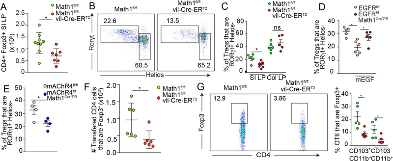
Tolerance to dietary antigens occurs in the SI and is mediated by CD4+ Foxp3+ pTregs that are generated in the draining LN. These pTregs subsequently traffic to and reside in the SI LP where they are maintained by continual stimulation by LP-APCs that have acquired the cognate antigen for these pTregs from the lumen 1,4,11,12. Accordingly, these pTregs may have a limited lifespan when their cognate antigen is withdrawn 13. In contrast, a substantial proportion of the pTregs residing in the colon LP differentiate in response to microbial stimuli and are longer-lived 13,39,40. A portion of these colonic pTregs can have specificity for gut bacterial antigens and their development requires GAPs in the proximal colon that are present for a defined period of time during a pre-weaning interval 41. This could suggest that luminal antigen delivery by GAP to LP-APCs might have a role in maintaining existing pTregs in the SI LP that have a more limited lifespan in the absence of continual stimulation. Indeed, we observed a decrease in the absolute number of SI LP Tregs when goblet cells/GAPs were deleted (Figure 4A). This decrease largely affected the Helios- pTregs in the SI LP (Figure 4B and C). We observed little change in the Helios-pTreg population in the colon LP(Figure 4C). The relative lack of an effect of goblet cell/GAP deletion on the colonic pTreg population could reflect that adherent bacteria, which can induce immune responses by GAP independent endocytosis via enterocytes, can drive pTreg development in the colon in the steady state 42–44. Consistent with the pTregs being gut pTregs39,40, almost all of these LP Helios- Tregs expressed the transcription factor RORγt (Figure 4B), which can be expressed by SI LP pTregs with specificity to dietary antigens13. Further we observed that the SI LP pTreg population was reduced with GAP inhibition by mEGF in a goblet cell intrinsic EGFR dependent manner (Figure 3A and 4D) and upon GAP inhibition by deletion of the mAChR4 in goblet cells (Figure 3F and 4E), demonstrating that the GAP function of goblet cells facilitated the maintenance of SI LP pTregs.
Figure 4: GAPs support the maintenance and induction of pTregs.
A) Absolute numbers of SI LP Tregs, and B) flow cytometry dot plots of Helios and RORγt expression by CD4+ Foxp3+ T cells in the SI LP, and C) quantification of RORγt+ Helios- pTregs populations in the SI and colon LP of goblet cell deficient mice (Math1f/f vil-Cre-ERT2 mice) and littermate controls. Quantification of SI LP RORγt+ Helios- pTreg populations in D) mice lacking EGFR in goblet cells (EGFRf/fMath1Cre*PR mice) and littermate controls treated with vehicle or mEGF, and in E) mice lacking mAChR4 in goblet cells (mAChR4f/f Math1Cre*PR mice) and littermate controls. F) Quantification of Foxp3 expression by MLN OTII T cells adoptively transferred into goblet cell deficient mice and littermate controls five days following i.v. injection of Ova. G) Representative flow cytometry dot plots and quantification of Foxp3 expression by OTII T cells cultured for 5 days with Ova and SI LP-APCs isolated from goblet cell deficient mice and littermate controls. * = P < 0.05, ns = not significant. Data is presented as the mean +/− SEM. Each data point represents an individual mouse.
Because the absence of GAPs impaired stimulation of Ova specific T cells to dietary antigen in the MLN, and by extension would impair their differentiation to effector T cells or pTregs, we injected Ova intravenously to mice following adoptive transfer of OTII T cells to evaluate naive Ova specific T cell differentiation in the absence of GAPs. We observed that in the absence of goblet cells, the in vivo induction of Tregs in the MLN in response to systemic Ova was impaired (Figure 4F). The impaired ability to induce Tregs in response to dietary Ova in mice lacking goblet cells/GAPs can in part be attributed to defects in the LP-APC population as LP-APCs isolated from mice lacking goblet cells were impaired at inducing antigen specific pTregs in ex-vivo cultures (Figure 4G).
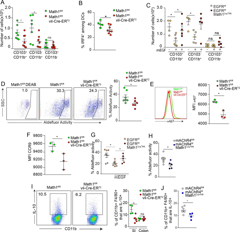
SI LP-APCs consist of IRF8 dependent CD103+ CD11b− DCs, IRF4 dependent CD103+ CD11b+ DCs, and CD103− CD11b+ DCs and macrophages, which can express, but are not dependent upon IRF4 6,7. We observed a reduction in the CD103+ CD11b+ and CD103− CD11b+ populations, but not the CD103+ CD11b− DCs in the absence of goblet cells and GAPs (Figure 5A). Accordingly, the absence of goblet cells and GAPs resulted in a decrease in the IRF4+ SI LP-APC population (Figure 5B). The decrease in CD103+ CD11b+ and CD103− CD11b+ LP-APCs was dependent upon the GAP function of goblet cells as these populations were reduced in response to mEGF in an EGFR goblet cell dependent manner (Figure 5C). CD103+ CD11b− and CD103+ CD11b+ SI LP-DCs can have aldehyde dehydrogenase activity 7,45, which facilitates the production of all-trans retinoic acid, a factor promoting the differentiation of and imprinting of pTregs with gut homing molecules 46–48. We observed that in the absence of goblet cells, SI LP-DCs had reduced aldehyde dehydrogenase (ALDH) activity (Figure 5D) and an impaired ability to induce the expression of the gut homing molecules α4β7 and CCR9 on responding T cells in in vitro co-cultures (Figure 5 E and F). Similar to the maintenance of pre-existing LP pTregs, SI LP-DC ALDH activity was facilitated by the GAP function of goblet cells, as this was impaired by GAP inhibition by mEGF in a goblet cell intrinsic EGFR dependent manner (Figure 5G) and by GAP inhibition via the deletion of mAChR4 in goblet cells (Figure 5H).
Figure 5: GAPs support the imprinting of LP-APCs.
Quantification of A) SI LP-APC subsets and B) IRF4+ SI LP-DCs in goblet cell deficient mice (Math1f/f vil-Cre-ERT2 mice) and littermate controls. C) Quantification of SI LP-APC subsets in mice lacking EGFR in goblet cells (EGFRf/fMath1Cre*PR mice) and littermate controls treated with mEGF. D) Flow cytometry dot plots and quantification of aldehyde dehydrogenase (ALDH) activity in LP CD11c+ MHCII+ SI APCs in goblet cell deficient mice (Math1f/f vil-Cre-ERT2 mice) and littermate controls. Expression of gut homing molecules E) α4β7 and F) CCR9 on OTII T cells following three days of in vitro culture with Ova and CD103+ DCs isolated from goblet cell deficient mice or littermate controls. Quantification of SI LP-APC with ALDH activity in G) mice lacking EGFR in goblet cells (EGFRf/fMath1Cre*PR mice) and littermate controls treated with mEGF and in SI LP-APCs from H) mice lacking mAChR4 in goblet cells (mAChR4f/f Math1Cre*PR mice) and littermate controls. I) Flow cytometry plots of SI LP macrophages and quantification of IL-10 expression by LP CD45+ CD11c+ MHCII+ F480+ cells from mice lacking goblet cells and their littermate controls. J) Quantification of IL-10 expression by SI LP macrophages from mice lacking mAChR4 in goblet cells and their littermate controls. * = P < 0.05, ns = not significant. Data is presented as the mean +/− SEM. Each data point represents and individual mouse, with the exception of E and F, where LP-APCs were pooled from three goblet cell deficient mice or three littermate controls.
LP macrophages have been implicated in SI LP Treg maintenance through the production of IL-10 and stimulation of pre-existing pTregs with their cognate antigen acquired from the lumen 11. We found that mice lacking goblet cells and GAPs as well as mice with goblet cells but lacking GAPs had impaired IL-10 production by SI, but not colonic, CD11c+ MHCII+ CD11b+ F4/80+ LP-APCs (Figure 5I and J), consistent with GAPs having a role in imprinting this LP-APC subtype in the SI. Thus, goblet cells and GAPs might contribute to multiple facets of oral tolerance including antigen delivery and imprinting LP-APCs for the induction and maintenance of pTregs specific for dietary antigens.
Goblet cells and GAPs support tolerance to dietary antigen
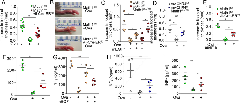
To directly evaluate the role for goblet cells and GAPs in tolerance to dietary antigens, mice lacking goblet cells and GAPs, their littermate controls, and mice in which GAPs were transiently inhibited at the time of luminal antigen administration by intraluminal mEGF or deletion of mAChR4 in goblet cells, were gavaged with Ova, immunized with Ova, challenged with Ova in the footpad, and evaluated for footpad swelling 24 hours later. Goblet cell deficient mice and mice in which GAP formation was inhibited demonstrated significantly greater footpad swelling indicative of decreased tolerance to dietary antigen (Figure 6A–D).
Figure 6: GAPs support tolerance to dietary antigen and tolerance to luminal antigens in the distal colon.
A) Quantification and B) images of footpad swelling following the induction of oral tolerance by dietary Ova, Ova immunization, and Ova footpad challenge in mice lacking goblet cells (Math1f/f vil-Cre-ERT2 mice) and their littermate controls. Quantification of footpad swelling following dietary Ova, Ova immunization, and Ova footpad challenge in C) mice lacking EGFR in goblet cells (EGFRf/fMath1Cre*PR mice) and their littermate controls treated with mEGF and in D) mice lacking mAChR4 in goblet cells (mAChR4f/f Math1Cre*PR mice) and littermate controls. E) Quantification of footpad swelling following the induction of tolerance by Ova enema, Ova immunization, and Ova footpad challenge in mice lacking goblet cells (Math1f/f vil-Cre-ERT2 mice) and their littermate controls. F-I) Serum IFNγ in mice treated as in A-E 24 hours following footpad challenge. * = P < 0.05. Data is presented as the mean +/− SEM. Each data point represents an individual mouse.
Moreover, deletion of EGFR in goblet cells at the time of mEGF and oral Ova administration reversed the effects mEGF on impaired tolerance, consistent with the effect of mEGF being due to GAP inhibition (Figure 6C). Notably the Math1 Cre targets differentiated goblet cells, which turn over every 3–5 days, and therefore the inhibition of GAPs by deletion of mAChR4 and the reversal of effects of EGF by deletion of EGFR in goblet cells is largely limited to the time of luminal Ova administration, and not due to effects on goblet cells at the time of Ova immunization and challenge. While the mAChR4 independent formation of GAPs in the distal colon prevented us from directly assessing the role of the GAP function of goblet cells in tolerance to luminal antigens in the colon, we did observe that deletion of goblet cells impaired the ability to induce tolerance to Ova administered via enema (Figure 6E). Loss of goblet cells might induce inflammatory responses due to the deficient mucus barrier, which could affect the capture of luminal substances by resident LP-APCs and the induction of tolerance independent of the loss of GAPs. Indeed, we observed that deletion of goblet cells resulted in an increase in monocytes and neutrophils in the SI lamina propria (Figure S7A). However we did not see an increase in monocytes in the lamina propria when GAPs were inhibited and goblet cells remained intact (Figure S7B) suggesting that inflammatory responses alone do not account for the loss of tolerance when GAPs are inhibited. Mice with goblet cell and GAP manipulation had increased serum levels of interferon-γ (IFNγ) following immunization (Figure 6F–I), correlating with their loss of tolerance to dietary Ova. The impaired tolerance in the absence of goblet cells or GAPs was not as severe as that seen in the absence of luminal Ova exposure (Figure 6 A–E), suggesting the potential for other or compensatory routes of luminal antigen delivery in the absence of goblet cells and GAPs. However, in total these observations indicate that GAPs support the induction of tolerance to luminal antigens on multiple levels.
Discussion
The gut lumen contains trillions of microbes and abundant microbial products. Inducing and maintaining tolerance to innocuous substances originating from this potentially inhospitable environment is fundamental to maintaining homeostasis and health. Indeed, tolerance is so effectively induced to antigens originating from the gut lumen that oral tolerance regimens are being leveraged to treat extra-intestinal diseases 49–51. Accordingly, how tolerance is induced and maintained at this mucosal surface has been a topic of many studies.
The gut microenvironment has unique properties supporting tolerance. Tolerance to non-self antigens is largely mediated by the conversion of naive T cells into Foxp3 expressing pTregs 52,53, which is facilitated by a local environment containing all-trans retinoic acid (ATRA) and TGFβ 46,47,54,55. Within the gut CD103+ DCs and MLN stromal cells expressing retinaldehyde dehydrogenase, the enzyme necessary to convert retinal to the biologically active ATRA, are sources of ATRA supporting pTreg induction and imprinting gut homing molecules on lymphocytes 46,56–60. DC imprinting with retinaldehyde dehydrogenase activity is induced by luminal retinoids and by DC association with the intestinal epithelium 45,61,62. Moreover, the goblet cell protein, mucin 2, promotes tolerogenic properties in DCs inducing pTregs including the production of TGFβ and the expression of retinaldehyde dehydrogenase 63, and select members of the gut microbiota promote pTregs through bacterial products or metabolites 42,64–70. Thus, these unique properties contribute to the tolerogenic tone of the gut environment, yet how luminal antigens are acquired by the immune system for the induction of tolerance and whether this process contributes to tolerance beyond antigen capture have been unexplored.
How luminal antigens are encountered by the immune system may affect the phenotype of the subsequent immune response 71–73. A landmark discovery identified that LP-APCs had the ability to extend dendrites between epithelial cells to capture luminal bacteria without compromising the epithelial barrier 16,74, suggesting that this process might allow minimally disruptive direct capture of luminal substances. However, LP-APC extension of TEDs is absent in some mouse strains 24, suggesting that unlike oral tolerance, LP- APC TED extension is not a universal phenomenon and other pathways of luminal antigen capture inducing oral tolerance exist. In addition, while the extension of TEDs is impaired in the absence of CX3CR115, CD4+ T cell responses to luminal antigens are not 4,15, suggesting that the defect in oral tolerance in CX3CR1 deficient mice was unrelated to luminal antigen capture. We observed that the extension of TEDs by LP-APCs is very rare in the steady state but became more common after the removal of the luminal contents and mucus layer, occurring in a frequency similar to prior reports 15,25. Why removal of the luminal contents and mucus layer induces TED extension is unclear, but could be related to the release of lactate and pyruvate by stressed epithelial cells as these metabolites were recently identified to induce TED extension in CX3CR1+ LP-APCs75 and we have observed that TED extension occurs when mice expire while imaging under anesthesia in the absence of removal of the luminal contents and mucus layer (unpublished observation). We did not observe LP-APC TED extension in the duodenum, the site where gavaged antigen is acquired by CX3CR1+ LP-APCs 26, or in the distal colon, the site where tolerance to luminal antigen is induced in the colon 14. While it is impossible to exclude a contribution of LP-APC TED extension, combined with the above observations, these findings indicate that LP-APC TED extension is less likely to be a major route of steady state soluble luminal antigen capture for the induction of oral tolerance.
Early observations suggested that M cells were restricted to the epithelium overlying the Peyer’s patches, however subsequent studies identified M cells overlying the non-follicle bearing villous epithelium18. Villous M cells are rare in the steady state but can be induced by systemic treatment with TNF superfamily member receptor activator of NF-κβ ligand (RANKL), whose expression is normally restricted to subepithelial stromal cells restricted to the Peyer’s patches 76. These villous M cells can be closely associated with mononuclear cells and have the capacity to transcytose bacteria to induce immune responses to luminal bacteria 18,77. We also found villous M cells to be very rare in the steady state, suggesting they are not a major pathway for luminal antigens to traverse the epithelium to support oral tolerance. Similarly, barrier leak, as evidenced by the presence of luminally administered 4 kD dextran in the serum did not correlate with LP-APCs acquisition of luminal antigen. Why barrier leak is less effective at loading LP-APCs with antigen in a manner capable of inducing T cell responses is unclear but could be related to the size of substances delivered via paracellular leak relative to the size of proteins/polypeptides required to induce antigen specific T cell responses as we did not see an increase in 40 kD dextran in the serum following gavage in goblet cell deficient mice.
In contrast, the presence of intestinal epithelial cells filling with luminal antigen was common in the steady state. Consistent with a recent report 27, we observed enteroendocrine cells containing luminal antigen, however they were rare and limited to the duodenum in the steady state. We more commonly observed Paneth cells containing luminal antigen, but these were still relatively rare occurring on average in one Paneth cell in every two villus cross sections. Moreover, Paneth cells are less likely to be a major contributor to steady state luminal antigen delivery supporting tolerance as they are absent from the colon, and due to their longer life span, persist for weeks following deletion of Math1 in epithelial cells, which we observed results in significantly impaired luminal antigen delivery to LP-APCs. In contrast, goblet cells filling with luminal antigen were commonly observed in the regions of the gut where luminal antigens are acquired to induce tolerance, suggesting that goblet cells and GAPs may be pathways delivering dietary antigens to LP-APCs to support oral tolerance. Of note GAPs are present in strains of mice in which the extension of TEDs by LP-APCs are absent 20,24. Indeed, we observed that in the absence of goblet cells and GAPs luminal antigen capture by CD103+ CD11b+, CD103− CD11b+, and CD103+ CD11b− LP-APCs was impaired. Our initial observation of GAP mediated antigen delivery to LP-APCs reported that GAPs delivered antigen to CD103+ LP-DCs 20. These studies focused on the functional outcome of inducing antigen specific T cell responses to luminal antigen, which was largely limited to the CD103+ LP-DC population and was impaired in the absence of goblet cells20. However other APC populations could acquire luminal antigen and stimulate antigen specific T cell responses when GAPs were induced above baseline levels20, suggesting that GAPs deliver antigen to these APC populations as well. We have observed CD103− LP-APC populations interacting with GAPs in the SI and colon 78,79. The preferential ability of CD103+ LP-APCs over CD103− LP-APCs to induce T cell responses to luminal antigen may be related to their enhanced antigen presentation and stimulation capacities 4 or may be due to passage of antigen from CD103− LP-APCs to CD103+ LP-DCs 26. Irrespective of the pathway by which CD103+ LP-DCs acquire luminal antigen, either by direct capture from goblet cells, or from transfer from CD103− LP-APCs, our observations indicate that this process is supported by the GAP function of goblet cells. While luminal antigen capture by LP-APCs was nearly undetectable in the absence of GAPs, proliferation of dietary antigen specific T cells in the MLN and oral tolerance, as measured by DTH responses, were less dramatically impaired. This could be consistent with contributions of dietary antigen capture at other sites, such as the Peyer’s patches, to T cell responses in the MLN and tolerance to dietary antigens.
The findings presented here indicate that GAPs function beyond simple antigen delivery to promote oral tolerance. LP-DCs with ALDH activity produce all-trans retinoic acid 45, which promotes the induction of pTregs 4,46,47,63. Further, the production of IL-10 by resident LP macrophages supports the expansion and maintenance of pre-existing LP pTregs specific for dietary antigens 11. We found that in the absence of goblet cells/GAPs imprinting SI LP-APCs with ALDH activity and the production of IL-10 by SI macrophages were impaired. When CD103+ LP-DCs acquire luminal antigens from GAPs, they also acquire goblet cell proteins 20. Combined with observations that the goblet cell protein mucin 2 imprints DCs with ALDH activity 63, this suggests that CD103+ LP-DC imprinting by GAPs may occur during antigen acquisition and that GAPs may deliver tolerogenic signals in concert with luminal substances to support antigen specific tolerance induction. The mechanism of tolerance induction to luminal substance encountered in the distal colon differs from that of the SI and can utilize other APC populations 8,14. While we did not observe LP-APCs defects in the colon in the absence of goblet cells/GAPs, the relevant properties of the APCs inducing tolerance in the distal colon are not known, and accordingly whether GAPs in the distal colon play an analogous role influencing this APC phenotype remains to be investigated. Beyond this we noted that GAPs supported the maintenance of pre-existing pTregs in the SI LP. These pTregs regressed within days of GAP inhibition, a time course that is much faster than regression of SI LP pTregs when deprived of cognate antigen13. This suggests that GAPs may play additional yet to be identified roles beyond antigen delivery in shaping the immune landscape of the gut. Related to this, enteric viral infection abrogates oral tolerance and promotes Th1 immune responses to dietary antigen80, and enteric bacterial infection inhibits GAPs and shifts immune responses to dietary antigen away from tolerance toward Th17 responses 81. In the context of the findings presented here, this might suggest that GAP inhibition during enteric infection is a physiologic response facilitating inflammatory responses for pathogen clearance.
Immune tolerance to innocuous substances encountered in the gut lumen is a recognized phenomenon that is essential for gut health. How this process occurs is a fundamental question. Here we identify a role for goblet cells and GAPs as routes for luminal antigen encounter by the immune system for the induction of tolerance to dietary antigens in the steady state. Moreover, we observed that GAPs imprint LP-APCs with properties necessary for the induction and maintenance of pTregs. Combined with studies demonstrating that GAP formation is closely regulated to prevent inappropriate inflammatory responses to luminal substances encountered in hostile settings21,78,81, and that GAPs promote the induction of antigen specific tolerance to commensal bacteria during a defined pre-weaning interval 41, the observations presented here suggest that goblet cell and GAP dysfunction may contribute to the pathogenesis of intestinal inflammatory diseases resulting from the loss of tolerance to dietary and microbial antigens. Moreover, these observations suggest that restoring goblet cell and GAP function may be one component of approaches to restore gut immune homeostasis.
Methods
See online supplementary information for complete methods.
Mice
All mice were 10 or more generations on the C57BL/6 background, with the exception of the mAChR4fl/fl mice, which were 6–7 generations on the C57BL/6 background at the time of these studies. C57BL/6 mice, congenic CD45.1 B6SJL mice, OTII T-cell receptor transgenic mice82, CD11cYFP transgenic mice83, CX3CR1GFP mice84, Math1fl/fl mice33, FoxP3GFP mice85, were purchased from The Jackson Laboratory (Bar Harbor, ME) or The National Cancer Institute (Frederick, MD). Transgenic mice in which a tamoxifen-dependent Cre recombinase is expressed under the control of the villin promoter (vil-Cre-ERT2) mice86 were a gift from Sylvie Robine (Institut Curie, Paris, France). Math1fl/fl mice were bred to vil-Cre-ERT2 mice to generate mice with inducible depletion of goblet cells following deletion of Math1 in villin expressing cells. Math1fl/flvil-Cre-ERT2 mice and the injection protocol to induce goblet cell deletion have been previously described21. EGFRfl/fl mice87 were a gift from Dr. David Threadgill, University of North Carolina. mAChR4fl/fl mice 88 were a kind gift from Jurgen Wess (National Institute Health, Bethesda, MD). EGFRfl/fl mice and mAChR4fl/fl mice were bred to Math1Cre*PR mice 35 to generate mice with an inducible deletion of EGFR or mAChR4 in goblet cells. Mice were housed in a specific-pathogen-free facility and fed routine chow diet. Mice of both sexes were used in this study. Animal procedures and protocols were performed in accordance with the IACUC at Washington University School of Medicine.
Intravital two-photon (2P) microscopy
In vivo two-photon imaging was performed as previously described 20.
Evaluation of luminal antigen uptake by epithelial cells
Tetramethylrhodamine-labeled 10 kD dextran or Texas Red labeled ovalbumin was administered in the SI, proximal and distal colon of anesthetized mice. After 1 hour, mice were sacrificed, and tissues thoroughly washed with cold PBS before fixing in 10% formalin buffered solution. Tissues were embedded in optimal cutting temperature compound (Fisher Scientific, Pittsburgh, PA) and 6 μm sections prepared. For studies in Figure 1, sections were stained with wheat germ agglutinin (WGA), Ulex europaeus agglutinin I (UEA I), anti-lysozyme antibodies, or anti-chromogranin A antibodies to identify goblet cells in the SI, goblet cells in the colon, Paneth cells, and enteroendocrine cells respectively. Sections were then stained with 4’,6-diamidino-2-phenylindole (DAPI, Sigma-Aldrich, St Louis, MO) and imaged using an Axioskop 2 microscope with a Plan-Neofluar 20x/0.5 objective (Carl Zeiss Microscopy, Thornwood, NY).
Analysis of luminal fluorescent antigen uptake by LP-APCs
Mice were anesthetized and 200 μg of Alexa Fluor 647 labeled ovalbumin (Ova-A647), dissolved in phosphate buffered saline (PBS), or PBS alone (controls), was injected into the SI lumen, or given via enema using a 16G plastic cannula inserted 3cm transanal into the colon. In some experiments, anesthetized mice were treated intraluminally with 10 μg murine EGF (Shenandoah Biotechnology, Warwick, PA) dissolved in PBS, or PBS alone 20 minutes prior to Ova-A647 administration. Two hours later cellular populations were isolated from the non-Peyer’s patch bearing SI or distal colon as described previously45. The distal colon segment represents the last two cm of the colon. Isolated LP cells were stained for APC markers and evaluated for Ova-A647 positive staining by flow cytometry.
Analysis of luminal antigen delivery to LP-APCs and induction of T cell proliferation in vitro
Mice were anesthetized and 2mg of ovalbumin (Ova) dissolved in phosphate buffered saline (PBS), or PBS alone (controls), was injected intraluminally into the SI. For delivery of Ova by enema or a 16g plastic cannula was inserted 3 cm transanal into the colon. In some experiments, anesthetized mice were intraluminally treated with 10μg murine EGF (Shenandoah Biotechnology, Warwick, PA) 20 minutes prior to Ova administration. Two hours later cellular populations were isolated from the non-Peyer’s patch bearing SI LP. APC populations and Ova specific CD4+ OTII T cells were isolated with flow cytometric cell sorting and cultured at a ratio of 1:10 APCs (1×10⁴) to T-cells (1×10⁵). As a positive control, 10μg Ova was added to cultures of APC populations isolated from mice receiving luminal PBS. After 3 days, cultures were evaluated for the number of T-cells by flow cytometry and cell counting.
Adoptive T cell transfer and analysis of in vivo antigen specific T cell responses to luminal Ova
To evaluate the role of goblet cells and GAPs on delivery of luminal antigen and antigen specific T cell proliferation in the draining lymph nodes, single cell suspensions of Ova-specific T cells were prepared from spleens and MLNs of CD45.1+ OTII T cell receptor transgenic mice, and CD4 T cell enrichment was performed using magnetic beads (Stemcell Technology, Vancouver, BC). Enriched CD4+ T cells were labeled with 2μM CFSE (Invitrogen, Carlsbad, CA) and 2×10⁶ CFSE-labeled cells were i.v. transferred into sex matched recipient mice. Twenty-four hours after transfer, mice were orally gavaged with 15 mg Ova (Sigma-Aldrich, St. Louis, MO) or in some experiments mice were administered 25 mg of ovalbumin via enema using a 16G plastic cannula as above. EGFRfl/fl or EGFRfl/flMath1Cre*PR mice, were administered with 10μg of murine EGF 20 minutes prior to receiving 15 mg ovalbumin in saline orally. Two days later SI draining MLNs or distal colon draining caudal and iliac LNs were removed and single-cell suspensions were prepared and analyzed by flow cytometry for CD45.1, CD3, Vβ5, Vα2 and CSFE. To evaluate the effect of systemic antigen administration on transferred T cells, 24 hours post adoptive transfer 200 μg of Ova was administered i.v. and transferred T cells evaluated on the same schedule as described above.
pTreg generation in vivo and in vitro
To evaluate de novo induction of pTreg cells, single cell suspensions from spleen and MLNs from Ova-specific CD45.1+ Foxp3GFP OTII T cell receptor transgenic mice were flow cytometrically sorted for GFP−, Vβ5+, Vα2+, CD45.1+, CD62hi cells. 5×10⁵ cells were i.v. administered into recipient Math1fl/flvil-Cre-ERT2 or Math1fl/fl mice 7 days after start of tamoxifen treatment. Recipient mice were gavaged with 15 mg Ova, and SI draining MLNs were evaluated five days later for Foxp3GFP+ cells among the transferred cells. To evaluate the de novo generation of pTregs in vitro naive Foxp3GFP− CD45.1 OTII T cells were isolated as above and cultured with flow cytometrically sorted LP-APCs at a ratio of 10:1 with 40μg of exogenous Ova. Five days later cultures were harvested and evaluated for Foxp3GFP+ expression by T cells.
ALDH activity
To evaluate the expression of ALDH in DCs, intestinal LP cells were stained using ALDEFLUOR (StemCell Technologies, Vancouver, BC, Canada) per the manufacturer’s recommendations as previously described 45.
Analysis of CCR9 and α4β7 induction by T cell in vitro
Cellular populations were isolated from the non-Peyer’s patch bearing SI LP of mice lacking goblet cells and littermate controls. CD11c+MHCII+CD103+CD11b+ populations and Ova specific CD4+ OTII T cells were isolated with flow cytometric cell sorting. Cell were cultured at a ratio of 1:10 APCs (1×10⁴) to T-cells (1×10⁵) and 2μg Ova was added each well. After 3 days, cultures were evaluated for the expression of CCR9 and α4β7 on T-cells by flow cytometry.
Measurement of mucus thickness
To determine the thickness of mucus layer, SI tissue containing luminal matter were fixed in Carnoy’s fixative overnight. Subsequently, tissues were passed reducing concentration of methanol, before being embedded in OCT. Tissue sections were cut to a thickness of 6μm and slides were dried to room temperature before staining with Alcian Blue for mucus.
Oral tolerance and Delayed Type Hypersensitivity Responses
Mice were given Ova 20g/L in drinking water, or drinking water alone for two weeks, or alternatively were gavaged with 20mg Ova daily for two weeks concurrent with gavage of 10μg murine EGF or given 25 mg Ova via enema. Two weeks and four weeks following dietary Ova exposure mice were immunized subcutaneously with 100μg Ova in incomplete Freund’s Adjuvant (Sigma Aldrich). Two weeks after the last immunization mice were challenged with 20μg Ova in the footpad and the change in footpad thickness evaluated using measurements taken with micrometer calipers before and 24 hours after challenge. Blood was collected 24 hours after footpad challenge and serum levels of IFNγ were measured using Mouse IFNγ ELISA kit (eBioscience, San Diego, CA), according to manufacturer’s protocol.
Statistical Analysis
Data analysis using a two sided student’s t test for studies involving two groups or one way ANOVA with a Dunnett’s or Tukey’s posttest with correction for multiple comparisons for studies involving 3 or more groups was performed using GraphPad Prism (GraphPad Software Inc., San Diego, CA). A cut off of p<0.05 was used for significance.
Supplementary Material
Acknowledgements
Supported by grants: DK097317, AI131342, AI112626, DK109066, AI136515, AI 140755, and Crohn’s and Colitis Foundation Research Fellowship Award 348359 and Swedish Research Council International Postdoc Award 2014-00366. The authors wish to thank Mark J Miller for advice and assistance with in vivo two photon imaging. The Washington University Digestive Diseases Research Center Core, supported by NIH grant P30 DK052574 assisted with imaging. Two photon in vivo imaging was performed at the Washington University School of Medicine In Vivo Imaging Core. The High Speed Cell Sorter Core at the Alvin J. Siteman Cancer Center at Washington University School of Medicine and Barnes-Jewish Hospital in St. Louis, MO. provided flow cytometric cell sorting services. The Siteman Cancer Center is supported in part by NCI Cancer Center Support Grant P30 CA91842.
Footnotes
Declaration of Interests
RDN, KAK, and KGM are inventors on U.S. Nonprovisional Application Serial No. 15/880,658 Compositions And Methods For Modulation Of Dietary And Microbial Exposure.
References
- 1.Pabst O & Mowat AM Oral tolerance to food protein. Mucosal Immunol 5, 232–239, doi: 10.1038/mi.2012.4 (2012). [DOI] [PMC free article] [PubMed] [Google Scholar]
- 2.Bogunovic M et al. Origin of the lamina propria dendritic cell network. Immunity 31, 513–525, doi: 10.1016/j.immuni.2009.08.010 (2009). [DOI] [PMC free article] [PubMed] [Google Scholar]
- 3.Varol C et al. Intestinal Lamina Propria Dendritic Cell Subsets Have Different Origin and Functions. Immunity 31, 502–512, doi: 10.1016/j.immuni.2009.06.025 (2009). [DOI] [PubMed] [Google Scholar]
- 4.Schulz O et al. Intestinal CD103+, but not CX3CR1+, antigen sampling cells migrate in lymph and serve classical dendritic cell functions. J Exp Med 206, 3101–3114, doi: 10.1084/jem.20091925 (2009). [DOI] [PMC free article] [PubMed] [Google Scholar]
- 5.Persson EK et al. IRF4 transcription-factor-dependent CD103(+)CD11b(+) dendritic cells drive mucosal T helper 17 cell differentiation. Immunity 38, 958–969 (2013). [DOI] [PubMed] [Google Scholar]
- 6.Schlitzer A et al. IRF4 transcription factor-dependent CD11b+ dendritic cells in human and mouse control mucosal IL-17 cytokine responses. Immunity 38, 970–983, doi: 10.1016/j.immuni.2013.04.011 (2013). [DOI] [PMC free article] [PubMed] [Google Scholar]
- 7.Luda KM et al. IRF8 Transcription-Factor-Dependent Classical Dendritic Cells Are Essential for Intestinal T Cell Homeostasis. Immunity 44, 860–874, doi: 10.1016/j.immuni.2016.02.008 (2016). [DOI] [PubMed] [Google Scholar]
- 8.Esterhazy D et al. Classical dendritic cells are required for dietary antigen-mediated induction of peripheral Treg cells and tolerance. Nat Immunol 17, 545–555, doi: 10.1038/ni.3408 (2016). [DOI] [PMC free article] [PubMed] [Google Scholar]
- 9.Spahn TW et al. Mesenteric lymph nodes are critical for the induction of high-dose oral tolerance in the absence of Peyer’s patches. Eur J Immunol 32, 1109–1113, doi: (2002). [DOI] [PubMed] [Google Scholar]
- 10.Spahn TW et al. Induction of oral tolerance to cellular immune responses in the absence of Peyer’s patches. Eur J Immunol 31, 1278–1287, doi: (2001). [DOI] [PubMed] [Google Scholar]
- 11.Hadis U et al. Intestinal tolerance requires gut homing and expansion of FoxP3+ regulatory T cells in the lamina propria. Immunity 34, 237–246, doi: 10.1016/j.immuni.2011.01.016 (2011). [DOI] [PubMed] [Google Scholar]
- 12.Worbs T et al. Oral tolerance originates in the intestinal immune system and relies on antigen carriage by dendritic cells. J Exp Med 203, 519–527, doi: 10.1084/jem.20052016 (2006). [DOI] [PMC free article] [PubMed] [Google Scholar]
- 13.Kim KS et al. Dietary antigens limit mucosal immunity by inducing regulatory T cells in the small intestine. Science, doi: 10.1126/science.aac5560 (2016). [DOI] [PubMed] [Google Scholar]
- 14.Veenbergen S et al. Colonic tolerance develops in the iliac lymph nodes and can be established independent of CD103 dendritic cells. Mucosal Immunol, doi: 10.1038/mi.2015.118 (2015). [DOI] [PMC free article] [PubMed] [Google Scholar]
- 15.Chieppa M, Rescigno M, Huang AY & Germain RN Dynamic imaging of dendritic cell extension into the small bowel lumen in response to epithelial cell TLR engagement. J Exp Med 203, 2841–2852 (2006). [DOI] [PMC free article] [PubMed] [Google Scholar]
- 16.Rescigno M et al. Dendritic cells express tight junction proteins and penetrate gut epithelial monolayers to sample bacteria. Nat Immunol 2, 361–367, doi: 10.1038/86373 (2001). [DOI] [PubMed] [Google Scholar]
- 17.Shen L, Weber CR, Raleigh DR, Yu D & Turner JR Tight Junction Pore and Leak Pathways: A Dynamic Duo. Annual Review of Physiology 73, 283–309, doi: 10.1146/annurev-physiol-012110-142150 (2011). [DOI] [PMC free article] [PubMed] [Google Scholar]
- 18.Jang MH et al. Intestinal villous M cells: an antigen entry site in the mucosal epithelium. Proc Natl Acad Sci U S A 101, 6110–6115, doi: 10.1073/pnas.0400969101 (2004). [DOI] [PMC free article] [PubMed] [Google Scholar]
- 19.Terahara K et al. Comprehensive gene expression profiling of Peyer's patch M cells, villous M-like cells, and intestinal epithelial cells. Journal of immunology (Baltimore, Md. : 1950) 180, 7840–7846 (2008). [DOI] [PubMed] [Google Scholar]
- 20.McDole JR et al. Goblet cells deliver luminal antigen to CD103+ dendritic cells in the small intestine. Nature 483, 345–349, doi: 10.1038/nature10863 (2012). [DOI] [PMC free article] [PubMed] [Google Scholar]
- 21.Knoop KA, McDonald KG, McCrate S, McDole JR & Newberry RD Microbial sensing by goblet cells controls immune surveillance of luminal antigens in the colon. Mucosal Immunology 8, 198–210, doi: 10.1038/mi.2014.58 (2015). [DOI] [PMC free article] [PubMed] [Google Scholar]
- 22.Kulkarni DH & Newberry RD Intestinal Macromolecular Transport Supporting Adaptive Immunity. Cellular and molecular gastroenterology and hepatology (2019). [DOI] [PMC free article] [PubMed]
- 23.Rimoldi M et al. Monocyte-derived dendritic cells activated by bacteria or by bacteria-stimulated epithelial cells are functionally different. Blood 106, 2818–2826, doi: 10.1182/blood-2004-11-4321 (2005). [DOI] [PubMed] [Google Scholar]
- 24.Vallon-Eberhard A, Landsman L, Yogev N, Verrier B & Jung S Transepithelial pathogen uptake into the small intestinal lamina propria. Journal of immunology (Baltimore, Md. : 1950) 176, 2465–2469 (2006). [DOI] [PubMed] [Google Scholar]
- 25.Niess JH et al. CX3CR1-mediated dendritic cell access to the intestinal lumen and bacterial clearance. Science 307, 254–258, doi: 10.1126/science.1102901 (2005). [DOI] [PubMed] [Google Scholar]
- 26.Mazzini E, Massimiliano L, Penna G & Rescigno M Oral tolerance can be established via gap junction transfer of fed antigens from CX3CR1(+) macrophages to CD103(+) dendritic cells. Immunity 40, 248–261, doi: 10.1016/j.immuni.2013.12.012 (2014). [DOI] [PubMed] [Google Scholar]
- 27.Nagatake T, Fujita H, Minato N & Hamazaki Y Enteroendocrine cells are specifically marked by cell surface expression of claudin-4 in mouse small intestine. PLoS One 9, e90638, doi: 10.1371/journal.pone.0090638 (2014). [DOI] [PMC free article] [PubMed] [Google Scholar]
- 28.Noah TK et al. IL-13-induced Intestinal secretory epithelial cell antigen passages are required for IgE-mediated food-induced anaphylaxis. The Journal of allergy and clinical immunology, doi: 10.1016/j.jaci.2019.04.030 (2019). [DOI] [PMC free article] [PubMed] [Google Scholar]
- 29.Farache J et al. Luminal bacteria recruit CD103+ dendritic cells into the intestinal epithelium to sample bacterial antigens for presentation. Immunity 38, 581–595, doi: 10.1016/j.immuni.2013.01.009 (2013). [DOI] [PMC free article] [PubMed] [Google Scholar]
- 30.Kim KW et al. In vivo structure/function and expression analysis of the CX3C chemokine fractalkine. Blood 118, e156–167, doi: 10.1182/blood-2011-04-348946 (2011). [DOI] [PMC free article] [PubMed] [Google Scholar]
- 31.Hapfelmeier S et al. Microbe sampling by mucosal dendritic cells is a discrete, MyD88-independent step in DeltainvG S. Typhimurium colitis. J Exp Med 205, 437–450, doi: 10.1084/jem.20070633 (2008). [DOI] [PMC free article] [PubMed] [Google Scholar]
- 32.Cruickshank SM et al. Rapid dendritic cell mobilization to the large intestinal epithelium is associated with resistance to Trichuris muris infection. J Immunol 182, 30553062, doi: 10.4049/jimmunol.0802749 (2009). [DOI] [PMC free article] [PubMed] [Google Scholar]
- 33.Shroyer NF et al. Intestine-specific ablation of mouse atonal homolog 1 (Math1) reveals a role in cellular homeostasis. Gastroenterology 132, 2478–2488, doi: 10.1053/j.gastro.2007.03.047 (2007). [DOI] [PubMed] [Google Scholar]
- 34.Yang Q Requirement of Math1 for Secretory Cell Lineage Commitment in the Mouse Intestine. Science 294, 2155–2158, doi: 10.1126/science.1065718 (2001). [DOI] [PubMed] [Google Scholar]
- 35.Rose MF, Ahmad KA, Thaller C & Zoghbi HY Excitatory neurons of the proprioceptive, interoceptive, and arousal hindbrain networks share a developmental requirement for Math1. Proc Natl Acad Sci U S A 106, 22462–22467, doi: 10.1073/pnas.0911579106 (2009). [DOI] [PMC free article] [PubMed] [Google Scholar]
- 36.Ben-Arie N et al. Functional conservation of atonal and Math1 in the CNS and PNS. Development 127, 1039–1048 (2000). [DOI] [PubMed] [Google Scholar]
- 37.Ireland H, Houghton C, Howard L & Winton DJ Cellular inheritance of a Cre-activated reporter gene to determine Paneth cell longevity in the murine small intestine. Dev Dyn 233, 1332–1336, doi: 10.1002/dvdy.20446 (2005). [DOI] [PubMed] [Google Scholar]
- 38.Troughton WD & Trier JS Paneth and goblet cell renewal in mouse duodenal crypts. J Cell Biol 41, 251–268 (1969). [DOI] [PMC free article] [PubMed] [Google Scholar]
- 39.Sefik E et al. Individual intestinal symbionts induce a distinct population of RORgamma+ regulatory T cells. Science 349, 993–997, doi: 10.1126/science.aaa9420 (2015). [DOI] [PMC free article] [PubMed] [Google Scholar]
- 40.Ohnmacht C et al. The microbiota regulates type 2 immunity through RORgammat+ T cells. Science, doi: 10.1126/science.aac4263 (2015). [DOI] [PubMed] [Google Scholar]
- 41.Knoop KA et al. Microbial antigen encounter during a preweaning interval is critical for tolerance to gut bacteria. Sci Immunol 2, doi: 10.1126/sciimmunol.aao1314 (2017). [DOI] [PMC free article] [PubMed] [Google Scholar]
- 42.Chai JN et al. Helicobacter species are potent drivers of colonic T cell responses in homeostasis and inflammation. Sci Immunol 2, doi: 10.1126/sciimmunol.aal5068 (2017). [DOI] [PMC free article] [PubMed] [Google Scholar]
- 43.Xu M et al. c-MAF-dependent regulatory T cells mediate immunological tolerance to a gut pathobiont. Nature 554, 373–377, doi: 10.1038/nature25500 (2018). [DOI] [PMC free article] [PubMed] [Google Scholar]
- 44.Ladinsky MS et al. Endocytosis of commensal antigens by intestinal epithelial cells regulates mucosal T cell homeostasis. Science 363, doi: 10.1126/science.aat4042 (2019). [DOI] [PMC free article] [PubMed] [Google Scholar]
- 45.McDonald KG et al. Epithelial expression of the cytosolic retinoid chaperone cellular retinol binding protein II is essential for in vivo imprinting of local gut dendritic cells by lumenal retinoids. Am J Pathol 180, 984–997, doi: 10.1016/j.ajpath.2011.11.009 (2012). [DOI] [PMC free article] [PubMed] [Google Scholar]
- 46.Coombes JL et al. A functionally specialized population of mucosal CD103+ DCs induces Foxp3+ regulatory T cells via a TGF-beta and retinoic acid-dependent mechanism. J Exp Med 204, 1757–1764, doi: 10.1084/jem.20070590 (2007). [DOI] [PMC free article] [PubMed] [Google Scholar]
- 47.Sun CM et al. Small intestine lamina propria dendritic cells promote de novo generation of Foxp3 T reg cells via retinoic acid. J Exp Med 204, 1775–1785, doi: 10.1084/jem.20070602 (2007). [DOI] [PMC free article] [PubMed] [Google Scholar]
- 48.Mora JR & von Andrian UH Role of retinoic acid in the imprinting of gut-homing IgA-secreting cells. Semin Immunol 21, 28–35, doi: 10.1016/j.smim.2008.08.002 (2009). [DOI] [PMC free article] [PubMed] [Google Scholar]
- 49.Herzog RW et al. Oral Tolerance Induction in Hemophilia B Dogs Fed with Transplastomic Lettuce. Mol Ther 25, 512–522, doi: 10.1016/j.ymthe.2016.11.009 (2017). [DOI] [PMC free article] [PubMed] [Google Scholar]
- 50.Chen X et al. Oral administration of visceral adipose tissue antigens ameliorates metabolic disorders in mice and elevates visceral adipose tissue-resident CD4+CD25+Foxp3+ regulatory T cells. Vaccine, doi: 10.1016/j.vaccine.2017.07.014 (2017). [DOI] [PubMed] [Google Scholar]
- 51.Thota LN, Ponnusamy T, Philip S, Lu X & Mundkur L Immune regulation by oral tolerance induces alternate activation of macrophages and reduces markers of plaque destabilization in Apobtm2Sgy/Ldlrtm1Her/J mice. Sci Rep 7, 3997, doi: 10.1038/s41598-017-04183-w (2017). [DOI] [PMC free article] [PubMed] [Google Scholar]
- 52.Kretschmer K et al. Inducing and expanding regulatory T cell populations by foreign antigen. Nat Immunol 6, 1219–1227, doi: 10.1038/ni1265 (2005). [DOI] [PubMed] [Google Scholar]
- 53.Mucida D et al. Oral tolerance in the absence of naturally occurring Tregs. J Clin Invest 115, 1923–1933, doi: 10.1172/JCI24487 (2005). [DOI] [PMC free article] [PubMed] [Google Scholar]
- 54.Ostroukhova M et al. Tolerance induced by inhaled antigen involves CD4(+) T cells expressing membrane-bound TGF-beta and FOXP3. J Clin Invest 114, 28–38, doi: 10.1172/JCI20509 (2004). [DOI] [PMC free article] [PubMed] [Google Scholar]
- 55.Mucida D et al. Reciprocal TH17 and regulatory T cell differentiation mediated by retinoic acid. Science 317, 256–260, doi: 10.1126/science.1145697 (2007). [DOI] [PubMed] [Google Scholar]
- 56.Mora JR et al. Selective imprinting of gut-homing T cells by Peyer’s patch dendritic cells. Nature 424, 88–93, doi: 10.1038/nature01726 (2003). [DOI] [PubMed] [Google Scholar]
- 57.Mora JR et al. Generation of Gut-Homing IgA-Secreting B Cells by Intestinal Dendritic Cells. Science 314, 1157–1160, doi: 10.1126/science.1132742 (2006). [DOI] [PubMed] [Google Scholar]
- 58.Jaensson E et al. Small intestinal CD103+ dendritic cells display unique functional properties that are conserved between mice and humans. J Exp Med 205, 2139–2149, doi: 10.1084/jem.20080414 (2008). [DOI] [PMC free article] [PubMed] [Google Scholar]
- 59.Hammerschmidt SI et al. Stromal mesenteric lymph node cells are essential for the generation of gut-homing T cells in vivo. J Exp Med 205, 2483–2490, doi: 10.1084/jem.20080039 (2008). [DOI] [PMC free article] [PubMed] [Google Scholar]
- 60.Cording S et al. The intestinal micro-environment imprints stromal cells to promote efficient Treg induction in gut-draining lymph nodes. Mucosal Immunol 7, 359–368, doi: 10.1038/mi.2013.54 (2014). [DOI] [PubMed] [Google Scholar]
- 61.McDonald KG et al. CCR6 Promotes Steady State Intestinal Mononuclear Phagocyte Association with the Intestinal Epithelium, Imprinting, and Immune Surveillance. Immunology, doi: 10.1111/imm.12801 (2017). [DOI] [PMC free article] [PubMed] [Google Scholar]
- 62.Jaensson-Gyllenback E et al. Bile retinoids imprint intestinal CD103+ dendritic cells with the ability to generate gut-tropic T cells. Mucosal Immunology, 1–10, doi: 10.1038/mi.2010.91 (2011). [DOI] [PMC free article] [PubMed] [Google Scholar]
- 63.Shan M et al. Mucus enhances gut homeostasis and oral tolerance by delivering immunoregulatory signals. Science 342, 447–453, doi: 10.1126/science.1237910 (2013). [DOI] [PMC free article] [PubMed] [Google Scholar]
- 64.Atarashi K et al. Induction of colonic regulatory T cells by indigenous Clostridium species. Science 331, 337–341, doi: 10.1126/science.1198469 (2011). [DOI] [PMC free article] [PubMed] [Google Scholar]
- 65.Atarashi K et al. T induction by a rationally selected mixture of Clostridia strains from the human microbiota. Nature, doi: 10.1038/nature12331 (2013). [DOI] [PubMed] [Google Scholar]
- 66.Round JL & Mazmanian SK Inducible Foxp3+ regulatory T-cell development by a commensal bacterium of the intestinal microbiota. Proc Natl Acad Sci U S A 107, 12204–12209, doi: 10.1073/pnas.0909122107 (2010). [DOI] [PMC free article] [PubMed] [Google Scholar]
- 67.Mazmanian SK, Round JL & Kasper DL A microbial symbiosis factor prevents intestinal inflammatory disease. Nature 453, 620–625, doi: 10.1038/nature07008 (2008). [DOI] [PubMed] [Google Scholar]
- 68.Chu H et al. Gene-microbiota interactions contribute to the pathogenesis of inflammatory bowel disease. Science 352, 1116–1120, doi: 10.1126/science.aad9948 (2016). [DOI] [PMC free article] [PubMed] [Google Scholar]
- 69.Furusawa Y et al. Commensal microbe-derived butyrate induces the differentiation of colonic regulatory T cells. Nature 504, 446–450, doi: 10.1038/nature12721 (2013). [DOI] [PubMed] [Google Scholar]
- 70.Arpaia N et al. Metabolites produced by commensal bacteria promote peripheral regulatory T-cell generation. Nature 504, 451–455, doi: 10.1038/nature12726 (2013). [DOI] [PMC free article] [PubMed] [Google Scholar]
- 71.FOTI M & RICCIARDICASTAGNOLI P Antigen sampling by mucosal dendritic cells. Trends in Molecular Medicine 11, 394–396, doi: 10.1016/j.molmed.2005.07.001 (2005). [DOI] [PubMed] [Google Scholar]
- 72.Schulz O & Pabst O Antigen sampling in the small intestine. Trends Immunol 34, 155–161, doi: 10.1016/j.it.2012.09.006 (2013). [DOI] [PubMed] [Google Scholar]
- 73.Knoop KA, Miller MJ & Newberry RD Transepithelial antigen delivery in the small intestine: different paths, different outcomes. Current Opinion in Gastroenterology 29, 112–118, doi: 10.1097/MOG.0b013e32835cf1cd (2013). [DOI] [PMC free article] [PubMed] [Google Scholar]
- 74.Rescigno M, Rotta G, Valzasina B & Ricciardi-Castagnoli P Dendritic cells shuttle microbes across gut epithelial monolayers. Immunobiology 204, 572–581 (2001). [DOI] [PubMed] [Google Scholar]
- 75.Morita N et al. GPR31-dependent dendrite protrusion of intestinal CX3CR1(+) cells by bacterial metabolites. Nature, doi: 10.1038/s41586-019-0884-1 (2019). [DOI] [PubMed] [Google Scholar]
- 76.Knoop KA et al. RANKL Is Necessary and Sufficient to Initiate Development of Antigen-Sampling M Cells in the Intestinal Epithelium. The Journal of Immunology 183, 5738–5747, doi: 10.4049/jimmunol.0901563 (2009). [DOI] [PMC free article] [PubMed] [Google Scholar]
- 77.Hase K et al. Uptake through glycoprotein 2 of FimH(+) bacteria by M cells initiates mucosal immune response. Nature 462, 226–230, doi: 10.1038/nature08529 (2009). [DOI] [PubMed] [Google Scholar]
- 78.Knoop KA, McDonald KG, Kulkarni DH & Newberry RD Antibiotics promote inflammation through the translocation of native commensal colonic bacteria. Gut 65, 1100–U1160, doi: 10.1136/gutjnl-2014-309059 (2016). [DOI] [PMC free article] [PubMed] [Google Scholar]
- 79.Knoop KA et al. Antibiotics promote the sampling of luminal antigens and bacteria via colonic goblet cell associated antigen passages. Gut microbes, 1–12, doi: 10.1080/19490976.2017.1299846 (2017). [DOI] [PMC free article] [PubMed] [Google Scholar]
- 80.Bouziat R et al. Reovirus infection triggers inflammatory responses to dietary antigens and development of celiac disease. Science 356, 44–50, doi: 10.1126/science.aah5298 (2017). [DOI] [PMC free article] [PubMed] [Google Scholar]
- 81.Kulkarni DH et al. Goblet cell associated antigen passages are inhibited during Salmonella typhimurium infection to prevent pathogen dissemination and limit responses to dietary antigens. Mucosal Immunol, doi: 10.1038/s41385-018-0007-6 (2018). [DOI] [PMC free article] [PubMed] [Google Scholar]
- 82.Barnden MJ, Allison J, Heath WR & Carbone FR Defective TCR expression in transgenic mice constructed using cDNA-based alpha- and beta-chain genes under the control of heterologous regulatory elements. Immunol Cell Biol 76, 34–40. (1998). [DOI] [PubMed] [Google Scholar]
- 83.Lindquist RL et al. Visualizing dendritic cell networks in vivo. Nat Immunol 5, 1243–1250, doi: 10.1038/ni1139 (2004). [DOI] [PubMed] [Google Scholar]
- 84.Jung S et al. Analysis of fractalkine receptor CX(3)CR1 function by targeted deletion and green fluorescent protein reporter gene insertion. Mol Cell Biol 20, 4106–4114 (2000). [DOI] [PMC free article] [PubMed] [Google Scholar]
- 85.Kim JM, Rasmussen JP & Rudensky AY Regulatory T cells prevent catastrophic autoimmunity throughout the lifespan of mice. Nat Immunol 8, 191–197, doi: 10.1038/ni1428 (2007). [DOI] [PubMed] [Google Scholar]
- 86.el Marjou F et al. Tissue-specific and inducible Cre-mediated recombination in the gut epithelium. Genesis 39, 186–193, doi: 10.1002/gene.20042 (2004). [DOI] [PubMed] [Google Scholar]
- 87.Lee TC & Threadgill DW Generation and validation of mice carrying a conditional allele of the epidermal growth factor receptor. Genesis 47, 85–92, doi: 10.1002/dvg.20464 (2009). [DOI] [PMC free article] [PubMed] [Google Scholar]
- 88.Jeon J et al. A subpopulation of neuronal M4 muscarinic acetylcholine receptors plays a critical role in modulating dopamine-dependent behaviors. J Neurosci 30, 2396–2405, doi: 10.1523/JNEUROSCI.3843-09.2010 (2010). [DOI] [PMC free article] [PubMed] [Google Scholar]
Associated Data
This section collects any data citations, data availability statements, or supplementary materials included in this article.


