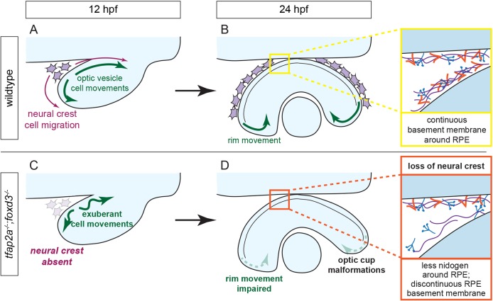
Fig. 8.
Model of optic cup morphogenesis in wild-type and tfap2a;foxd3 double-mutant zebrafish. (A,B) Optic cup morphogenesis in a wild-type embryo. Neural crest cells migrate around the optic vesicle and enable efficient movement of optic vesicle cells (A). Cells undergo rim movement and contribute to the neural retina, partially enabled by the presence of a continuous BM along the surface of the RPE (B). (C,D) Optic cup morphogenesis in a tfap2a;foxd3 double-mutant embryo. Most neural crest cells are absent, resulting in optic vesicle cells that move faster and farther than those in wild-type embryos (C). Rim movement is impaired in the absence of a complete, continuous BM around the RPE, resulting in optic cup malformations (D).

