

Abstract
There are very few reports in the literature about clavicular fractures being associated with a pneumothorax. With this combination of injuries, there are also minimal reports of a delayed presentation of a pneumothorax. This is the first report of a delayed pneumothorax followed by a recurrence of a pneumothorax due to the fractured ends of the clavicle. This case report describes a 49-year-old man who sustained a right-sided pneumothorax from a fractured clavicle several hours after a bus accident. His initial chest examination and radiographs showed no evidence of a pneumothorax. The pneumothorax resolved after 5 days of treatment with a thoracostomy tube. After removing the tube, the procedure was repeated later that day as he again developed a pneumothorax. Ten days later, the patient had surgical intervention of the clavicle due to the unresolved pneumothorax. The clavicle is usually managed conservatively in patients sustaining a clavicular fracture and pneumothorax, however, surgical intervention was mandatory based on failed conservative management. At 3 months follow up, the patient had normal shoulder function. Clinicians must be aware that fractured ends of the clavicle may cause repeated pulmonary damage resulting in a delayed and or a recurrent presentation of a pneumothorax.
Keywords: Clavicle, Delayed pneumothorax, Recurrent pneumothorax, Fracture, Thoracostomy tube, Novel
Highlights
-
•
This is the first reported case of a delayed pneumothorax and a recurrent pneumothorax following an ipsilateral clavicular fracture
-
•
chest X-ray is done in the presence of a clavicle fracture to ensure a potentially fatal pneumothorax is not present
-
•
ongoing monitoring of the respiratory system in patients who sustained a clavicle fracture especially with a high energy mechanism of injury is mandatory
-
•
the clavicle is usually successfully conservatively managed, operative management is mandatory in cases whereby the pneumothorax is not resolving despite appropriate thoracostomy tube placement
Introduction
Clavicle fractures represent approximately 4% of all fractures [1]. There are only a few reported cases of a fractured clavicle associated with a pneumothorax [2]. Additionally, there are even fewer cases of a delayed presentation of a pneumothorax secondary to a fractured clavicle. If a patient develops respiratory symptoms or signs after a clavicle fracture has been diagnosed, the treating clinician should have a high index of suspicion and investigate for a delayed pneumothorax. If this combination of injuries is missed and remains untreated, there may be lethal consequences [2]. We report a case of a delayed pneumothorax with an ipsilateral fracture of the clavicle which then recurred post removal of the initial thoracostomy tube. To our knowledge, there is no other reported case of this occurrence in the literature.
Case presentation
A 49-year-old male presented to the emergency room at the Saint Ann's Bay Regional Hospital after riding in a bus which had overturned. He sustained direct trauma to his right shoulder and chest while the bus was overturning. He complained of right shoulder pain. On examination, there was no evidence of clinical distress. Oxygen saturation was 95% on room air. Breath sounds were normal bilaterally. There was a deformity over the right clavicle with intact overlying skin. There were no upper limb neurovascular deficits.
Initial X-Rays demonstrated a fracture of the middle third of the right clavicle (see Fig. 1). The lung markings extended to the peripheries bilaterally. The following day while on the ward, the patient had a gradual onset of chest pain with associated shortness of breath. Radiographs demonstrated an obvious right sided apical pneumothorax (see Fig. 2). A right sided thoracostomy tube was placed (see Fig. 3). This was left in situ for five days and the patient was extubated after repeat radiographs confirmed full lung expansion. Later that day, the patient had significant shortness of breath. Examination was consistent with a repeat pneumothorax and a right sided thoracostomy tube was again placed. Radiographs later that day was in keeping with a pneumothorax and confirmed an appropriately placed thoracostomy tube. Ten days later, the pneumothorax persisted and the patient was referred to the Orthopaedic team.
Fig. 1.

Initial Chest X-Ray demonstrated no pneumothorax.
Fig. 2.

Chest X-Ray demonstrating delayed pneumothorax.
Fig. 3.

Post Thoracostomy tube placement for pneumothorax.
The patient was taken to the operating theatre after informed consent was obtained (see Fig. 4). An oblique incision was made just inferior to the clavicle. Sharp dissection through the platysma followed by dissection through the clavipectoral fascia was performed. Upon exposure of the fracture, a clamp was utilised to retract the lateral fragment from the pleura. The lateral fragment was found to be inferiorly displaced and easily mobilised. The fracture ends were reduced and maintained with a 6-hole reconstruction plate and screw construct. Closure was performed through the thick skin flaps. The post-operative X-ray taken on Day One demonstrated resolution of the pneumothorax and an anatomical reduction (see Fig. 5, Fig. 6).
Fig. 4.
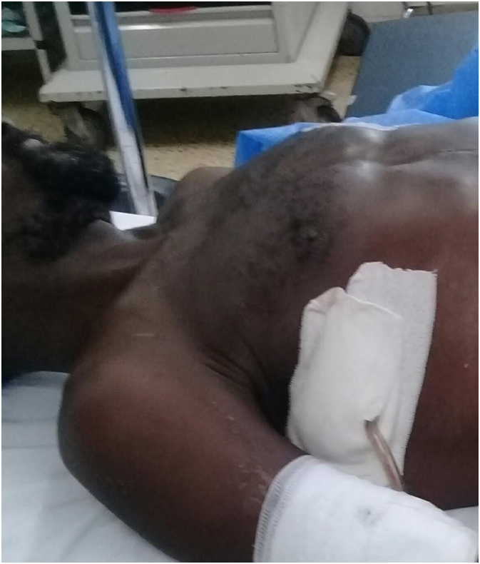
Patient on operating table, claviclular fracture and thorocostomy tube can be seen.
Fig. 5.

Clavicular X-ray Day 1 post operation.
Fig. 6.

Day 1 post operation Chest X-Ray demonstrating resolution of the pneumothorax and the plate and screw construct for clavicle fixation.
Three months post injury; he had full painless range of motion of his right shoulder. There were also no subsequent respiratory symptoms. Radiographs at that time demonstrated a healed fracture with no evidence of a pneumothorax.
Discussion
The clavicle has an S-shape whereby the cross sectional anatomy along its course changes from flat to tubular to prismatic, from laterally to medially. The majority of fractures of the clavicle occur in the middle third due to a stress riser at the junction of the flat and tubular regions [3]. The lung apex, brachial plexus, subclavian vessels and the clavicle are all in close proximity to each other however, it is very rare to have a fractured clavicle associated with a pneumothorax [4]. Kao, et al. [5] stated that the fracture ends directed at the lungs may penetrate the pleural cavity leading to a pneumothorax. When the clavicle is fractured, an associated pneumothorax must be ruled out clinically and radiographically and hence a chest X-ray ought to be mandatory in order to avoid a fatal outcome, especially with a high energy mechanism of injury [6]. If the patient has normal clinical and radiological features, they must be counselled about the possibility of this delayed complication which may easily be missed by inexperienced physicians [6]. Our index case had a high energy mechanism of injury and had developed respiratory signs while he was being monitored.
There have been few case reports in the literature describing the combination of a clavicle fracture and pneumothorax. Ahmed, et al. [6] described nine adult cases with this combination of injuries and only one of those patients had an undisplaced fracture. Steenvorde, et al. [7] found only five such cases in the English speaking literature. He also noted that in all of those cases, the clavicle fracture was managed conservatively while the pneumothorax was always treated with a thoracostomy tube. His report was the first case of both pathologies being managed conservatively. The majority of fractures of the clavicle have been successfully treated conservatively [3]. DeAngelis, et al. [8] found only one case which was operatively managed. In general, some of the accepted indications for surgery of the clavicle are impending skin perforation, neurovascular complications and psuedoarthrosis [3].
Surgery should be strongly considered in patients involved in a high energy mechanism of injury with a widely displaced fracture with multiple associated injuries [8]. DeAngelis, et al. [8] reported a case in which computer tomographic images demonstrated pulmonary alveolar tissue interposed in the fracture site of the clavicle. They felt that this would lead to a non-union of the clavicle and more importantly, would not allow the pneumothorax to resolve. Those authors also felt that in a highly active patient, the possibility of residual disability from either a malunion or non-union would be unacceptable. Our case had developed a pneumothorax after the initial evaluation, and recurred after the first thoracostomy tube was removed. The second pneumothorax was not resolving with the thoracostomy tube in situ for a prolonged period. These factors all played a role in our decision to undergo operative management. The mobile lateral fragment was likely responsible for the delayed, as well as the recurrent pneumothorax. The authors suggest that the injuries were likely to be dynamic in nature. This accounts for the delayed and recurrent presentations. Once the clavicle was surgically stabilised the dynamic repetitive insult was treated. Of note, the pneumothorax resolved the day after the clavicle was fixed.
Conclusion
Patients presenting with a fractured clavicle must be evaluated for a concomitant pneumothorax especially if they are involved in a high energy mechanism of injury, have displaced fragments or have delayed respiratory symptoms. Although the majority of these cases do not require fixation of the clavicle, surgical management should be considered mandatory for cases with an associated recurrent pneumothorax or if the pneumothorax is taking longer than usual to resolve during treatment with a thoracostomy tube.
Authors contributions
-
1)
Cary Fletcher: visualised, conceptualised and writing – review and editing, approval of final manuscript.
-
2)
Kaye Lambert Fletcher: writing – review and editing, approval of final manuscript.
Declaration of competing interest
The authors declare no conflict of interest relevant to this article.
Acknowledgments
Acknowledgements
None.
Funding
This research did not receive any specific grant from funding agencies in the public, commercial, or not-for-profit sectors, and no material support of any kind was received.
Footnotes
The authors confirm that informed consent for the patient was taken for publication of this case report.
References
- 1.Nordqvist A., Petersson C. The incidence of fractures of the clavicle. ClinOrthop. 1994;300:127–132. [PubMed] [Google Scholar]
- 2.Gandham S. 2013. BMJ Case Rep. [DOI] [PMC free article] [PubMed] [Google Scholar]
- 3.Feriani N., BenGhezala H., Snouda S. Pneumothorax caused by an isolated midshaft clavicle fracture. Case Reports in Emergency Medicine. 2016;Volume doi: 10.1155/2016/2409894. [DOI] [PMC free article] [PubMed] [Google Scholar]
- 4.Hani R., Ennaciri B., Jeddi I., Bardouni A.E., Mahfoud M., Berrada M.S. Pneumothorax complicating isolated clavicle fracture. Pan Afr. Med. J. 2015;21:202. doi: 10.11604/pamj.2015.21.202.6796. [DOI] [PMC free article] [PubMed] [Google Scholar]
- 5.Yao Y.H., Goh S.H. Isolated clavicle fracture with secondary pneumothorax: a case report. Hong Kong J. Emerg. Med. 2006;13:113–115. [Google Scholar]
- 6.Ahmed I., Niaz Z., Kassem W., Nabeel M. Eyes can’t see what the mind doesn’t know - a case of pneumothorax complicating clavicle fracture. J. Clin. Rev. Case Rep. 2018;3(1):1–3. [Google Scholar]
- 7.Steenvoorde P., Vanlieshout A.P.W., Oskam J. Conservative treatment of a closed fracture of the clavicle complicated by pneumothorax- a case report. Acta Orthop. Belg. 2005;71:481–483. [PubMed] [Google Scholar]
- 8.DeAngelis R.D., Graf K.W., Mashru R.P. Intrapleural penetration of a clavicle fracture: an indication for operative fixation. Journal of Orthopaedic Case Reports. 2017;7(4):17–20. doi: 10.13107/jocr.2250-0685.830. Jul–Aug. [DOI] [PMC free article] [PubMed] [Google Scholar]