PLOS ONE logoLink to PLOS ONE
. 2020 Feb 27;15(2):e0229238. doi: 10.1371/journal.pone.0229238

Changes in airway diameter and mucus plugs in patients with asthma exacerbation

Yuki Yoshida 1,2,#, Yotaro Takaku 1,*,#, Yasuo Nakamoto 1, Noboru Takayanagi 1, Tsutomu Yanagisawa 1, Hajime Takizawa 2, Kazuyoshi Kurashima 1,#
Editor: Josué Sznitman3
PMCID: PMC7046273  PMID: 32106233

Abstract

Background

Airway obstruction due to decreased airway diameter and increased incidence of mucus plugs has not been directly observed in asthma exacerbation. We studied the changes in the inner diameter of the airway (Din) and the frequency of mucus plugs by airway generation in patients with asthma exacerbation. We compared these patients to those in a stable phase using high-resolution computed tomography (HRCT).

Methods and findings

Thirteen patients with asthma were studied by HRCT during asthma exacerbation and in a stable period. The HRCT study was performed on patients who could safely hold their breath for a short while in a supine position 1 hour after initial treatment for asthma exacerbation. Using a curved multiplanar reconstruction (MPR) software, we reconstructed the longitudinal airway images and the images exactly perpendicular to the airway axis to measure the Din and mucus plugs from the second- (segmental) to sixth-generation bronchi in all segments of the lungs.The ratios of Din (exacerbation/stable) were 0.91(P = 0.016), 0.88 (P = 0.002), 0.83 (P = 0.001), 0.80 (P = 0.001), and 0.87 (NS) in the second-, third-, fourth-, fifth-, and sixth-generation bronchi, respectively. The percentages of airway obstruction due to mucus plugs were notably higher in the fourth- and fifth-generation bronchi (17.9%/18.1% in stable phase and 43.2%/45.9% in the exacerbation phase, respectively) than in the other generations of bronchi.

Conclusions

Among the bronchi examined, the fourth- and fifth-generation bronchi were significantly obstructed during asthma exacerbation compared with the stable phase in terms of a decreased airway diameter and mucus plugs.

Introduction

In asthma exacerbation, the Global Initiative for Asthma (GINA) guideline recommends inhalation treatments with a short-acting beta-2 agonist (SABA) and ipratropium bromide in addition to systemic corticosteroids [1]. However, most inhaled therapies do not reach the small airways (airways with internal diameter < 2 mm) that comprise multiple aspects of asthma [28]. Also, mucus plugs are considered to be one of the mechanisms of airway obstruction in fatal asthma [9,10]. If the inner diameter of the airway (Din) and mucus plugs could be directly observed in acute asthma, it would help us understand how inhalers act on airways in asthmatics. The mucus plugs, as well as narrowed airways, could be the target of inhaled corticosteroids and bronchodilators during asthma exacerbation.

Recently, a quantitative image analysis of airway obstruction due to mucus plugs has been explored in severe asthma [11]. Quantitative computed tomography (CT) imaging can provide structural and functional information on asthma [1216]. The recently developed CT technique known as curved multiplanar reconstruction (MPR) can visualize longitudinal airway images and accurately analyze short-axis images of small airways that cannot be recognized in standard high-resolution CT (HRCT) images. Using such a technique, we have shown that small airways with a 1.5-mm Din on a conventional multidetector HRCT or a 0.8-mm Din on ultra-HRCT can be measured in clinical situations [17, 18]. Airway analysis of MPR images can be used to measure mucus plugs quantitatively as well.

The purpose of this study is to evaluate the changes in Din and the frequency of airway occlusion by mucus plugs from the segmental bronchus (second generation) to the sixth-generation bronchi by comparing HRCT data during asthma exacerbation with those obtained in the stable phase. The findings might provide a new dimension in quantifying asthma severity and asthma heterogeneity.

Materials and methods

Study design and participants

The Institutional Review Board of Saitama Cardiovascular and Respiratory Center approved this study (IRB No. 2012 033), and written informed consent was obtained from all study patients. CT scans of the subjects were obtained during asthma exacerbation and in the stable phase during the follow-up period. There was over 6 months between the CT scans in the exacerbation phase and the recovery phase. The asthma severity in the stable phase and exacerbation severity were classified according to the GINA guidelines [1]. We evaluated asthma control according to the consensus-based GINA symptom control tool. All patients during the asthma exacerbation phase visited our clinic due to the worsening of wheezing with dyspnea and were diagnosed with asthma exacerbation. The severity of asthma exacerbation in this study’s patients was determined according to the descriptions in the section “Management of asthma exacerbations in the emergency department” in the GINA guidelines. We used objective assessment and other measurements, such as respiratory rate, pulse rate, O2 saturation, and accessory muscles being used, for the determination. The patients with asthma exacerbation with life-threatening signs, as assessed by the doctor in charge, were excluded from this study. The CT study was performed 1 hour after initial treatments to evaluate the airway obstruction and screen for other comorbidities that mimic asthma exacerbation, such as bronchopneumonia or heart failure. In the stable phase of asthma, a CT study was performed to assess airway remodeling and airway dimensions. Stable asthma was defined as the absence of clinic visits for asthma exacerbation, unchanged use of asthma medication for maintenance therapy, and the stable use of rescue medication (no more than four puffs per day of a short-acting bronchodilator) during the previous month.

CT data acquisition and image analyses

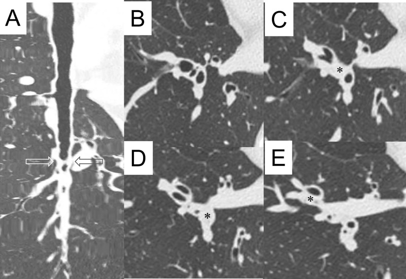
A 256-slice CT scanner (Brilliance iCT; Philips Healthcare, Cleveland, OH, USA) was used as previously described [17]. The CT scans were obtained from the suspended end-inspiratory volume at baseline and during exacerbation. We also checked lung size and lung architecture in the same slice to confirm they were obtained at the end-inspiratory volume. All CT row data sets were analyzed using an MPR software program (SYNAPSE3D, Fujifilm Co. Tokyo, Japan), where the Din and mucus plugs were measured per airway generation for all 18 segments of the lung. We first selected a target airway and a point in the airway lumen at the level of the segmental bronchus, that is, the second-generation bronchus according to the Japanese Cancer Society [19]. From this point, the airway tree was identified using a trained detector and a spanning tree algorithm, and we obtained the selected bronchial pathway to the point of the sixth-generation bronchus. If a mucus plug obstructed the bronchus before the sixth-generation bronchus, we could trace the bronchus manually to the sixth-generation bronchus. The bronchus was selected on the principle that it should be more centered and well recognized in one segment. The same bronchial trees were used for analysis during the stable phase and at exacerbation. We were then able to obtain short-axis images that were exactly perpendicular to the long axis of the airway at each generation bronchus. From the centroid point of the lumen, rays fanning out over 360 degrees were examined to determine the inner airway walls along the rays using the full width with the half-maximum principle. We were then able to obtain the mean values for the Din. Mucus plug (mucus occlusion) was defined as the complete occlusion of a bronchus, contiguous with patent airway lumen on the longitudinal airway image (Fig 1). Mucus plugs were distinguished from focal opacified airways, such as airway collapse or adjacent blood vessels, identifying no focal decrease of outer airway caliber and recognizing adjacent blood vessels between opened proximal and distal airways from the mucous area. We checked whether mucus plugs were associated with bronchiectasis, defined as a bronchoarterial ratio of greater than 1.5. We noted the presence of mucus plugs as “yes” or “no” from the second- to sixth-generation bronchial compartments in one selected airway in one segment. We could then calculate the percentage of mucus plug in a given bronchial generation in a given lung segment. The validation of these CT measurements has been described by [17].

Fig 1. Representative view of mucus plugs.

Fig 1

A: Longitudinal view of right B10 bronchus. Peripheral parts of the bronchus (bottom of the panel) are open. B: Conventional HRCT images of right B7, B8, B9, and B10. C: Conventional HRCT image just below the level of panel B. Occluded segmental bronchus (second generation, marked with an asterisk) of right B9 is shown. D: Conventional HRCT image just below the level of panel C (indicated level in panel A by arrows). Occluded segmental bronchus (second generation, marked with an asterisk) of right B10 is shown. E: Conventional HRCT image just below the level of panel D. Occluded subsegmental bronchus (third generation, marked with an asterisk) of right B8 is shown.

Pulmonary function tests

Pulmonary function tests (PFTs) were performed within two weeks of obtaining the HRCT scans in the stable phase. PFTs were performed according to the guidelines of the American Thoracic Society [20]. Spirometry parameters (forced expiratory volume in 1 second (FEV1) and FEV1/forced vital capacity (FVC)) were measured with a CHESTAC8800 (Chest Inc., Tokyo, Japan). Spirometry was done before and 20–30 minutes after the inhalation of 200 g of salbutamol, and the values after the bronchodilator were used for baseline data.

Statistical analyses

Mean Din and the number of mucus-plug-positive bronchial compartments were calculated in the second- to sixth-generation bronchial compartments in all 18 segments of the lung in 13 subjects. These values were compared by total, by bronchial generations, or by lobes between the stable phase and acute phase. The mean Din of the total compartments and the total number of mucus-positive bronchial compartments in the stable phase were analyzed for correlation to spirometry valuables in the stable phase. Data are expressed as the mean ± standard deviation (SD) or as the median (range), as appropriate. A paired t test was used to compare the Din in the stable and acute phase. Statistical analyses between groups were first performed with the Kruskal–Wallis test followed by Dunn’s test for comparisons between groups. A value of P < 0.05 was considered to be significant. The Prism 5 software program (GraphPad Software, Inc., La Jolla, CA, USA) was used for the analyses.

Results

Seventeen patients who had emergency visits to our center with asthma attacks were included. Of these, three were excluded because of their life-threatening condition. One did not consent, and the remaining 13 patients were enrolled. The patient characteristics are reported in Table 1. There was no notable difference in the number of men and women in the subjects. Events of asthma exacerbation were classified as mild/moderate and severe, and four patients were hospitalized for further treatment. When CT studies were performed after initial treatments, no patients showed any life-threatening signs or orthopnea. All patients could hold full inspiration for a while. The patients’ asthma was well (61.5%) or partly controlled (38.5%) in the stable phase.

Table 1. Patient characteristics.

Number of subjects 13
Age (years) 56 (± 9.8)
Gender, F:M 7:6
Smoking history (number) Never: 7
Ex: 6
Current: 0
Asthma duration (years) 3.5 (1.6–13.4)
Asthma medication SABA: 3
ICS: 1
ICS/LABA: 3
ICS/LABA/LTRA: 1
ICS/LABA/LTRA/TP: 2
ICS/LABA/LTRA/TP/OCS: 2
ICS/LABA/LTRA/TP/OCS/anti-IgE: 1
Asthma severity at stable (number) Well-controlled: 8
Partly controlled: 5
Exacerbation severity (number) Mild or moderate: 9
Severe: 4
Life-threatening: 0
Body mass index (kg/m2) 24.3 (±4.3)
Baseline FEV1(% predicted) 82.2 (±16.4)
Baseline FVC (% predicted) 87.1 (±17.4)
Baseline FEV1/FVC (% predicted) 85.3 (±14.9)
Bronchodilator reversibility (%) 5.4 (±4.7)
Baseline FeNO (ppb) 47.2 (±32.6)
Baseline eosinophils in blood (×103 μ/L) 0.54 (±0.60)
Exacerbation eosinophils in blood (×103 μ/L) 0.83 (±0.99)

FeNO, fractional exhaled nitric oxide; FEV1; forced expiratory volume 1 second; FVC, forced vital capacity; ICS, inhaled corticosteroid; LABA, long-acting beta-2 agonist; LTRA, leukotriene receptor antagonist; OCS, oral corticosteroid; SABA, short-acting beta-2 agonist; TP, theophylline

In 13 patients, five bronchial compartments from segmental (second generation) to the sixth-generation bronchus of the representative airway for one segment were analyzed in 18 segments of the lung in the stable phase and during an asthma exacerbation. As shown in Fig 1, Din was not measured in mucus-occluded parts.

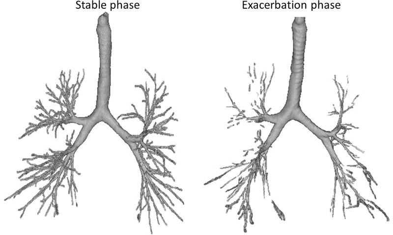
Fig 2 shows a representative view of the bronchial trees of the same patient in the stable phase and during exacerbation. The tracheal smooth muscles constricted, and the tracheal cartilage is visible from the upper to the middle part of the trachea during asthma exacerbation. Parts of the peripheral airways could not be traced, and middle-zone airways were thin or occluded during exacerbation.

Fig 2. Representative view of bronchial trees by HRCT of the same asthma patient in the stable phase and asthma exacerbation.

Fig 2

During the exacerbation phase, the tracheal smooth muscles are constricted, and the tracheal cartilage is visible. Parts of the peripheral airways could not be traced, and middle-zone airways are thin or occluded at exacerbation.

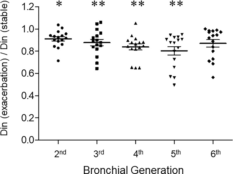
Fig 3 shows ratios of Din during exacerbation and the stable phase. Each point represents the mean Din ratio of 13 subjects for each segment. The mean Din for all segments of 13 patients were 3.6 ± 0.5, 2.6 ± 0.3, 2.2 ± 0.3, 1.8 ± 0.2, and 1.8 ± 0.2 mm, respectively, in the second- to sixth-generation bronchi at asthma exacerbation. The ratios of the Din were notably reduced in the second-, third-, fourth-, fifth-, and sixth-generation bronchi (0.91 ± 0.08, P = 0.016; 0.88 ± 0.13, P = 0.002; 0.83 ± 0.11, P = 0.001; 0.80 ± 0.15, P = 0.001; 0.87 ± 0.13, NS, respectively).

Fig 3. The ratios of Din during asthma exacerbation and in the stable phase observed from the second- (segmental) to sixth-generation bronchi.

Fig 3

Din was measured per airway generation in all segments of the lung by HRCT for each subject at suspended end-inspiratory volume using curved MPR software. Each point represents the mean Din ratio of 13 subjects for each segment. * denotes P < 0.05 and ** denotes P < 0.01.

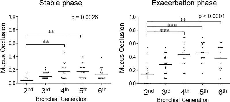
The ratios of mucus-occluded bronchial compartments in 13 subjects by airway generation during asthma exacerbation and the stable phase are shown in Fig 4. Each point represents the ratio of mucus-occluded compartments in 13 patients for each segment.

Fig 4. The ratio of mucus-occluded bronchial compartments in 13 subjects by airway generation during asthma exacerbation and the stable phase.

Fig 4

Mucus occlusion (mucus plug) was measured by HRCT using curved MPR software. Mucus plugs are defined as the complete occlusion of a bronchus, contiguous with patent airway lumen on longitudinal airway image. Mucus occlusions are counted per airway generation in all segments of the lung for each subject. Each point represents the ratio of mucus-occluded compartments in 13 patients for each segment.

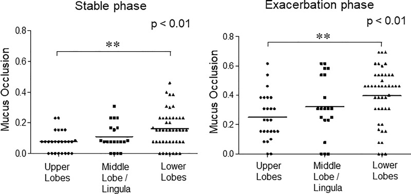
In the stable phase of asthma, mucus occlusion was observed in 17.9% of the fourth-generation bronchi and 18.1% in fifth-generation bronchi. Mucus plugs are most notable in the fourth- and fifth-generation bronchi and in the lower lobes (left panels of Fig 4 and Fig 5). During the exacerbation phase of asthma, mucus plugs are observed in 43.2% of the fourth-generation bronchi and 45.9% in fifth-generation bronchi. Mucus plugs are most notable in the fourth- and fifth-generation bronchi and in the lower lobes (right panels of Fig 4 and Fig 5). Interestingly, the levels of airway where the mucus occlusion is most frequently observed are different among the lung lobes, especially during asthma exacerbation. In the upper lobes, the frequency of mucus plugs is higher in the fifth- and sixth-generation bronchi, but in the lower lobes, the frequency of mucus plugs is higher in the fourth- and fifth-generation bronchi than in others (S1 Fig and S2 Fig). These mucus plugs occurred in the absence of bronchiectasis.

Fig 5. The ratio of mucus-occluded bronchial compartments in 13 subjects during asthma exacerbation and the stable phase observed in the upper, middle/lingula, and lower lobes.

Fig 5

Mucus occlusion (mucus plug) was measured by HRCT using curved MPR software. Mucus occlusions were counted per airway generation in all segments of the lung for each subject. Airway segment data were then grouped by lobes for analysis. Each point represents the ratio of mucus-occluded bronchial compartments in 13 patients for each bronchial generation/segment.

The relationship between CT image parameters and airflow obstruction by spirometry could be analyzed in the stable phase (Table 2). The mean Din for all compartments or by airway generation (data not shown) did not significantly correlate with the predicted percentage of FEV1 or FEV1/FVC, but the total number of mucus-occluded bronchial compartments for all compartments significantly correlated with the predicted percentage for FEV1 (P = 0.0162) and FEV1/FVC (P = 0.0064).

Table 2. The relationship between the airway diameter, mucus score, and spirometry measures.

Spirometry value Total bronchial area Din (mm) Total bronchial area mucus plug (number)
FEV1 (% predicted) NS −1.1 (−1.9, −0.3)
R2 = 0.42, P = 0.0162
FVC (% predicted) NS NS
FEV1/FVC NS −0.6 (−1.1, −0.2)
R2 = 0.51, P = 0.0064

NS, not significant

The linear regression model reports a coefficient (95% CI) for spirometry values. FEV1 and FVC denote forced expiratory volume for 1 second and forced vital capacity, respectively. The inner airway diameter (Din) and presence of mucus plugs are measured in the second- to sixth-generation bronchial compartments in the selected airway (see text) in each of the 18 segments of the lung. The total bronchial area Din represents the mean Din of the total bronchial compartments, and the total bronchial area mucus plug represents the total number of mucus-plug-positive compartments.

Regarding exacerbation severity, nine cases were classified as mild/moderate exacerbation, and four cases were classified as severe exacerbation. The mean Din for all compartments in mild/moderate exacerbation cases and severe cases were not different in the stable phase (2.6 ± 0.8 mm versus 2.5 ± 0.8 mm, NS), but the mean Din was smaller in patients with severe exacerbation (2.5 ± 0.8 mm2 versus 2.3 ± 0.7 mm, P = 0.042). The average percentage of mucus plugs in the total measurement points in mild/moderate exacerbation cases and severe cases were 35.1% ± 19.6% versus 29.8% ± 27.5%, respectively, during the exacerbation phase (NS).

Discussion

In this study, we explored the Din and mucus plugs from the segmental bronchi (second generation) to the sixth-generation bronchi during asthma exacerbation and in the stable phase. During asthma exacerbation, the most significant change in the Din was observed at the fifth-generation bronchi, and its mean Din was below 2 mm. Also, more than 40% of the airways were occluded by mucus plugs at the fourth- (sub-subsegmental) and fifth-generation bronchi during asthma exacerbation. Because of the decreased Din and mucus plugs, considerable parts of the airways were severely occluded in the middle zone in acute asthma. These findings may help us to understand the pathophysiology of acute asthma and may provide useful information in quantifying asthma severity.

CT is useful for detecting underlying comorbidities and assessing the severity of airway obstruction in acute asthma. With the analysis of HRCT images, we were able to exclude bronchopneumonia, heart failure, and other diseases that mimic asthma exacerbation.

In quantitative CT imaging, the airway parameters of wall thickness percentage (WT%), wall area percent (WA%), and low attenuation area have been well studied concerning lung function or asthma severity [1216]. Mucus plugs have only been recently recognized as another CT parameter for the severity of asthma [11]. In this study, we focused on Din and mucus plugs that would directly influence the effectiveness of inhalation therapy during asthma exacerbation. Mucus plugs were more frequently observed than we expected during asthma exacerbation on longitudinal images of the airway. The definition of mucus plugs by CT image was the same as that of the report by Dunican et al. [11]. However, a plugged airway in a cross section and longitudinal section could not necessarily be seen by conventional HRCT images. In this study, precise cross-sectional and longitudinal-sectional images could be obtained by MPR for each bronchial segment. Also, we analyzed the peripheral airway to the sixth-generation bronchi.

In a pathological model of bronchial trees, small airways were reported to compromise the airway generations 8–23 [21]. Since airways with a diameter of < 2 mm are classified as small airways, this study covers small airways. Changes of airway lumen area on HRCT and airway reactivity were first described by Herold et al. [22]. Okazawa et al. [23] identified airway narrowing of intermediate-sized airways produced by inhaled methacholine in asthmatic patients. Shimizu et al. reported that the WA% of the fourth- and fifth-generation bronchi correlated well with airflow limitation in older asthmatics [24], and we previously reported that the airway diameter of the fifth-generation bronchi was most significantly correlated with FEV1 in patients with stable asthma [17]. In the present study, because of the small size of the data, we could not show the relationship between Din and the predicted percent of FEV1 or FEV1/FVC. However, we did show that the total number of mucus plugs correlated with the predicted percent of FEV1 and FEV1/FVC during the stable phase. It supports the previous observation [11] that mucus plugs are associated with airflow limitation in asthma.

In this study, we found that the Din in the fifth-generation bronchi reduced notably during asthma exacerbation. Also, mucus plugs were predominantly observed in sub-subsegmental (fourth-generation) and the fifth-generation bronchi during asthma exacerbation as well as in the stable phase. These observations are partly in accordance with the findings of a previous study that reported that most mucus plugs were in subsegmental bronchi in patients with severe asthma [11]. These data suggest that the fourth- and fifth-generation bronchi are functionally critical areas in bronchial asthma. Further studies on the severity of airway inflammation per airway generation are needed to answer why morphometrical changes are most significant in these levels of bronchi in patients with asthma.

The airway mucus in healthy people is normally a lightly cross-linked gel that does not form plugs [2526], and why mucus plugs are so common in asthma and why mucus plugs are less frequently observed in more distal airways than in more proximal ones remain to be determined. It is noteworthy that, if there is no surfactant on the airway surface, a small quantity of liquid would move to a narrower part of the airways and eventually block the airway lumen with surface tension [27]. It is reported that the stability of the small airway is maintained by the airway surfactant [28, 29], and the surface activity of the sputum is disrupted in conditions of acute asthma [30, 31]. Therefore, the airway surfactant from the alveolus might partly explain why there are more mucus plugs in the fourth- and fifth-generation bronchi than in sixth-generation bronchi.

There are several limitations to this study that should be noted.

Accuracy of measurements

It is well accepted that recent CT scanners can correctly measure 1.5-mm airways [32]. In our previous study, differences in phantom tubes (Din, 1.5–4.9 mm) and CT data were 0.1–0.2 mm for Din, and their coefficient of variation (CV) was 1.14%–2.14% [17]. Because of the small differences in repeated measurements, we considered that it was enough to compare the Din in stable phase asthma and asthma exacerbation. However, we avoided using the airway luminal area and WA% to minimize the CV.

Other limitations

First, the number of study patients was limited. Performing a CT study under the same CT scanner/condition was not possible with a multicenter study, and only part of the asthma exacerbations were studied because of the priority of treatments. To compare the Din at the stable phase and exacerbation, patients had to hold their breath at full inspiration for a while. Therefore, the patients who could not hold their breath at full inspiration could not enter this study. For this reason, the interpretation of the data according to the severity of the asthma attack is limited. Second, the Din during an asthma exacerbation should be much smaller than those presented because we measured the Din 1 hour after the initiation of treatment. Specifically, Din in the fourth- and fifth-generation bronchi should be much smaller because the Din in the compartments with full mucus plugs could not be measured. Because of these limitations, the changes in the Din during an asthma attack should have only been moderately assessed. However, it would be enough to show the relative site of airway obstruction and how severe the airway obstruction is during asthma exacerbation. Finally, our data do not exclude the possible pathological significance in the more distal part of the sixth-generation bronchi.

Our data provide evidence that severe airway obstruction occurs at the fourth- and fifth-generation bronchi in acute asthma. During asthma exacerbation, these airways may become less than 2 mm in diameter or occluded by mucus plugs. Nonetheless, it should be important to note that the first-line treatment for asthma exacerbation is the use of inhalers [1], and they act directly on obstructed airways. This study shows considerable mucus plugs in acute asthma, but all the patients in this study were successfully relieved by inhaled bronchodilators and systemic corticosteroids. It might be that even partial responses to the inhalers were effective to relieve dyspnea in asthmatics. Mucus plugs can be another target of therapy in acute asthma, as well as in the stable phase.

Supporting information

S1 Fig. Ratio of mucus-occluded bronchial compartments in 13 subjects during asthma exacerbation and the stable phase observed from the second- (segmental) to sixth-generation bronchi in the upper, middle/lingula, and lower lobes.

Mucus occlusions were measured by HRCT using curved MPR software. Mucus occlusions were counted per airway generation in all segments of the lung for each subject. Airway segment data were grouped by lobes and by airway generation for analysis. Each point represents the ratio of mucus-occluded compartments in 13 patients for each segment.

(TIF)

S2 Fig. Ratio of mucus-occluded bronchial compartments in 13 subjects during asthma exacerbation and in the stable phase by lung segments.

Mucus occlusion (mucus plug) was measured by HRCT for each subject at suspended end-inspiratory volume using curved MPR software. Airway segment data per airway generation are expressed. The black bar represents the ratio of mucus occlusion in the stable phase, and the black bar plus gray bar represents the ratio of mucus occlusion during asthma exacerbation.

(TIF)

Acknowledgments

We wish to thank the Saitama Cardiovascular and Respiratory Center medical staff who cared for the patients.

Data Availability

All relevant data are within the paper and its Supporting Information files.

Funding Statement

The authors received no specific funding for this work.

References

  • 1.Global Initiative for Asthma (GINA). Global strategy for asthma management and prevention, updated 2018. Available from:www.ginasthma.org. Accessed: May 10, 2018.
  • 2.Lavorini F, Pedersen S, Usmani OS. Dilemmas, confusion, and misconceptions related to small airways directed therapy. Chest. 2017;151:1345–55. 10.1016/j.chest.2016.07.035 [DOI] [PubMed] [Google Scholar]
  • 3.Usmani OS. Small-airway disease in asthma: pharmacological considerations. Curr Opin Pulm Med. 2015;21:55–67. 10.1097/MCP.0000000000000115 [DOI] [PubMed] [Google Scholar]
  • 4.Usami OS. Small airways dysfunction in asthma: evaluation and management to improve asthma control. Allergy Asthma Immunol Res. 2014;6:376–88. 10.4168/aair.2014.6.5.376 [DOI] [PMC free article] [PubMed] [Google Scholar]
  • 5.In’t Veen JC, Beekman AJ, Bel EH, Sterk PJ. Recurrent exacerbation in severe asthma are associated with enhanced airway closure during stable episodes. Am J Respir Crit Care Med. 2000;161:1902–6. 10.1164/ajrccm.161.6.9906075 [DOI] [PubMed] [Google Scholar]
  • 6.Kraft M, Pak J, Martin RJ, Kaminsky D, Irvin CG. Distal lung dysfunction at night in nocturnal asthma. Am J Respir Crit Care Med. 2001;163;1551–6. [DOI] [PubMed] [Google Scholar]
  • 7.Kaminsky DA, Irvin CG, Gurka DA, Feldsien DC, Wagner EM, Liu MC, et al. Peripheral airways responsiveness to cool, dry air in normal and asthmatic individuals. Am J Respir Crit Care Med. 1995;152:1784–90. [DOI] [PubMed] [Google Scholar]
  • 8.Usmani OS, Singh D, Spinola M, Bizzi A, Barnes PJ. The prevalence of small airways in adult asthma: a systemic literature review. Respir Med. 2016;116:19–27. 10.1016/j.rmed.2016.05.006 [DOI] [PubMed] [Google Scholar]
  • 9.Huber HC, Koessler KK. Pathology of bronchial asthma. Arch Intern Med. 1992;30:689–760. [Google Scholar]
  • 10.Dunnill MS. The pathology of asthma, with special reference to changes in the bronchial mucosa. J Clin Pathol. 1960;13:27–33. 10.1136/jcp.13.1.27 [DOI] [PMC free article] [PubMed] [Google Scholar]
  • 11.Dunican EM, Elicker BM, Gierada DS, Nagle SK, Schiebler ML, Newell JD, et al. Mucus plugs in patients with asthma linked to eosinophilia and airflow obstruction. J Clin Invest. 2018;128:997–1009. 10.1172/JCI95693 [DOI] [PMC free article] [PubMed] [Google Scholar]
  • 12.Montaudon M, Lederlin M, Reich S, Begueret H, Tunon-de-Lara JM, Marthan R, et al. Bronchial measurements in patients with asthma: comparison of quantitative thin-section CT findings with those in healthy subjects and correlation with pathologic findings. Radiology. 2009;253:844–53. 10.1148/radiol.2533090303 [DOI] [PubMed] [Google Scholar]
  • 13.de Jong PA, Müller NL, Coxson HO. Computed tomographic imaging of the airways: relationship to structure and function. Eur Respir J. 2005;26:140–52. 10.1183/09031936.05.00007105 [DOI] [PubMed] [Google Scholar]
  • 14.DeBoer EM, Spielberg DR, Brody AS. Clinical potential for imaging in patients with asthma and other lung disorders. J Allergy Clin Immunol. 2017;139:21–8. 10.1016/j.jaci.2016.11.004 [DOI] [PubMed] [Google Scholar]
  • 15.Choi S, Hoffman EA, Wenzel SE, Castro M, Fain S, Jarjour N, et al. Quantitative computed tomographic imaging-based clustering differentiates asthmatic subgroups with distinctive clinical phenotypes. J Allergy Clin Immunol. 2017;140:690–700. 10.1016/j.jaci.2016.11.053 [DOI] [PMC free article] [PubMed] [Google Scholar]
  • 16.Shim SS, Schiebler ML, Evans MD, Jarjour N, Sorkness RL, Denlinger LC et al. Lumen area change (Delta Lumen) between inspiratory and expiratory CT as a measure of severe asthma. J Allergy Clin Immunol. 2018;142:1773–80. 10.1016/j.jaci.2017.12.1004 [DOI] [PMC free article] [PubMed] [Google Scholar]
  • 17.Kurashima K, Hoshi T, Takayanagi N, Takaku Y, Kagiyama N, Ohta C, et al. Airway dimensions and pulmonary function in chronic pulmonary disease and bronchial asthma. Respiology. 2012;17:79–86. [DOI] [PubMed] [Google Scholar]
  • 18.Kurashima K, Hoshi T, Takaku Y, Kanauchi T, Nakamoto K, Ueda M, et al. Changes in the airway lumen and surrounding parenchyma in chronic obstructive pulmonary disease. Int J chronic Obstruct Pulmon Dis. 2013;8:523–32. [DOI] [PMC free article] [PubMed] [Google Scholar]
  • 19.The Japan Lung Cancer Society. General Rule for Clinical and Pathological Record of Lung Cancer. Kanehara Ltd, Tokyo, 1999. [Google Scholar]
  • 20.Standardization of Spirometry, 1994 Update. American Thoracic Society. Am J Respir Crit Care Med. 1995;152:1107–36. 10.1164/ajrccm.152.3.7663792 [DOI] [PubMed] [Google Scholar]
  • 21.Weibel ER. Morphometry of the Human Lung. Berlin, Germany: Springer-Verlag; 1963, 4–9. [Google Scholar]
  • 22.Herold CJ, Brown RH, Mitzner W, Links JM, Hirshman CA, Zerhouni EA. Assessment of pulmonary airway reactivity with high-resolution CT. Radiology. 1991;181:369–74. 10.1148/radiology.181.2.1924774 [DOI] [PubMed] [Google Scholar]
  • 23.Okazawa M, Müller N, McNamara AE, Child S, Verburgt L, Paré PD. Human airway narrowing measured using high resolution computed tomography. Am J Respir Crit Care Med. 1996;154:1557–62. 10.1164/ajrccm.154.5.8912780 [DOI] [PubMed] [Google Scholar]
  • 24.Shimizu K, Hasegawa M, Makita H, Nasuhara Y, Konno S, Nishimura M. Airflow limitation and airway dimensions assessed per bronchial generation in older asthmatics. Respir Med. 2010;104:1809–16. 10.1016/j.rmed.2010.06.013 [DOI] [PubMed] [Google Scholar]
  • 25.Innes AL, Carrington SD, Thornton DJ, Kirkham S, Rousseau K, Dougherty RH, et al. Ex vivo sputum analysis reveals impairment of protease-dependent mucus degradation by plasma proteins in acute asthma. Am J Respir Crit Care Med. 2009;180:203–10. 10.1164/rccm.200807-1056OC [DOI] [PMC free article] [PubMed] [Google Scholar]
  • 26.Fahy JV, Dckey BF. Airway mucus function and dysfunction. N Eng J Med. 2010;363:2233–47. [DOI] [PMC free article] [PubMed] [Google Scholar]
  • 27.Enhorning G, Holm BA. Disruption of pulmonary surfactant's ability to maintain openness of a narrow tube. J Appl Physiol. 1993;74:2922–7. 10.1152/jappl.1993.74.6.2922 [DOI] [PubMed] [Google Scholar]
  • 28.Macklem PT, Proctor DF, Hogg JC. The stability of peripheral airways. Resp Physiol. 1970;8:191–203. [DOI] [PubMed] [Google Scholar]
  • 29.Enhorning G, Duffy LC, Welliver RC. Pulmonary surfactant maintains patency of the rat's conducting airways. Am J Respir Crit Care Med. 1995;151:554–6. 10.1164/ajrccm.151.2.7842219 [DOI] [PubMed] [Google Scholar]
  • 30.Liu M, Wang L, Enhorning G. Surfactant dysfunction develops when the immunized guinea-pig is challenged with ovalbumin aerosol. Clin Exp Allergy. 1995;25:1053–60. 10.1111/j.1365-2222.1995.tb03251.x [DOI] [PubMed] [Google Scholar]
  • 31.Kurashima K, Fujimura M, Matsuda T, Kobayashi T. Surface activity of sputum from acute asthmatic patients. Am J Respir Crit Care Med. 1997;155:1254–9. 10.1164/ajrccm.155.4.9105063 [DOI] [PubMed] [Google Scholar]
  • 32.Hasegawa M, Nasuhara Y, Onodera Y, Makita H, Nagai K, Fuke S, et al. Airflow limitation and airway dimensions in chronic obstructive pulmonary disease. Am J Respir Crit Care Med. 2006;173:1309–15. 10.1164/rccm.200601-037OC [DOI] [PubMed] [Google Scholar]

Decision Letter 0

Josué Sznitman

30 Oct 2019

PONE-D-19-25209

Changes in airway dimensions and mucus plugs in patients with asthma exacerbation

PLOS ONE

Dear Dr Takaku,

Thank you for submitting your manuscript to PLOS ONE. After careful consideration, we feel that it has merit but does not fully meet PLOS ONE’s publication criteria as it currently stands. Therefore, we invite you to submit a revised version of the manuscript that addresses the points raised during the review process.

The Reviewers have pointed to a number of issues that should be amended prior to resubmission.

We would appreciate receiving your revised manuscript by Dec 14 2019 11:59PM. When you are ready to submit your revision, log on to https://www.editorialmanager.com/pone/ and select the 'Submissions Needing Revision' folder to locate your manuscript file.

If you would like to make changes to your financial disclosure, please include your updated statement in your cover letter.

To enhance the reproducibility of your results, we recommend that if applicable you deposit your laboratory protocols in protocols.io, where a protocol can be assigned its own identifier (DOI) such that it can be cited independently in the future. For instructions see: http://journals.plos.org/plosone/s/submission-guidelines#loc-laboratory-protocols

Please include the following items when submitting your revised manuscript:

  • A rebuttal letter that responds to each point raised by the academic editor and reviewer(s). This letter should be uploaded as separate file and labeled 'Response to Reviewers'.

  • A marked-up copy of your manuscript that highlights changes made to the original version. This file should be uploaded as separate file and labeled 'Revised Manuscript with Track Changes'.

  • An unmarked version of your revised paper without tracked changes. This file should be uploaded as separate file and labeled 'Manuscript'.

Please note while forming your response, if your article is accepted, you may have the opportunity to make the peer review history publicly available. The record will include editor decision letters (with reviews) and your responses to reviewer comments. If eligible, we will contact you to opt in or out.

We look forward to receiving your revised manuscript.

Kind regards,

Josué Sznitman

Academic Editor

PLOS ONE

Journal Requirements:

1. When submitting your revision, we need you to address these additional requirements.

Please ensure that your manuscript meets PLOS ONE's style requirements, including those for file naming. The PLOS ONE style templates can be found at

http://www.journals.plos.org/plosone/s/file?id=wjVg/PLOSOne_formatting_sample_main_body.pdf and http://www.journals.plos.org/plosone/s/file?id=ba62/PLOSOne_formatting_sample_title_authors_affiliations.pdf

2. We noticed you have some minor occurrence(s) of overlapping text with the following previous publication(s), which needs to be addressed:

https://doi.org/10.1111/j.1440-1843.2011.02052.x

doi: 10.1097/LBR.0000000000000020

In your revision ensure you cite all your sources (including your own works), and quote or rephrase any duplicated text outside the Methods section. Further consideration is dependent on these concerns being addressed.

3. Please include in your Methods section the date ranges over which you recruited participants to this study.

4. Please amend either the title on the online submission form (via Edit Submission) or the title in the manuscript so that they are identical.

[Note: HTML markup is below. Please do not edit.]

Reviewers' comments:

Reviewer's Responses to Questions

Comments to the Author

1. Is the manuscript technically sound, and do the data support the conclusions?

The manuscript must describe a technically sound piece of scientific research with data that supports the conclusions. Experiments must have been conducted rigorously, with appropriate controls, replication, and sample sizes. The conclusions must be drawn appropriately based on the data presented.

Reviewer #1: Yes

Reviewer #2: Yes

Reviewer #3: Yes

**********

2. Has the statistical analysis been performed appropriately and rigorously?

Reviewer #1: I Don't Know

Reviewer #2: Yes

Reviewer #3: Yes

**********

3. Have the authors made all data underlying the findings in their manuscript fully available?

The PLOS Data policy requires authors to make all data underlying the findings described in their manuscript fully available without restriction, with rare exception (please refer to the Data Availability Statement in the manuscript PDF file). The data should be provided as part of the manuscript or its supporting information, or deposited to a public repository. For example, in addition to summary statistics, the data points behind means, medians and variance measures should be available. If there are restrictions on publicly sharing data—e.g. participant privacy or use of data from a third party—those must be specified.

Reviewer #1: Yes

Reviewer #2: No

Reviewer #3: No

**********

4. Is the manuscript presented in an intelligible fashion and written in standard English?

PLOS ONE does not copyedit accepted manuscripts, so the language in submitted articles must be clear, correct, and unambiguous. Any typographical or grammatical errors should be corrected at revision, so please note any specific errors here.

Reviewer #1: No

Reviewer #2: No

Reviewer #3: No

**********

5. Review Comments to the Author

Please use the space provided to explain your answers to the questions above. You may also include additional comments for the author, including concerns about dual publication, research ethics, or publication ethics. (Please upload your review as an attachment if it exceeds 20,000 characters)

Reviewer #1: In this paper, Yoshida et al. investigated the utility of using Chest CT scans to evaluate airway diameter and mucus plugs in a small cohort of patients with asthma both during and after an exacerbation. This paper builds on growing evidence from several groups that CT scans provide useful information in quantifying asthma severity, and can possibly provide a new dimension of asthma heterogeneity (e.g. the CT-derived “mucus score”, recently reported by Dunican et al.).

Yoshida et al. studied 13 asthmatics one hour after initial presentation for an asthma exacerbation, as long as they were able to lie flat and sustain a breath hold during CT imaging. Asthma diameter was quantified using curved multiplanar reconstruction software, and mucus plugs were identified as areas of airway opacification. The main findings were that multiple airways were narrower and more occluded during an exacerbation, as compared to steady state. Although these findings are predictable and not surprising in and of themselves, they add further support to the idea that CT scans can be useful adjuncts to other measures of asthma severity.

The paper is succinctly written with clear figures and results. The manuscript would benefit from careful proofreading and correction for English grammar (starting with the introductory sentence of the Background).

A few additions and modifications would strengthen the impact of the paper.

1) Please provide the length of time between the exacerbation phase and the recovery phase CT scans.

2) Please explain how their quantification of airway occlusion from mucus plugs distinguished between opacified airways, adjacent blood vessels, or possibly airway collapse.

3) How does their method of counting mucus plugs compare or contrast with the “mucus score” reported by Dunican et al (reference 11)?

4) Please provide more information about the subjects studied, including: (i) medication use, (ii) timing of lung function tests relative to CT imaging, (iii) other lung function data (e.g. RV/TLC ratio or bronchodilator reversibilty), and (iv) any data on blood eosinophils or FeNO?

5) It would be more informative to represent the data using individual data points as well as averages, and not just summary bar graphs.

6) Was there any correlation between their CT indices of airway narrowing, or mucus plugs, with lung function measurements, or severity of the asthma exacerbation?

Reviewer #2: Yoshida et al. present a study of airway obstruction measured by two different indices on CT, in patients with asthma at time of exacerbation and at stable follow-up. The first main finding of the paper is that internal diameters (Din) of the 2nd to 6th generation airways are smaller during asthma exacerbation than at follow-up and that these differences are greater in progressively smaller airways up to the 5th generation. The second main finding of the paper is that mucus plugs were more frequent on CT scan up to the 5th generation in patients during asthma exacerbation than at follow-up . The authors conclude that the greatest obstruction at time of exacerbation was seen in the 4-5th generations and that this obstruction was seen in both decreased Din (from smooth muscle bronchoconstriction?, though authors do not state this directly) and increased mucus plugging. There is a lack of published data in the literature comparing patients longitudinally during exacerbation and at stability, which this paper goes some way to address, though in small numbers.

Major comments

By way of background in the abstract, the authors state that “decreased airway diameter and increased mucus plugs … influence the effectiveness of inhaled drugs”. The reviewer is not aware of data supporting this statement. In the introduction, the authors present a good unbiased review of the literature in this area. At the conclusion of the introduction, the authors refer to “the pitfall of inhalation therapy at asthma exacerbation” which needs to be supported by a reference. The conclusion in the abstract accurately reflect and do not overstate the findings of the paper and do not

In the methodology, the authors describe an automated airway tree segmentation using a spanning tree algorithm. I think this needs to be more descriptive. Is this fully automated or is there any manual component to the measurement as there is with many segmenting algorithms? Are the mucus plug measured or counted manually on the segmented CT scans? Counting is alluded to in a figure legend but not in the methods text. How are the same airways/plugs compared longitudinally across scans? How did the authors handle multiple mucus plugs in the same segment? A more detailed description in the on the algorithm for generating the CT measurements and mucus measurements would be welcome in the main text or supplementary data.

In the statistics section there is no reference to adjusting for any confounding variables.

In the results, the authors claim that the decrease in Din and presence of mucus plugs contribute to airway obstruction during an asthma exacerbation but, although pulmonary function was described in the methods, no PFT data is reported in the results. This is likely because it is not standard practice to perform PFT;s during exacerbation, but it would be nice to show a correlation between FEV1 or FEV1/FVC and 4-5th airway obstruction by DIN, by plugs and by a combination of both. In this way, the authors claim that these CT findings are particularly important for airway obstruction would be supported by the data. There is not data in this study to support the hypothesis that the changes in Din and mucus plugs are influencing the pharmacodynamics of inhalation therapy.

In the discussion, the authors report that mucus plugs were less frequently observed in the more distal airways but this is not reflected in their data. The authors discuss the possible role of surfactant in the aetiology of mucus plugging in the small airway but most airways analysed in this study were not considered small airways (<2 mm).

Minor comments:

The text would benefit from review by a native English speaker. In general, it was well written and there were only a few places in the text where minor changes should be made to the language.

Please clarify what is meant by “The exacerbation severity … classified according to GINA guideline”. Authors should clarify whether they mean severity based on symptoms, peak flow, vital signs on presentation etc.

Number of patients recruited should be reported at the beginning of the results section. A consort diagram showing the number of patients screened, excluded, did not consent etc., could be included.

Asthma control is referred to in the results (well- or partially-controlled) but not defined in the methods.

How was bronchiectasis measured? It is referred to in the results but not the methods.

The authors overstate the importance of these finding in suggesting that patients should be made aware that the limited effectiveness of their inhalers during an acute exacerbation and that they should use a systemic corticosteroid instead. This goes agains current guidelines and is not a message we would want to emphasise at this stage. Rewording should be considered.

Reviewer #3: The manuscript titled “Changes in Airway Diameter and Mucus Plugs in Patients with Asthma Exacerbation” by Yoshida et al. is focused on identifying airway diameter changes and presence of mucus plugs immediately following an asthma exacerbation. To do this, the authors followed a small cohort of stable and unstable asthmatics. High resolution CT (HRCT) images were acquired at baseline and one-hour following the exacerbation. In addition, spirometry tests were collected at baseline. While the study is unique, mainly because of the imaging performed immediately following an exacerbation, there are a few points that should be addressed prior to publication:

My main concern lies in the identification of mucus plugs during the exacerbation phase. How are mucus plus distinguished from airway closure due to hyper-constricted airways? Specifically, why would there be more mucus plugs during an exacerbation than during the stable phase?

Why were the PFTs taking two weeks after the HRCT scans were acquired during the stable phase? Should they have been collected at the same time? Were PFTs attempted during or immediately following the exacerbation?

The sentence on line 193 is confusing: “This study clearly showed that small airway shifted toward proximal at asthma exacerbation.” Is this because the small airways are solely defined based on their diameter and the diameters become smaller because of airway constriction? Should classification of which airways are considered small should be done at baseline to allow for direct comparison?

The CT scans were collected at the end of inspiration – which I assume is FRC + TV? Were the lung volumes verified? Specifically, did the authors verify that the images were acquired at the same lung volume during both the baseline and exacerbation scans?

While the manuscript is clear, and it is easy to understand the data collection and interpretation of the results, I believe that the manuscript could benefit from a careful grammar analysis. For example, the first sentence of the abstract should read something like: “Airway obstruction, due to decreased airway diameters and an increased incidence of mucus plugs, are two structural variables that influences the effectiveness of inhaled drugs during an asthma exacerbation.”

**********

6. PLOS authors have the option to publish the peer review history of their article (what does this mean?). If published, this will include your full peer review and any attached files.

If you choose “no”, your identity will remain anonymous but your review may still be made public.

Do you want your identity to be public for this peer review? For information about this choice, including consent withdrawal, please see our Privacy Policy.

Reviewer #1: No

Reviewer #2: No

Reviewer #3: No

[NOTE: If reviewer comments were submitted as an attachment file, they will be attached to this email and accessible via the submission site. Please log into your account, locate the manuscript record, and check for the action link "View Attachments". If this link does not appear, there are no attachment files to be viewed.]

While revising your submission, please upload your figure files to the Preflight Analysis and Conversion Engine (PACE) digital diagnostic tool, https://pacev2.apexcovantage.com/. PACE helps ensure that figures meet PLOS requirements. To use PACE, you must first register as a user. Registration is free. Then, login and navigate to the UPLOAD tab, where you will find detailed instructions on how to use the tool. If you encounter any issues or have any questions when using PACE, please email us at figures@plos.org. Please note that Supporting Information files do not need this step.

PLoS One. 2020 Feb 27;15(2):e0229238. doi: 10.1371/journal.pone.0229238.r002

Author response to Decision Letter 0


20 Dec 2019

Response to the reviewers’ comments

Reviewer #1

1) Please provide the length of time between the exacerbation phase and the recovery phase CT scans.

Answer: Materials and Methods section, Study design and participants in revised text. (lines 76-77)

Thank you for your comment. We added the following:

  “There was over 6 months between the CT scans in the exacerbation phase and the recovery phase.”

2) Please explain how their quantification of airway occlusion from mucus plugs distinguished between opacified airways, adjacent blood vessels, or possibly airway collapse.

Answer: Materials and Methods section, CT data acquisition and image analyses in revised text. (lines 118-121)

Thank you for the comment. We added the following sentence in the text.

“Mucus plugs were distinguished from focal opacified airways, such as airway collapse or adjacent blood vessels, identifying no focal decrease of outer airway caliber and recognizing adjacent blood vessels between opened proximal and distal airways from the mucous area.”

3) How does their method of counting mucus plugs compare or contrast with the “mucus score” reported by Dunican et al. (reference 11)?

Answer: Discussion section in revised text. (lines 286-291)

Thank you for the comment. We added the following sentence in the text.

“The definition of mucus plugs by CT image was the same as that of the report by Dunican et al. [11]. However, a plugged airway in a cross section and longitudinal section could not necessarily be seen by conventional HRCT images. In this study, precise cross-sectional and longitudinal-sectional images could be obtained by MPR for each bronchial segment. Also, we analyzed the peripheral airway to the sixth-generation bronchi.”

4) Please provide more information about the subjects studied, including (i) medication use, (ii) timing of lung function tests relative to CT imaging, (iii) other lung function data (e.g., RV/TLC ratio or bronchodilator reversibility), and (iv) any data on blood eosinophils or FeNO.

Answer: Table 1 and Materials and Methods section in revised text. (lines 139-140)

Thank you for the comment. The matters that you pointed out about (i) medication use, (iii) other lung function data on bronchodilator reversibility, and (iv) any data on blood eosinophils are now included in Table 1. The matter that you pointed out about (ii) timing of lung function tests relative to CT imaging is now addressed in the revised text as follows: “Pulmonary function tests (PFTs) were performed within two weeks of obtaining the HRCT scans in the stable phase.”

5) It would be more informative to represent the data using individual data points as well as averages, and not just summary bar graphs.

Answer: Figures 3, 4, 5, and S1 as well as Figure Legend in revised text.

Thank you for the comment. We changed the figures by using summary bar graphs and individual data points as well as averages. Each point represents the mean Din ratio of each segment or the percentage of mucus plugs in each segment of 13 subjects.

6) Was there any correlation between their CT indices of airway narrowing, or mucus plugs, with lung function measurements, or severity of the asthma exacerbation?

Answer: Table 2, Result, Discussion section in revised text.

Thank you very much for this important comment.

Lung function and CT indices were analyzed in the stable phase and added to Table 2, the results (second paragraph from the last: lines 240-245), and the discussion section (third paragraph: lines 301-305). The results of the CT indices and severity of asthma exacerbation are presented in the last paragraph of the results section (lines 259-265).

Response to the reviewers’ comments

Reviewer #2

Major comments

By way of background in the abstract, the authors state that “decreased airway diameter and increased mucus plugs … influence the effectiveness of inhaled drugs”. The reviewer is not aware of data supporting this statement. In the introduction, the authors present a good unbiased review of the literature in this area. At the conclusion of the introduction, the authors refer to “the pitfall of inhalation therapy at asthma exacerbation” which needs to be supported by a reference. The conclusion in the abstract accurately reflect and do not overstate the findings of the paper and do not

Answer: Abstract, Introduction section in revised text.

Thank you for this comment.

We changed the background section in the abstract as follows: (line 19)

“Airway obstruction, due to decreased airway diameter and increased incidence of mucus plugs, has not been directly observed at asthma exacerbation.”

We also changed the introduction section as follows: (lines: 54-55)

“Thus, the airway dimeter and mucus plug are two critical factors to limit the effectiveness of inhaled corticosteroids (ICS) and bronchodilators at asthma exacerbations. The mucus plugs, as well as narrowed airways, could be the target of inhaled corticosteroids and bronchodilators during asthma exacerbation.”

In the methodology, the authors describe an automated airway tree segmentation using a spanning tree algorithm. I think this needs to be more descriptive. Is this fully automated or is there any manual component to the measurement as there is with many segmenting algorithms? Are the mucus plug measured or counted manually on the segmented CT scans? Counting is alluded to in a figure legend but not in the methods text. How are the same airways/plugs compared longitudinally across scans? How did the authors handle multiple mucus plugs in the same segment? A more detailed description in the on the algorithm for generating the CT measurements and mucus measurements would be welcome in the main text or supplementary data.

Answer: Materials and methods section, CT data acquisition and image analyses in revised text.

Thank you for the comment.

The trace of bronchial trees was half manually. As described in the text, one bronchus was selected on the principle that it should be more centered and well recognized in one segment in the stable and acute phase. If the bronchus was obstructed by a mucus plug before the sixth-generation bronchus, we traced the bronchus manually to the sixth-generation bronchus. We recorded the mucus plugs as “yes” or “no” from the second- to the sixth-generation bronchial compartments in a single segment. We then calculated the percentage of mucus plugs among the 13 subjects in a given bronchial generation in a given lung segment.

In the statistics section there is no reference to adjusting for any confounding variables.

 Answer: Materials and methods section, CT data acquisition and image analyses in   

revised text. (lines: 122-125)

We added the following comments in materials and methods section to show how we evaluated the Din and mucus plug.

“We noted the presence of mucus plugs as “yes” or “no” from the second- to

sixth-generation bronchial compartments in one selected airway in one segment. We could then calculate the percentage of mucus plug among the 13 subjects in a given bronchial generation in a given lung segment.”

In the results, the authors claim that the decrease in Din and presence of mucus plugs contribute to airway obstruction during an asthma exacerbation but, although pulmonary function was described in the methods, no PFT data is reported in the results. This is likely because it is not standard practice to perform PFT;s during exacerbation, but it would be nice to show a correlation between FEV1 or FEV1/FVC and 4-5th airway obstruction by DIN, by plugs and by a combination of both. In this way, the authors claim that these CT findings are particularly important for airway obstruction would be supported by the data. There is not data in this study to support the hypothesis that the changes in Din and mucus plugs are influencing the pharmacodynamics of inhalation therapy.

 Answer: Results and Discussion section in revised text.

Thank you for the comment.

We did not perform a PFT in patients with acute asthma. We performed PFT in patients in the stable phase, and the data were used only for patient characteristics, Table 1. It is well known that Din correlates with PFT data, but we did not analyze the correlation in this study because the size of the study was too small.

In the revised manuscript, we analyzed the relationship between PFT data and CT image data. To our surprise, the mean Din of all bronchial compartments nor the mean Din of the second to the sixth airway was not correlated with the predicted percentage FEV1 or FEV1/FVC, however, the total number of mucus-plug-positive bronchial compartments were correlated with the predicted percentage FEV1 and FEV1/FVC (Table 2 was added). The data further supports the results of Dunican’s paper (reference 11).

In the discussion, the authors report that mucus plugs were less frequently observed in the more distal airways but this is not reflected in their data. The authors discuss the possible role of surfactant in the aetiology of mucus plugging in the small airway but most airways analysed in this study were not considered small airways (<2 mm).

Answer: Results and Discussion section in revised text. (line: 323-325)

Thank you for the comment.

We discussed the surfactant to explain the results of Figs. 4 and S2. We changed the section in the discussion as follows:

 “Therefore, the airway surfactant from the alveolus might partly explain why there are more mucus plugs in the fourth- and fifth-generation bronchi than in sixth-generation bronchi.”

Minor comments:

The text would benefit from review by a native English speaker. In general, it was well written and there were only a few places in the text where minor changes should be made to the language.

Answer:

Thank you for the comment.

The revised text was reviewed by a native English speaker.

Please clarify what is meant by “The exacerbation severity … classified according to GINA guideline”. Authors should clarify whether they mean severity based on symptoms, peak flow, vital signs on presentation etc.

Answer: Materials and methods section, Study design and participants in revised text.

(lines: 81-86)

Thank you for the comment. We added the text as follows:

“The severity of asthma exacerbation in this study’s patients was determined according to the descriptions in the section “Management of asthma exacerbations in the emergency department” in the GINA guidelines. We used objective assessment and other measurements, such as respiratory rate, pulse rate, O2 saturation, and accessory muscles being used, for the determination.”

Number of patients recruited should be reported at the beginning of the results section. A consort diagram showing the number of patients screened, excluded, did not consent etc., could be included.

Answer: Result section in revised text. (lines: 161-163)

Thank you for the comment. We added the following sentence to the result section:

“Seventeen patients who had emergency visits to our center with asthma attacks were included. Of these, three were excluded because of their life-threatening condition. One did not consent, and the remaining 13 patients were enrolled.”

Asthma control is referred to in the results (well- or partially-controlled) but not defined in the methods.

Answer: Materials and methods section, Study design and participants in revised text.

(lines: 78-79)

Thank you for the comment. We added the following sentence in the methods section,

“We evaluated asthma control according to the consensus-based GINA symptom control tool.”

How was bronchiectasis measured? It is referred to in the results but not the methods.

Answer: Materials and methods section, CT data acquisition and image analyses in revised text. (lines: 121-122)

Thank you for the comment.

We added the following sentence in the materials and methods section, CT data acquisition and image analysis.

“We checked whether mucus plugs were associated with bronchiectasis, defined as a bronchoarterial ratio of greater than 1.5.”

The authors overstate the importance of these finding in suggesting that patients should be made aware that the limited effectiveness of their inhalers during an acute exacerbation and that they should use a systemic corticosteroid instead. This goes agains current guidelines and is not a message we would want to emphasise at this stage. Rewording should be considered.

Answer: Discussion section in revised text.

Thank you very much for the important comment.

“Nonetheless, it should be important to note that the first-line treatment for asthma exacerbation is the use of inhalers [1], and they act directly on obstructed airways. This study shows considerable mucus plugs in acute asthma, but all the patients in this study were successfully relieved by inhaled bronchodilators and systemic corticosteroids. It might be that even partial responses to the inhalers were effective to relieve dyspnea in asthmatics.”

We changed the first paragraph of the introduction and added the above comment to the conclusion part of the discussion (lines: 351-357).

Response to the reviewers’ comments

Reviewer #3

My main concern lies in the identification of mucus plugs during the exacerbation phase. How are mucus plus distinguished from airway closure due to hyper-constricted airways? Specifically, why would there be more mucus plugs during an exacerbation than during the stable phase?

Answer: Materials and methods section, CT data acquisition and image analyses in revised text.

Thank you for the comment.

We added the following sentence to distinguish mucus plugs from airway closure.

“Mucus plugs were distinguished from focal opacified airways, such as airway collapse or adjacent blood vessels, identifying no focal decrease of outer airway caliber and recognizing adjacent blood vessels between opened proximal and distal airways from the mucous area.” (lines: 118-121)

Increased mucus plugs were reported in fatal asthma [9, 10], and its mechanisms are considered as type 2 inflammation, as noted in the discussion [11].

Why were the PFTs taking two weeks after the HRCT scans were acquired during the stable phase? Should they have been collected at the same time? Were PFTs attempted during or immediately following the exacerbation?

Answer:

Thank you very much for the comment.

It is desirable to test CT and PFTs on the same day, and they were done on the same day in five patients. However, we also considered it acceptable if they were done within two weeks.

The sentence on line 193 is confusing: “This study clearly showed that small airway shifted toward proximal at asthma exacerbation.” Is this because the small airways are solely defined based on their diameter and the diameters become smaller because of airway constriction? Should classification of which airways are considered small should be done at baseline to allow for direct comparison?

Answer: Discussion section, third paragraph in revised text.

Thank you for the comment.

To our knowledge, the original definition of “small airways” is solely defined based on their diameter. However, what we would like to say is that sixth-generation bronchi are less than 2 mm in diameter in the stable phase and during exacerbation. We changed the section as follows:

“In a pathological model of bronchial trees, small airways were reported to comprise

the airway generations 8–23 [21]. Since airways with a diameter of < 2 mm are classified as small airways, this study covers small airways”. the zone of small airways may shift according to the changes of airway diameter that occur in asthma. This study clearly showed that small airway shifted toward proximal at asthma exacerbation.”

The CT scans were collected at the end of inspiration – which I assume is FRC + TV? Were the lung volumes verified? Specifically, did the authors verify that the images were acquired at the same lung volume during both the baseline and exacerbation scans?

 Answer: Materials and methods section, CT data acquisition and image analyses in   

revised text.

Thank you for the comment.

The CT images were acquired at the same lung volume during the baseline and exacerbation at the end of inspiration. We checked the lung volume by the lung size and lung architecture in the same lung slice in a CT image.

We changed the sentence as follows: (lines 99-101)

“A 256-slice CT scanner (Brilliance iCT; Philips Healthcare, Cleveland, OH, USA) was used as previously described [17]. The CT scans were obtained from the suspended end-inspiratory volume at baseline and during exacerbation. We also checked lung size and lung architecture in the same slice to confirm they were obtained at the end-inspiratory volume.”

While the manuscript is clear, and it is easy to understand the data collection and interpretation of the results, I believe that the manuscript could benefit from a careful grammar analysis. For example, the first sentence of the abstract should read something like: “Airway obstruction, due to decreased airway diameters and an increased incidence of mucus plugs, are two structural variables that influences the effectiveness of inhaled drugs during an asthma exacerbation.”

Answer:

Thank you very for the comment.

The revised text was reviewed by a native English speaker.

Attachment

Submitted filename: Response to reiewers.docx

Decision Letter 1

Josué Sznitman

3 Feb 2020

Changes in airway diameter and mucus plugs in patients with asthma exacerbation

PONE-D-19-25209R1

Dear Dr. Takaku,

We are pleased to inform you that your manuscript has been judged scientifically suitable for publication and will be formally accepted for publication once it complies with all outstanding technical requirements.

Within one week, you will receive an e-mail containing information on the amendments required prior to publication. When all required modifications have been addressed, you will receive a formal acceptance letter and your manuscript will proceed to our production department and be scheduled for publication.

Shortly after the formal acceptance letter is sent, an invoice for payment will follow. To ensure an efficient production and billing process, please log into Editorial Manager at https://www.editorialmanager.com/pone/, click the "Update My Information" link at the top of the page, and update your user information. If you have any billing related questions, please contact our Author Billing department directly at authorbilling@plos.org.

If your institution or institutions have a press office, please notify them about your upcoming paper to enable them to help maximize its impact. If they will be preparing press materials for this manuscript, you must inform our press team as soon as possible and no later than 48 hours after receiving the formal acceptance. Your manuscript will remain under strict press embargo until 2 pm Eastern Time on the date of publication. For more information, please contact onepress@plos.org.

With kind regards,

Josué Sznitman

Academic Editor

PLOS ONE

Additional Editor Comments (optional):

Reviewers' comments:

Reviewer's Responses to Questions

Comments to the Author

1. If the authors have adequately addressed your comments raised in a previous round of review and you feel that this manuscript is now acceptable for publication, you may indicate that here to bypass the “Comments to the Author” section, enter your conflict of interest statement in the “Confidential to Editor” section, and submit your "Accept" recommendation.

Reviewer #1: All comments have been addressed

Reviewer #3: All comments have been addressed

**********

2. Is the manuscript technically sound, and do the data support the conclusions?

The manuscript must describe a technically sound piece of scientific research with data that supports the conclusions. Experiments must have been conducted rigorously, with appropriate controls, replication, and sample sizes. The conclusions must be drawn appropriately based on the data presented.

Reviewer #1: Yes

Reviewer #3: Yes

**********

3. Has the statistical analysis been performed appropriately and rigorously?

Reviewer #1: Yes

Reviewer #3: Yes

**********

4. Have the authors made all data underlying the findings in their manuscript fully available?

The PLOS Data policy requires authors to make all data underlying the findings described in their manuscript fully available without restriction, with rare exception (please refer to the Data Availability Statement in the manuscript PDF file). The data should be provided as part of the manuscript or its supporting information, or deposited to a public repository. For example, in addition to summary statistics, the data points behind means, medians and variance measures should be available. If there are restrictions on publicly sharing data—e.g. participant privacy or use of data from a third party—those must be specified.

Reviewer #1: Yes

Reviewer #3: No

**********

5. Is the manuscript presented in an intelligible fashion and written in standard English?

PLOS ONE does not copyedit accepted manuscripts, so the language in submitted articles must be clear, correct, and unambiguous. Any typographical or grammatical errors should be corrected at revision, so please note any specific errors here.

Reviewer #1: Yes

Reviewer #3: Yes

**********

6. Review Comments to the Author

Please use the space provided to explain your answers to the questions above. You may also include additional comments for the author, including concerns about dual publication, research ethics, or publication ethics. (Please upload your review as an attachment if it exceeds 20,000 characters)

Reviewer #1: The authors adequately addressed all of the comments raised during the initial review. The inclusion of individual data points and new data about patients strengthens the impact of the manuscript.

Reviewer #3: (No Response)

**********

7. PLOS authors have the option to publish the peer review history of their article (what does this mean?). If published, this will include your full peer review and any attached files.

If you choose “no”, your identity will remain anonymous but your review may still be made public.

Do you want your identity to be public for this peer review? For information about this choice, including consent withdrawal, please see our Privacy Policy.

Reviewer #1: No

Reviewer #3: Yes: Jessica M Oakes

Acceptance letter

Josué Sznitman

13 Feb 2020

PONE-D-19-25209R1

Changes in airway diameter and mucus plugs in patients with asthma exacerbation

Dear Dr. Takaku:

I am pleased to inform you that your manuscript has been deemed suitable for publication in PLOS ONE. Congratulations! Your manuscript is now with our production department.

If your institution or institutions have a press office, please notify them about your upcoming paper at this point, to enable them to help maximize its impact. If they will be preparing press materials for this manuscript, please inform our press team within the next 48 hours. Your manuscript will remain under strict press embargo until 2 pm Eastern Time on the date of publication. For more information please contact onepress@plos.org.

For any other questions or concerns, please email plosone@plos.org.

Thank you for submitting your work to PLOS ONE.

With kind regards,

PLOS ONE Editorial Office Staff

on behalf of

Prof. Josué Sznitman

Academic Editor

PLOS ONE

Associated Data

    This section collects any data citations, data availability statements, or supplementary materials included in this article.

    Supplementary Materials

    S1 Fig. Ratio of mucus-occluded bronchial compartments in 13 subjects during asthma exacerbation and the stable phase observed from the second- (segmental) to sixth-generation bronchi in the upper, middle/lingula, and lower lobes.

    Mucus occlusions were measured by HRCT using curved MPR software. Mucus occlusions were counted per airway generation in all segments of the lung for each subject. Airway segment data were grouped by lobes and by airway generation for analysis. Each point represents the ratio of mucus-occluded compartments in 13 patients for each segment.

    (TIF)

    S2 Fig. Ratio of mucus-occluded bronchial compartments in 13 subjects during asthma exacerbation and in the stable phase by lung segments.

    Mucus occlusion (mucus plug) was measured by HRCT for each subject at suspended end-inspiratory volume using curved MPR software. Airway segment data per airway generation are expressed. The black bar represents the ratio of mucus occlusion in the stable phase, and the black bar plus gray bar represents the ratio of mucus occlusion during asthma exacerbation.

    (TIF)

    Attachment

    Submitted filename: Response to reiewers.docx

    Data Availability Statement

    All relevant data are within the paper and its Supporting Information files.


    Articles from PLoS ONE are provided here courtesy of PLOS

    RESOURCES