PLOS ONE logoLink to PLOS ONE
. 2020 Feb 28;15(2):e0229409. doi: 10.1371/journal.pone.0229409

miR-1-5p targets TGF-βR1 and is suppressed in the hypertrophying hearts of rats with pulmonary arterial hypertension

Martin Connolly 1, Benjamin E Garfield 1,2, Alexi Crosby 3, Nick W Morrell 3, Stephen J Wort 2, Paul R Kemp 1,*
Editor: Fabio Martelli4
PMCID: PMC7048278  PMID: 32109943

Abstract

The microRNA miR-1 is an important regulator of muscle phenotype including cardiac muscle. Down-regulation of miR-1 has been shown to occur in left ventricular hypertrophy but its contribution to right ventricular hypertrophy in pulmonary arterial hypertension are not known. Previous studies have suggested that miR-1 may suppress transforming growth factor-beta (TGF-β) signalling, an important pro-hypertrophic pathway but only indirect mechanisms of regulation have been identified. We identified the TGF-β type 1 receptor (TGF-βR1) as a putative miR-1 target. We therefore hypothesized that miR-1 and TGF-βR1 expression would be inversely correlated in hypertrophying right ventricle of rats with pulmonary arterial hypertension and that miR-1 would inhibit TGF-β signalling by targeting TGF-βR1 expression. Quantification of miR-1 and TGF-βR1 in rats treated with monocrotaline to induce pulmonary arterial hypertension showed appropriate changes in miR-1 and TGF-βR1 expression in the hypertrophying right ventricle. A miR-1-mimic reduced enhanced green fluorescent protein expression from a reporter vector containing the TGF-βR1 3’- untranslated region and knocked down endogenous TGF-βR1. Lastly, miR-1 reduced TGF-β activation of a (mothers against decapentaplegic homolog) SMAD2/3-dependent reporter. Taken together, these data suggest that miR-1 targets TGF-βR1 and reduces TGF-β signalling, so a reduction in miR-1 expression may increase TGF-β signalling and contribute to cardiac hypertrophy.

Introduction

Pulmonary arterial hypertension (PAH) is characterised by high, pre-capillary pulmonary vascular resistance caused by remodelling of pulmonary arterioles [1,2]. PAH can be, heritable, idiopathic, related to drug or toxin exposure or associated with an underlying condition [3].

Across all aetiologies of PAH, the pathology remains largely consistent and involves significant restriction and remodelling of the pulmonary vasculature, leading to a reduction in cross-sectional area available for pulmonary blood flow [2]. The elevated vascular resistance promotes right ventricular (RV) hypertrophy [1] as a consequence of increased afterload and can lead to right heart failure and premature death [2].

As adult myocardial cells do not divide, increased RV mass is a consequence of cell hypertrophy rather than proliferation. Increased activity of a number of signalling pathways has been demonstrated to drive cardiomyocyte hypertrophy. One pathway shown to be increased in a range of different hypertrophic conditions is the transforming growth factor-beta (TGF-β) signalling pathway. For example, TGF-β expression is elevated in models of myocardial infarction and remodelling, where it is known to associate with fibrosis and hypertrophy [4]. To stimulate the pathway, TGF-β binds to a complex of type I and type II receptors increasing the serine/threonine kinase activity of the receptors with the consequent activation of the (mothers against decapentaplegic homolog) SMAD signalling pathway and activation of TGF-β activated kinase 1 (TAK1) [4]. Activation of the TGF-β signalling pathway is important in fibrosis and the synthesis of matrix components in the heart in response to myocardial infarction as well as to the inflammatory response of the heart [5,6]. TGF-β also contributes to cardiac myocyte hypertrophy in particular in response to increased angiotensin II [7]. Factors that modulate TGF- β signalling are therefore likely to contribute to hypertrophy.

MicroRNAs (miRNAs) are key regulators of cell phenotype that fine-tune the proteome by inhibiting the translation or promoting the degradation of target mRNAs. A number of miRNAs have been shown to modulate the expression of components of the TGF-β signalling pathway, both activating and inhibiting the pathway. miRNA activators of the pathway target inhibitory components and include miR-424-5p which targets SMAD7 and miR-542-5p, which targets SMAD7 and SMURF1 (SMAD specific E3 ubiquitin protein ligase 1) [8,9]. Inhibitors of the pathway include miR-101a which targets TGF-β receptor type I (TGF-βR1), and miR-422a which targets SMAD4, the co-SMAD required for both bone morphogenic protein (BMP) and TGF-β signalling [10,11]. Basal expression of these miRNAs will contribute to setting the level of protein synthesis and thereby the capacity of the cell to signal under basal conditions. To contribute to changes in cell phenotype, expression of these miRNAs also needs to change in response to the disease process. Consistent with increased TGF-β signalling contributing to hypertrophy the expression of miR-424-5p is increased in some models of cardiac hypertrophy, whereas the expression of miR-101a is suppressed post-coronary occlusion [12,13].

miR-1, one of the most highly expressed miRNAs in the heart, contributes to myocyte phenotype. miR-1 expression is suppressed in hypertrophy, where TGF-β signalling is increased [14]. Similarly, miR-1 is suppressed in the skeletal muscle of patients with intensive care unit acquired weakness (ICUAW) who have increased nuclear pSMAD2/3 [15] suggesting reduced miR-1 contributes to the increase in TGF-β signalling. To date studies have shown an indirect effect of miR-1 on TGF-β signalling as the miRNA targets and suppresses histone deacetylase 4 (HDAC4) expression. This suppression of HDAC4 protein content increases follistatin expression, thereby reducing the amount of free TGF-β ligand and suppressing TGF-β signalling [16]. However, bioinformatic analysis predicts that miR-1 will target the TGF-β type I receptor ALK5 (activin A receptor type II-like kinase) suggesting a more direct effect on the pathway. We therefore determined the expression of miR-1 and ALK5 in the right ventricle of rats with pulmonary hypertension as a consequence of monocrotaline treatment. This model allows us to investigate factors contributing to myocyte hypertrophy. We hypothesized that RV hypertrophy would be associated with a reduction in miR-1 and increased TGF-βR1 protein levels. We also hypothesized that miR-1 would suppress ALK5 and TGF-β signalling in cells in culture.

Methods

In silico analysis of miR-1

Potential targets in the TGF-β signalling pathway for miR-1 were identified via the miRWalk 2.0 database, [17]. Putative targets were identified but screening the data base for targets identified by all 10 of the available algorithms. Putative targets were chosen as those identified by 5 or more algorithms. TGF-βR1/ALK-5 was predicted as a target by 6 algorithms including miTRWalk, miRanda and Targetscan. As TGF-β signalling has previously been shown to be important in regulating cardiac size, it was chosen as the basis of an enquiry into miR-1 regulation of cardiac hypertrophy.

Monocrotaline rat model

Monocrotaline (MCT, Sigma; C2401) or phosphate buffered saline (PBS) was administered to 6–8 week-old rats via s.c. injection as previously described [18]. Eighteen male Sprague Dawley rats bought from Charles River Laboratories, Harlow, UK were included in the study and were randomly assigned to monocrotaline or PBS treatment. The study was reviewed and approved by the Cambridge University Animal Welfare and Ethical Review Body under the Home office license number 80/2460 19b/Section E No 7. Animals were housed and monitored according to local guidelines in the Centre for Biomedical Resources at Cambridge University. Animals were housed in groups (2–4 per cage) in temperature-controlled accommodation with free access to water and food. A day night cycle was maintained throughout the course of treatment. A number of quality improvement projects, aimed at increasing the animals’ welfare, were conducted during the project, from which the rats benefitted. Animals were checked for health by staff at least once per day and were weighed every 2–3 days during treatment. Any animal that showed signs of distress of moderate severity or more, or who lost 20% of body weight was managed as per protocol and humanely killed. Animals who reached the end of the treatment course were anaesthetised with isoflurane, to render them unconscious, before right heart catheterisation was performed. They were subsequently humanely killed, without regaining consciousness. Consequently, animals did not survive to require post-operative care. Animals were euthanised by exsanguination, by transecting the femoral vessels, under terminal anaesthesia, as per the project license. The primary outcomes for this part of the study were the expression of miR-1 and of Alk5.

RNA extraction & RT-qPCR

Ventricular RNA was extracted as previously described [19]. Cell RNA extractions were performed using the TaKaRa CellAmp Direct RNA kit (Clontech) as per manufacturer’s protocol. miRNA expression was quantified by reverse transcription followed by EXPRESS SyBr PCR using the Agilent Genomics kit for polyA addition and first strand synthesis according to the manufacturer’s instructions. mRNA was quantified by reverse transcription (RT) followed by quantitative polymerase chain reaction (qPCR) using SYBR green detection as previously described [20] and the primer sequences as shown in Table 1. The reverse primer for the PCR of miR-1 and U6 was the reverse primer provided with the polyadenylation and first strand synthesis kit.

Table 1. Primer list for qPCR.

Gene Forward sequence Reverse sequence
miR-1-5p CCGGTGGAATGTAAAGAAGTATGTAT Agilent Universal reverse primer
U6 (housekeeper) CTCGCTTCGGCAGCACA Agilent Universal reverse primer
TGF-βR1 GAACTCCCAACTACAGAAAAGCA GCAGACTGGACCAGCAATGA
GAPDH (housekeeper) GGTGGTCTCCTCTGACTTCAACA GTTGCTGTAGCCAAATTCGTTGT

Cell culture

LHCN-M2 human skeletal myoblasts obtained from Vincent Mouly (Sorbonne Unitversité) in 2014 and were cultured in Skeletal muscle growth medium (PromoCell) supplemented with 20% FCS were transfected with miRNA mimic (miRVana) and Lipofectamine 2000 (Invitrogen) as previously described [21].

Protein isolation from cells & western blotting

Cells were lysed in 1X cell lysis buffer (Cell Signalling Technologies) supplemented with protease inhibitor cocktail (Sigma) (1/100) and quantified as previously described [19]. Protein expression was analysed by western blotting after electrophoresis through a 10% SDS polyacrylamide gel as previously described [22]. Blots were probed for ALK5 protein using anti-TGF-βR1 (Santa Cruz, sc-9048) diluted 1:500 in PBS supplemented with 5% milk overnight at 4°C, washed 3 times for 5 mins per wash in PBS Tween20 (0.05%) and detected with Horseradish peroxidase conjugated anti-rabbit IgG (1:3000 in PBS +5% milk) for 90 mins. Uncropped original blots are shown in S1 and S2 Figs.

EGFP reporter cloning & assay

A section of the 3’UTR of TGF-βR1 containing the putative miR-1 binding site was amplified from human cDNA using primers: forward (5’–3’), GGAGATCTGGGTGTTTGATATTTCTTCAT reverse: (5’-3’) GGGGGATCCGGACATTTTCTGTACATATCTTA and ligated into pGEM-T Easy (Promega). After sequencing to ensure the correct DNA had been amplified the insert was removed by BglII and BamHI digestion and ligated into a pCAGGS-enhanced green fluorescent protein (EGFP) vector down-stream of the coding region. Final plasmid sequences were confirmed by sequencing.

To quantify the effect of miR-1 on protein expression cells were transfected with the miR-mimic or control as previously described [21]. Twenty four hours later, the cells were transfected with this pCAGGS-EGFP-reporter either with or without the 3’UTR inset, cells were then lysed 24h later and EGFP expression was quantified by fluorescence with excitation at 480nm and emission detected at 510nm in a Cytofluor plate reader (Applied Biosystems).

Luciferase assay

24 hours following miRNA transfection, cells were transfected with a 3:1 ratio of luciferase reporter vectors (CAGA)12 (firefly luciferase) and pRLTK (renilla luciferase) as previously described; the (CAGA)12 vector contains a SMAD binding element (SBE) specific to SMAD3 and SMAD4 [23]. 24 hours following this transfection cells were left untreated or treated with 5ng or 1ng of TGF-β1 ligand for 6 hours before harvesting for luciferase assay as previously described [9].

Statistics

All statistical analyses were performed in GraphPad PRISM and no samples were excluded as outliers and data were analysed using a between samples design. Animal experiments were conducted in two groups for a total of 9 MCT- and 9 PBS-treated animals. Differences in miRNA and mRNA expression between animal groups and left and right ventricles were calculated via Kruskal-Wallis test (ANOVA) for non-parametric data with post-hoc analysis using Dunn’s multiple test correction. All other comparisons calculated using Student’s t-test for normally distributed data or by Mann-Whitney U test for non-parametric data. In vitro mRNA expression and luciferase data shown were produced in three independent experiments, each consisting of six independent transfections; mRNA measures assayed in duplicate. Box-plots expressed as median with min-max bars. In vitro protein expression data shown via western blot are three independent experiments from 6-well plates. All tests were based on two tailed analysis and differences were taken to be significant at p<0.05.

Results

miR-1-5p & TGF-βR1 are inversely expressed in the RV of the MCT treated rat

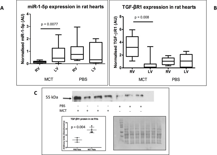
4 weeks after MCT or PBS-treatment, RV weight (RV / ((left ventricle) LV + septum)) and right ventricular systolic pressure (RVSP) were found to be higher in the MCT-treated rats compared to controls confirming increased vascular resistance, as previously published [18]. miR-1-5p expression was decreased 7-fold (p = 0.0077) in the RV of MCT compared to PBS treated rats (Fig 1A). There was no significant difference in expression of miR-1-5p in the left ventricles of the MCT and PBS-treated animals. Median expression of miR-1-5p was higher in the LV than RV of MCT treated animals but this difference did not reach statistical significance (p = 0.089). TGF-βR1 mRNA expression was higher in MCT-treated rat RVs compared to PBS controls (2.5-fold, p = 0.008), whereas no change in expression was noted between PBS-treated RVs to LVs (Fig 1B). TGF-βR1 protein expression was also higher (2.5-fold, p = 0.004) (Fig 1C) in the RVs of monocrotaline treated rats.

Fig 1. miR-1-5p and TGF-βR1 are inversely expressed in the RV of MCT-treated rats with PAH.

Fig 1

miR-1 and transforming growth factor-beta receptor 1 (TGF-βR1) were quantified in RNA extracted from left (LV) and right ventricles (RVs) of monocrotaline (MCT)-treated rats by qPCR. A. miR-1-5p expression was significantly reduced in MCT-treated rat RVs compared to phosphate buffered saline (PBS)-treated (9 animals in each group). Mean expression of miR-1-5p was lower in the MCT RV compared to the MCT LV but this difference was not statistically significant. B. TGF-βR1 expression was significantly increased in MCT-treated rat RVs compared to LVs and to PBS-treated RVs. C. Western blot showing increased TGF-βR1 protein in the MCT-treated RVs compared to PBS treated RVs. Quantification of TGF-βR1 (bottom left) normalised to total protein in each lane as determined by Ponceau S staining (bottom right).

miR-1-5p targets TGF-βR1

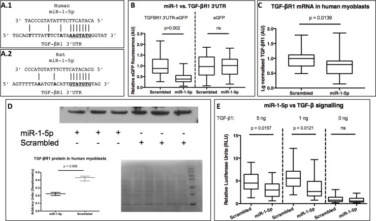
In silico analysis predicted one binding-site for miR-1-5p in the 3’UTR TGF-βR1 (ALK5) of both humans and rats (Fig 2A). The region of the 3’ UTR of human ALK-5 containing this sequence was amplified by PCR and cloned into the 3’-UTR of EGFP in the vector pCAGGS-EGFP to generate pCAGGS-EGFP-3T and the effect of miR-1-5p on EGFP expression was determined in LHCN-M2 cells. Transfection of miR-1-5p reduced EGFP expression from pCAGGS-EGFP-3T compared to control miR-mimic but did not affect the expression of EGFP from pCAGGS-EGFP (Fig 2B).

Fig 2. miR-1 targets TGF-βR1 and inhibits TGF-β signalling.

Fig 2

A. Putative binding side of miR-1- 5p in the transforming growth factor-beta receptor 1 (TGF-βR1) 3’- untranslated region (3’UTR) in humans (A.1) and rats (A.2). Transfection of LHCN-M2 cells with miR1-5p significantly reduced enhanced green fluorescent protein (EGFP) expression from a reporter gene containing the TGF-βR1 3’UTR binding site (B). TGF-βR1 mRNA (C) and TGF-βR1 protein (D) are reduced following miR-1 transfection. E. Transfection of LHCN-M2 cells with miR-1-5p reduced TGF-β1 stimulated luciferase reporter gene expression.

miR-1 reduces TGF-βR1 mRNA & protein expression and signalling in vitro

To determine the effect of miR-1-5p on TGF-βR1 mRNA and protein, LHCN-M2 cells were transiently transfected with miR-1-5p mimic or control mimic. Transfection with the miR-1-5p mimic significantly reduced both TGF-βR1 mRNA and protein levels compared to transfection with the control mimic (Fig 2C and 2D). To determine whether TGF-β signalling was suppressed by miR-1-5p, a luciferase reporter with a TGF-β responsive promoter (CAGA)12 was used. Transfection with miR-1-5p reduced TGF-β stimulated reporter expression in response to both 1 and 5 ng/mL TGF-β, compared to control mimic transfection. However, there was no effect of the miRNA on basal reporter activity (Fig 2E).

Discussion

Our data identify miR-1 as a regulator of TGF-β signalling by suppressing the expression of ALK-5. Furthermore, the data show that miR-1-5p is suppressed in the RV of MCT rats coincident with increased ALK-5. This observation suggests that under normal conditions miR-1 reduces the sensitivity of cardiomyocytes to TGF-β by reducing the expression of ALK-5. Consequently, this reduction in miR-1-5p in hypertrophy leads to an increase in ALK-5 protein levels and thereby an increase in TGF-β signalling.

Given the effects of TGF-β on cardiac myocytes such an increase in TGF-β signalling is likely to contribute to cardiac hypertrophy. However, there are a number of limitations to our study that preclude us from confirming that the observed reduction in miR-1 promotes cardiac hypertrophy by relieving miR-1 suppression of TGF-β signalling. Firstly, we cannot determine whether the suppression in miR-1-5p preceded or came in the early phase of the hypertrophic response, which would be required for the reduction in miR-1 to be a key regulator of the response rather than an epiphenomenon. Secondly, we cannot be sure that the increase in ALK-5 protein occurred in the cardiac myocytes rather than in fibroblasts where it would contribute to fibrosis. For example, in pressure overload hypertrophy neutralizing antibodies to TGF-β 1 inhibit the fibrotic response but do not inhibit the hypertrophy of the myocytes [24]. This lack of effect may be due to a limited effect of TGF-β 1 in cardiac myocyte hypertrophy or may be due to the inability of the antibodies to interact with the pool of TGF-β 1 that acts on myocytes. Similarly, it may be a consequence of alternative TGF-β ligands (e.g. TGF-β 3) which also signal via TGF- βR1 contributing to the hypertrophic response and are not neutralized by the antibody, but which would be inhibited by over-expression of a dominant inhibitory TGF-β type II receptor (TGFBR2). Finally, miR-1 has been shown to affect the expression of a number of different proteins that regulate cardiac cell physiology in a manner consistent with the suppression of growth. Consequently, we cannot determine the relative importance of the interaction between miR-1 and TGF-βR1 in the hypertrophic response. These proteins include the sodium calcium exchanger (NCX1) [25] cyclin-dependent kinase 9 (Cdk9) and fibronectin [14] in the heart. Further evidence for a role for miR-1 in the development of the RV hypertrophy in response to PAH would require the delivery of miR-1 to the RV (e.g. by adenovirus) and demonstration that this slowed or prevented the development of hypertrophy. However, these experiments are beyond the current scope of our study and whilst they have not been performed in PAH similar experiments have shown that over-expression of miR-1-5p reverses insulin-like growth factor-1 (IGF1) induced hypertrophy in vitro [26,27] and that adenoviral delivery of miR-1 to the myocardium has been shown to reverse overload induced LV hypertrophy [28]. Furthermore, other studies have shown increasing SERCA2a reverses hypertrophy and restores miR-1 expression [25]. Similarly, determination of the relative contribution of TGF-β signalling in the development of the same hypertrophy could be determined by the delivery of TGF-β neutralizing antibodies or more selectively by over-expression of a dominant inhibitory form of the TGFBR2 in the RV in a model of PAH.

miR-1-5p expression is not restricted to cardiomyocytes as it is also highly expressed in vascular and skeletal muscle cells. Consequently, it seems likely that miR-1 may also contribute to the TGF-β responses of these cells and thereby to TGF-β driven pathology in the associated tissues. Increased TGF-β signalling is suggested to promote the pulmonary artery smooth muscle cell (PASMC) proliferation that contributes to PAH [29]. Indeed, ALK-5 is thought to mediate abnormal PASMC proliferation in patients with familial PAH and inhibition of ALK-5 has been shown to inhibit the progression of PAH [30]. Similarly over-expression of a dominant inhibitory TGFBR2 has been shown to inhibit the development of PAH [31]. Consequently, it is possible that reduced expression of miR-1 in PASMCs contributes to this phenotype but appropriate changes in miR-1 expression in the PASMCs have yet to be demonstrated so such a contribution remains to be determined. In skeletal muscle, signalling through ALK-5 is a major contributor to skeletal muscle atrophy with both TGF-β promoting atrophy via ALK-5 and the TGF-βR2 and activin and myostatin promoting atrophy through ALK-5 and the activin type 2B receptor. Consistent with a role for the expression of miR-1-5p contributing to the maintenance of muscle mass, reduced miR-1 expression has been observed in the quadriceps of patients with intensive care unit-acquired weakness (ICUAW) and those with chronic obstructive pulmonary disease (COPD) [15,19]. In both conditions there is muscle atrophy and increased signalling through ALK-5.

In conclusion we have shown that miR-1-5p binds to the 3’UTR of the TGF-β1 receptor ALK-5, suppressing the expression of the protein and the strength of signals induced by the ligand. These data suggest that under basal conditions high miR-1 expression reduces TGF-βR1 and lowers the response of the cell to TGF-β ligands. However, in the RV of rats with PAH the reduction in miR-1 contributes to hypertrophy by increasing ALK-5 protein levels.

Supporting information

S1 Fig. The full western blot from Fig 1C.

(TIFF)

S2 Fig. Full blot for Fig 2D.

(TIFF)

Acknowledgments

We are grateful to Dr Vincent Mouly (UPMC, University of Paris) for the gift of the LHCN‐M2 cell line.

Data Availability

All relevant data are within the manuscript, full blots are provided as supplementary material.

Funding Statement

Funded by Award recipient: Paul Kemp. Grant number: FS/14/71/31038 Funder: British Heart Foundation (BHF). Funder website: https://www.bhf.org.uk/. The funders had no role in study design, data collection and analysis, decision to publish, or preparation of the manuscript.

References

  • 1.Fourie PR, Coetzee AR, Bolliger CT. Pulmonary artery compliance: its role in right ventricular-arterial coupling. Cardiovasc Res. 1992. September;26(9):839–44. 10.1093/cvr/26.9.839 [DOI] [PubMed] [Google Scholar]
  • 2.Farber HW, Loscalzo J. Pulmonary arterial hypertension. N Engl J Med. 2004. October 14;351(16):1655–65. 10.1056/NEJMra035488 [DOI] [PubMed] [Google Scholar]
  • 3.Simonneau G, Gatzoulis MA, Adatia I, Celermajer D, Denton C, Ghofrani A, et al. Updated clinical classification of pulmonary hypertension. J Am Coll Cardiol. 2013. December 24;62(25 Suppl):D34–41. [DOI] [PubMed] [Google Scholar]
  • 4.Dobaczewski M, Chen W, Frangogiannis NG. Transforming growth factor (TGF)-β signalling in cardiac remodeling. J Mol Cell Cardiol. 2011. October;51(4):600–6. 10.1016/j.yjmcc.2010.10.033 [DOI] [PMC free article] [PubMed] [Google Scholar]
  • 5.Bujak M, Ren G, Kweon HJ, Dobaczewski M, Reddy A, Taffet G, et al. Essential role of Smad3 in infarct healing and in the pathogenesis of cardiac remodeling. Circulation. 2007. November 6;116(19):2127–38. 10.1161/CIRCULATIONAHA.107.704197 [DOI] [PubMed] [Google Scholar]
  • 6.Leask A. Targeting the TGFbeta, endothelin-1 and CCN2 axis to combat fibrosis in scleroderma. Cell Signal. 2008. August;20(8):1409–14. 10.1016/j.cellsig.2008.01.006 [DOI] [PubMed] [Google Scholar]
  • 7.Gray MO, Long CS, Kalinyak JE, Li HT, Karliner JS. Angiotensin II stimulates cardiac myocyte hypertrophy via paracrine release of TGF-beta 1 and endothelin-1 from fibroblasts. Cardiovasc Res. 1998. November;40(2):352–63. 10.1016/s0008-6363(98)00121-7 [DOI] [PubMed] [Google Scholar]
  • 8.Li Y, Li W, Ying Z, Tian H, Zhu X, Li J, et al. Metastatic heterogeneity of breast cancer cells is associated with expression of a heterogeneous TGFβ-activating miR424-503 gene cluster. Cancer Res. 2014. November 1;74(21):6107–18. 10.1158/0008-5472.CAN-14-0389 [DOI] [PubMed] [Google Scholar]
  • 9.Garros RF, Paul R, Connolly M, Lewis A, Garfield BE, Natanek SA, et al. MicroRNA-542 Promotes Mitochondrial Dysfunction and SMAD Activity and Is Elevated in Intensive Care Unit-acquired Weakness. Am J Respir Crit Care Med. 2017. 01;196(11):1422–33. 10.1164/rccm.201701-0101OC [DOI] [PMC free article] [PubMed] [Google Scholar]
  • 10.Zhao X, Wang K, Liao Y, Zeng Q, Li Y, Hu F, et al. MicroRNA-101a inhibits cardiac fibrosis induced by hypoxia via targeting TGFβRI on cardiac fibroblasts. Cell Physiol Biochem. 2015;35(1):213–26. 10.1159/000369689 [DOI] [PubMed] [Google Scholar]
  • 11.Paul R, Lee J, Donaldson AV, Connolly M, Sharif M, Natanek SA, et al. miR-422a suppresses SMAD4 protein expression and promotes resistance to muscle loss. J Cachexia Sarcopenia Muscle. 2018;9(1):119–28. 10.1002/jcsm.12236 [DOI] [PMC free article] [PubMed] [Google Scholar]
  • 12.Tijsen AJ, van der Made I, van den Hoogenhof MM, Wijnen WJ, van Deel ED, de Groot NE, et al. The microRNA-15 family inhibits the TGFβ-pathway in the heart. Cardiovasc Res. 2014. October 1;104(1):61–71. 10.1093/cvr/cvu184 [DOI] [PubMed] [Google Scholar]
  • 13.Pan Z, Sun X, Shan H, Wang N, Wang J, Ren J, et al. MicroRNA-101 inhibited postinfarct cardiac fibrosis and improved left ventricular compliance via the FBJ osteosarcoma oncogene/transforming growth factor-β1 pathway. Circulation. 2012. August 14;126(7):840–50. 10.1161/CIRCULATIONAHA.112.094524 [DOI] [PubMed] [Google Scholar]
  • 14.Sayed D, Hong C, Chen I-Y, Lypowy J, Abdellatif M. MicroRNAs play an essential role in the development of cardiac hypertrophy. Circ Res. 2007. February 16;100(3):416–24. 10.1161/01.RES.0000257913.42552.23 [DOI] [PubMed] [Google Scholar]
  • 15.Bloch SA, Lee JY, Syburra T, Rosendahl U, Griffiths MJD, Kemp PR, et al. Increased expression of GDF-15 may mediate ICU-acquired weakness by down-regulating muscle microRNAs. Thorax. 2015. March;70(3):219–28. 10.1136/thoraxjnl-2014-206225 [DOI] [PMC free article] [PubMed] [Google Scholar]
  • 16.Sun Y, Ge Y, Drnevich J, Zhao Y, Band M, Chen J. Mammalian target of rapamycin regulates miRNA-1 and follistatin in skeletal myogenesis. J Cell Biol. 2010. June 28;189(7):1157–69. 10.1083/jcb.200912093 [DOI] [PMC free article] [PubMed] [Google Scholar]
  • 17.Sticht C, Torre CDL, Parveen A, Gretz N. miRWalk: An online resource for prediction of microRNA binding sites. PLOS ONE. 2018. October 18;13(10):e0206239 10.1371/journal.pone.0206239 [DOI] [PMC free article] [PubMed] [Google Scholar]
  • 18.Connolly M, Garfield BE, Crosby A, Morrell NW, Wort SJ, Kemp PR. miR-322-5p targets IGF-1 and is suppressed in the heart of rats with pulmonary hypertension. FEBS Open Bio. 2018;8(3):339–48. 10.1002/2211-5463.12369 [DOI] [PMC free article] [PubMed] [Google Scholar]
  • 19.Lewis A, Riddoch-Contreras J, Natanek SA, Donaldson A, Man WD-C, Moxham J, et al. Downregulation of the serum response factor/miR-1 axis in the quadriceps of patients with COPD. Thorax. 2012. January;67(1):26–34. 10.1136/thoraxjnl-2011-200309 [DOI] [PMC free article] [PubMed] [Google Scholar]
  • 20.Ellis PD, Smith CWJ, Kemp P. Regulated tissue-specific alternative splicing of enhanced green fluorescent protein transgenes conferred by alpha-tropomyosin regulatory elements in transgenic mice. J Biol Chem. 2004. August 27;279(35):36660–9. 10.1074/jbc.M405380200 [DOI] [PubMed] [Google Scholar]
  • 21.Favot L, Hall SM, Haworth SG, Kemp PR. Cytoplasmic YY1 is associated with increased smooth muscle-specific gene expression: implications for neonatal pulmonary hypertension. Am J Pathol. 2005. December;167(6):1497–509. 10.1016/S0002-9440(10)61236-9 [DOI] [PMC free article] [PubMed] [Google Scholar]
  • 22.Connolly M, Paul R, Farre‐Garros R, Natanek SA, Bloch S, Lee J, et al. miR‐424‐5p reduces ribosomal RNA and protein synthesis in muscle wasting. J Cachexia Sarcopenia Muscle. 2018. April;9(2):400–16. 10.1002/jcsm.12266 [DOI] [PMC free article] [PubMed] [Google Scholar]
  • 23.Jonk LJC, Itoh S, Heldin C-H, Dijke P ten, Kruijer W. Identification and Functional Characterization of a Smad Binding Element (SBE) in the JunB Promoter That Acts as a Transforming Growth Factor-β, Activin, and Bone Morphogenetic Protein-inducible Enhancer. J Biol Chem. 1998. August 14;273(33):21145–52. 10.1074/jbc.273.33.21145 [DOI] [PubMed] [Google Scholar]
  • 24.Koitabashi N, Danner T, Zaiman AL, Pinto YM, Rowell J, Mankowski J, et al. Pivotal role of cardiomyocyte TGF-β signalling in the murine pathological response to sustained pressure overload. J Clin Invest. 2011. June 1;121(6):2301–12. 10.1172/JCI44824 [DOI] [PMC free article] [PubMed] [Google Scholar]
  • 25.Kumarswamy R, Lyon AR, Volkmann I, Mills AM, Bretthauer J, Pahuja A, et al. SERCA2a gene therapy restores microRNA-1 expression in heart failure via an Akt/FoxO3A-dependent pathway. Eur Heart J. 2012. May 1;33(9):1067–75. 10.1093/eurheartj/ehs043 [DOI] [PMC free article] [PubMed] [Google Scholar]
  • 26.Yu X-Y, Song Y-H, Geng Y-J, Lin Q-X, Shan Z-X, Lin S-G, et al. Glucose induces apoptosis of cardiomyocytes via microRNA-1 and IGF-1. Biochem Biophys Res Commun. 2008. November 21;376(3):548–52. 10.1016/j.bbrc.2008.09.025 [DOI] [PubMed] [Google Scholar]
  • 27.Hua Y, Zhang Y, Ren J. IGF-1 deficiency resists cardiac hypertrophy and myocardial contractile dysfunction: role of microRNA-1 and microRNA-133a. J Cell Mol Med. 2012. January;16(1):83–95. 10.1111/j.1582-4934.2011.01307.x [DOI] [PMC free article] [PubMed] [Google Scholar]
  • 28.Karakikes I, Chaanine AH, Kang S, Mukete BN, Jeong D, Zhang S, et al. Therapeutic cardiac-targeted delivery of miR-1 reverses pressure overload-induced cardiac hypertrophy and attenuates pathological remodeling. J Am Heart Assoc. 2013. April 23;2(2):e000078 10.1161/JAHA.113.000078 [DOI] [PMC free article] [PubMed] [Google Scholar]
  • 29.Morrell NW, Yang X, Upton PD, Jourdan KB, Morgan N, Sheares KK, et al. Altered growth responses of pulmonary artery smooth muscle cells from patients with primary pulmonary hypertension to transforming growth factor-beta(1) and bone morphogenetic proteins. Circulation. 2001. August 14;104(7):790–5. 10.1161/hc3201.094152 [DOI] [PubMed] [Google Scholar]
  • 30.Thomas M, Docx C, Holmes AM, Beach S, Duggan N, England K, et al. Activin-like kinase 5 (ALK5) mediates abnormal proliferation of vascular smooth muscle cells from patients with familial pulmonary arterial hypertension and is involved in the progression of experimental pulmonary arterial hypertension induced by monocrotaline. Am J Pathol. 2009. February;174(2):380–9. 10.2353/ajpath.2009.080565 [DOI] [PMC free article] [PubMed] [Google Scholar]
  • 31.Chen Y-F, Feng J-A, Li P, Xing D, Zhang Y, Serra R, et al. Dominant negative mutation of the TGF-beta receptor blocks hypoxia-induced pulmonary vascular remodeling. J Appl Physiol. 2006. February;100(2):564–71. 10.1152/japplphysiol.00595.2005 [DOI] [PubMed] [Google Scholar]

Decision Letter 0

Fabio Martelli

20 Nov 2019

PONE-D-19-27153

miR-1-5p targets TGF-βR1 and is suppressed in the hypertrophying hearts of rats with pulmonary arterial hypertension

PLOS ONE

Dear Dr. Kemp,

Thank you for submitting your manuscript to PLOS ONE. After careful consideration, we feel that it has merit but does not fully meet PLOS ONE’s publication criteria as it currently stands. Therefore, we invite you to submit a revised version of the manuscript that addresses the points raised during the review process.

Specifically, referee #1 correctly pointed out that to demonstrate miR-1 involvement in the development of cardiac hypertrophy with pulmonary arterial hypertension, miR-1 gain or loss of function experiments in vivo are indicated. These experiments are strongly recommended. Should the authors being unable to perform them, an accurate and detailed discussion of the consequent study limitations is required.

The authors are also requested to address all the other issues raised by both referees.

We would appreciate receiving your revised manuscript by 19/02/2020. When you are ready to submit your revision, log on to https://www.editorialmanager.com/pone/ and select the 'Submissions Needing Revision' folder to locate your manuscript file.

If you would like to make changes to your financial disclosure, please include your updated statement in your cover letter.

To enhance the reproducibility of your results, we recommend that if applicable you deposit your laboratory protocols in protocols.io, where a protocol can be assigned its own identifier (DOI) such that it can be cited independently in the future. For instructions see: http://journals.plos.org/plosone/s/submission-guidelines#loc-laboratory-protocols

Please include the following items when submitting your revised manuscript:

  • A rebuttal letter that responds to each point raised by the academic editor and reviewer(s). This letter should be uploaded as separate file and labeled 'Response to Reviewers'.

  • A marked-up copy of your manuscript that highlights changes made to the original version. This file should be uploaded as separate file and labeled 'Revised Manuscript with Track Changes'.

  • An unmarked version of your revised paper without tracked changes. This file should be uploaded as separate file and labeled 'Manuscript'.

Please note while forming your response, if your article is accepted, you may have the opportunity to make the peer review history publicly available. The record will include editor decision letters (with reviews) and your responses to reviewer comments. If eligible, we will contact you to opt in or out.

We look forward to receiving your revised manuscript.

Kind regards,

Fabio Martelli

Academic Editor

PLOS ONE

Journal Requirements:

1. When submitting your revision, we need you to address these additional requirements.

Please ensure that your manuscript meets PLOS ONE's style requirements, including those for file naming. The PLOS ONE style templates can be found at

http://www.journals.plos.org/plosone/s/file?id=wjVg/PLOSOne_formatting_sample_main_body.pdf and http://www.journals.plos.org/plosone/s/file?id=ba62/PLOSOne_formatting_sample_title_authors_affiliations.pdf

2. Please provide additional information about the LHCN-M2 cell line used in this work, including source, history, culture conditions and any quality control testing procedures (authentication, characterisation, and mycoplasma testing). For more information, please see http://journals.plos.org/plosone/s/submission-guidelines#loc-cell-lines.

3. At this time, we ask that you please provide additional details regarding the monocrotaline drug used in this study. Specifically, please provide the source, product number and lot number of the drug. Thank you for your attention this request.

4. To comply with PLOS ONE submission guidelines, in your Methods section, please provide additional information regarding your statistical analyses. For more information on PLOS ONE's expectations for statistical reporting, please see https://journals.plos.org/plosone/s/submission-guidelines.#loc-statistical-reporting.

5. Your ethics statement must appear in the Methods section of your manuscript. If your ethics statement is written in any section besides the Methods, please move it to the Methods section and delete it from any other section. Please also ensure that your ethics statement is included in your manuscript, as the ethics section of your online submission will not be published alongside your manuscript.

6. PLOS ONE now requires that authors provide the original uncropped and unadjusted images underlying all blot or gel results reported in a submission’s figures or Supporting Information files. This policy and the journal’s other requirements for blot/gel reporting and figure preparation are described in detail at https://journals.plos.org/plosone/s/figures#loc-blot-and-gel-reporting-requirements and https://journals.plos.org/plosone/s/figures#loc-preparing-figures-from-image-files. When you submit your revised manuscript, please ensure that your figures adhere fully to these guidelines and provide the original underlying images for all blot or gel data reported in your submission. See the following link for instructions on providing the original image data: https://journals.plos.org/plosone/s/figures#loc-original-images-for-blots-and-gels.

In your cover letter, please note whether your blot/gel image data are in Supporting Information or posted at a public data repository, provide the repository URL if relevant, and provide specific details as to which raw blot/gel images, if any, are not available. Email us at plosone@plos.org if you have any questions.

[Note: HTML markup is below. Please do not edit.]

Reviewers' comments:

Reviewer's Responses to Questions

Comments to the Author

1. Is the manuscript technically sound, and do the data support the conclusions?

The manuscript must describe a technically sound piece of scientific research with data that supports the conclusions. Experiments must have been conducted rigorously, with appropriate controls, replication, and sample sizes. The conclusions must be drawn appropriately based on the data presented.

Reviewer #1: Partly

Reviewer #2: Yes

**********

2. Has the statistical analysis been performed appropriately and rigorously?

Reviewer #1: Yes

Reviewer #2: Yes

**********

3. Have the authors made all data underlying the findings in their manuscript fully available?

The PLOS Data policy requires authors to make all data underlying the findings described in their manuscript fully available without restriction, with rare exception (please refer to the Data Availability Statement in the manuscript PDF file). The data should be provided as part of the manuscript or its supporting information, or deposited to a public repository. For example, in addition to summary statistics, the data points behind means, medians and variance measures should be available. If there are restrictions on publicly sharing data—e.g. participant privacy or use of data from a third party—those must be specified.

Reviewer #1: Yes

Reviewer #2: Yes

**********

4. Is the manuscript presented in an intelligible fashion and written in standard English?

PLOS ONE does not copyedit accepted manuscripts, so the language in submitted articles must be clear, correct, and unambiguous. Any typographical or grammatical errors should be corrected at revision, so please note any specific errors here.

Reviewer #1: Yes

Reviewer #2: Yes

**********

5. Review Comments to the Author

Please use the space provided to explain your answers to the questions above. You may also include additional comments for the author, including concerns about dual publication, research ethics, or publication ethics. (Please upload your review as an attachment if it exceeds 20,000 characters)

Reviewer #1: The study is of putative interest as it shows that miR-1-5p binds and suppress the TGF-β1 receptor ALK-5 expression thereby reduce TGF-β signalling, which may regulate cardiac hypertrophy. In an animal model of pulmonary arterial hypertension which is associated with right ventricle hypertrophy, the authors showed that miR-1 was reduced while TGF-βR1 protein levels increased. Using in vitro cell culture experiments they demonstrated that miR-1 suppress ALK5 and TGF β signalling.

The manuscript is logical and well-structured and the experiments have been described in sufficient detail. Nevertheless, although potentially interesting, the information brought forward by this study is too preliminary and there are some issues that must be addressed before being published.

Major issues:

This is a rather elementary and indirect demonstration of miR-1 in the development of cardiac hypertrophy caused by pulmonary arterial hypertension. In order the truly demonstrate that this axis is directly involved in the development of cardiac hypertrophy, animal studies using inhibition or overexpression of miR-1 (using Antagomirs or miR mimics) are needed. Moreover, histologically measured cardiomyocyte hypertrophy should be also performed.

Minor points:

1. The method section does not describe the pCAGGS-GFP assay.

2. Figures are reversed (Fig 1 si fig 2 and vice versa)

3. The connection between TGFbR1 inactivation data and the luciferase reporter assay is not clearly explained. There should be better stated that (CAGA)12-luciferase reporter plasmid is exclusively activated by TGF-β-induced complex between Smad3 and Smad4.

4. Statistical data for ALK-5 protein expression quantification should be included. Aslo the graph in 2D should be presented as Box-plot like all the others.

Reviewer #2: In this paper, the authors reported that miR-1 expression is reduced in the hypertrophic right ventricle of monocrotaline-treated rats. The authors identified by bioinformatic analysis that TGF-βR1 is a potential target for miR-1, the expression of which being increased in rat right ventricles. Finally, the authors used miR-1-mimic to reduce EGFP expression from a reporter vector containing the ALK5 3’-UTR and also knocked down endogenous TGF-βR1. The data confirm miR-1 targets TGF-βR1 thereby reduce TGF-β signaling, which may regulate cardiac hypertrophy. The article is interesting and original, with a good experimental design and bioinformatics approach. However, there are some issues to be solved that could improve the manuscript.

General comments:

1. Abstract need a clear aim or a motivation for the study presented; also, all abbreviations in the abstract must be detailed or avoided.

2. The authors should provide the “Supporting Information files” according to their statement: “All relevant data are within the manuscript and its Supporting Information files.”

3. Overall in the manuscript, the authors should provide all the abbreviations used when they are used for the first time. Many abbreviations are not detailed (see TGFβ, SMAD7, SMURF1, BMP, HDAC4, ALK5 at pages 3-4). Of special significance are the missing definition for RV and LV (pages 4 and 7) and MCT (pages 6-7 and later).

4. Methods, page 5: “RNA extraction and qPCR”. The authors should provide either the commercial ID or the custom sequences for all the primers used here for the SYBR green-based PCR for miRNA and mRNA expression (maybe in a Supplemental file, also missed) since these sequences are absent in the given reference #19.

5. Methods section missed the description of the in silico analysis approach performed for miR-1-5p and details about TGFβR1 3’UTR cloning approach (both results being also described at page 7).

6. The Figures 1 and 2 are given in the reverse order; please correct this.

7. When talking about data in Fig. 2A at page 7, it should be better to provide both Fig. 2A.1 and Fig. 2A.2 panels (there is no Fig. 2A there).

8. A minor typographical correction is needed to separate the third subtitle from the next paragraph from Results section. Also, at page 9, a correction is necessary for the references 15, 18 in the first paragraph.

9. Figure 1 legend needs a correction for its title: “miR-1-5p and TGF-βR1 are inversely expressed in the RV of PAH” could probably become “miR-1-5p and TGF-βR1 are inversely expressed in the RV of MCT-treated rats with PAH”.

10. Both Figure legends must be self-explanatory when describing the panels, with all abbreviations detailed, independently of the text manuscript.

**********

6. PLOS authors have the option to publish the peer review history of their article (what does this mean?). If published, this will include your full peer review and any attached files.

If you choose “no”, your identity will remain anonymous but your review may still be made public.

Do you want your identity to be public for this peer review? For information about this choice, including consent withdrawal, please see our Privacy Policy.

Reviewer #1: No

Reviewer #2: Yes: Loredan S. Niculescu

[NOTE: If reviewer comments were submitted as an attachment file, they will be attached to this email and accessible via the submission site. Please log into your account, locate the manuscript record, and check for the action link "View Attachments". If this link does not appear, there are no attachment files to be viewed.]

While revising your submission, please upload your figure files to the Preflight Analysis and Conversion Engine (PACE) digital diagnostic tool, https://pacev2.apexcovantage.com/. PACE helps ensure that figures meet PLOS requirements. To use PACE, you must first register as a user. Registration is free. Then, login and navigate to the UPLOAD tab, where you will find detailed instructions on how to use the tool. If you encounter any issues or have any questions when using PACE, please email us at figures@plos.org. Please note that Supporting Information files do not need this step.

PLoS One. 2020 Feb 28;15(2):e0229409. doi: 10.1371/journal.pone.0229409.r002

Author response to Decision Letter 0


2 Jan 2020

Response to Referees’ comments

Specifically, referee #1 correctly pointed out that to demonstrate miR-1 involvement in the development of cardiac hypertrophy with pulmonary arterial hypertension, miR-1 gain or loss of function experiments in vivo are indicated. These experiments are strongly recommended. Should the authors being unable to perform them, an accurate and detailed discussion of the consequent study limitations is required.

Reviewer #1: The study is of putative interest as it shows that miR-1-5p binds and suppress the TGF-β1 receptor ALK-5 expression thereby reduce TGF-β signalling, which may regulate cardiac hypertrophy. In an animal model of pulmonary arterial hypertension which is associated with right ventricle hypertrophy, the authors showed that miR-1 was reduced while TGF-βR1 protein levels increased. Using in vitro cell culture experiments they demonstrated that miR-1 suppress ALK5 and TGF β signalling.

The manuscript is logical and well-structured and the experiments have been described in sufficient detail. Nevertheless, although potentially interesting, the information brought forward by this study is too preliminary and there are some issues that must be addressed before being published.

Major issues:

This is a rather elementary and indirect demonstration of miR-1 in the development of cardiac hypertrophy caused by pulmonary arterial hypertension. In order the truly demonstrate that this axis is directly involved in the development of cardiac hypertrophy, animal studies using inhibition or overexpression of miR-1 (using Antagomirs or miR mimics) are needed. Moreover, histologically measured cardiomyocyte hypertrophy should be also performed.

We accept the point that the reviewer raises and that it would be ideal to use gain and loss of function studies to demonstrate the relative importance of miR-1 in pulmonary hypertrophy as a consequence of PAH. Unfortunately we do not have either the funding or the licences required to carry out this experiment within the timeframe. However, as the editor has allowed us to detail the study limitations we have included a section describing the limitations of our study and the need to carry out similar experiments in a PAH model.

This section reads:

Given the effects of TGF-β on cardiac myocytes such an increase in TGF-β signaling is likely to contribute to cardiac hypertrophy. However, there are a number of limitations to our study that preclude us from confirming that the observed reduction in miR-1 promotes cardiac hypertrophy by relieving miR-1 suppression of TGF- β signaling. Firstly, we cannot determine whether the suppression in miR-1-5p preceded or came in the early phase of the hypertrophic response, which would be required for the reduction in miR-1 to be a key regulator of the response rather than an epiphenomenon. Secondly, we cannot be sure that the increase in ALK-5 protein occurred in the cardiac myocytes rather than in fibroblasts where it would contribute to fibrosis. For example, in pressure overload hypertrophy neutralizing antibodies to TGF-β 1 inhibit the fibrotic response but do not inhibit the hypertrophy of the myocytes (24). This lack of effect may be due to a limited effect of TGFβ 1 in cardiac myocyte hypertrophy or may be due to the inability of the antibodies to interact with the pool of TGF-β 1 that acts on myocytes. Similarly it may be a consequence of alternative TGFβ ligands (e.g. TGFβ 3) which also signal via TGFBR1 contributing to the hypertrophic response and are not neutralized by the antibody, but which would be inhibited by over-expression of a dominant inhibitory TGFβ type II receptor (TGFBR2). Finally, miR-1 has been shown to affect the expression of a number of different proteins that regulate cardiac cell physiology in a manner consistent with the suppression of growth. Consequently, we cannot determine the relative importance of the interaction between miR-1 and TGFβR1 in the hypertrophic response. These proteins include the sodium calcium exchanger (NCX1)(25) cyclin-dependent kinase 9 (Cdk9) and fibronectin (14) in the heart. Further evidence for a role for miR-1 in the development of the RV hypertrophy in response to PAH would require the delivery of miR-1 to the RV (e.g. by adenovirus) and demonstration that this slowed or prevented the development of hypertrophy. However, these experiments are beyond the current scope of our study and whilst they have not been performed in PAH similar experiments have shown that over-expression of miR-1-5p reverses insulin-like growth factor-1 (IGF1) induced hypertrophy in vitro (26,27) and that adenoviral delivery of miR-1 to the myocardium has been shown to reverse overload induced LV hypertrophy (28). Furthermore, other studies have shown increasing SERCA2a reverses hypertrophy and restores miR-1 expression (25). Similarly determination of the relative contribution of TGF-β signaling in the development of the same hypertrophy could be determined by the delivery of TGF-β neutralizing antibodies or more selectively by over-expression of a dominant inhibitory form of the TGFBR2 in the RV in a model of PAH.

Minor points:

1. The method section does not describe the pCAGGS-GFP assay.

We apologise for this omission which has been corrected and the added section reads

EGFP reporter cloning & assay - A section of the 3’UTR of TGF-�R1 containing the putative miR-1 binding site was amplified from human cDNA using primers: forward (5’–3’), GGAGATCTGGGTGTTTGATATTTCTTCAT reverse: (5’-3’) GGGGGATCCGGACATTTTCTGTACATATCTTA and ligated into pGEM-T Easy (Promega). After sequencing to ensure the correct DNA had been amplified the insert was removed by BglII and BamHI digestion and ligated into a pCAGGS-enhanced green fluorescent protein (EGFP) vector down-stream of the coding region. Final plasmid sequences were confirmed by sequencing.

To quantify the effect of miR-1 on protein expression cells were transfected with the miR-mimic or control as previously described (21). Twenty four hours later, the cells were transfected with this pCAGGS-EGFP-reporter either with or without the 3’UTR inset, cells were then lysed 24h later and EGFP expression was quantified by fluorescence with excitation at 480nm and emission detected at 510nm in a Cytofluor plate reader (Applied Biosystems).

2. Figures are reversed (Fig 1 is fig 2 and vice versa)

The figures have now been placed in the correct order.

3. The connection between TGFbR1 inactivation data and the luciferase reporter assay is not clearly explained. There should be better stated that (CAGA)12-luciferase reporter plasmid is exclusively activated by TGF-β-induced complex between Smad3 and Smad4.

24 hours following miRNA transfection, cells were transfected with a 3:1 ratio of luciferase reporter vectors (CAGA)12 (firefly luciferase) and pRLTK (renilla luciferase) as previously described; the (CAGA)12 vector contains a SMAD binding element (SBE) specific to SMAD3 and SMAD4 (23).

4. Statistical data for ALK-5 protein expression quantification should be included. Also the graph in 2D should be presented as Box-plot like all the others.

The graph needs to be presented as it stands because there are only 3 data points in the Western blots. Consequently we can only show min/max and the median bar. This is the standard presentation of box plots with this number of data points in GraphPad.

Reviewer #2: In this paper, the authors reported that miR-1 expression is reduced in the hypertrophic right ventricle of monocrotaline-treated rats. The authors identified by bioinformatic analysis that TGF-βR1 is a potential target for miR-1, the expression of which being increased in rat right ventricles. Finally, the authors used miR-1-mimic to reduce EGFP expression from a reporter vector containing the ALK5 3’-UTR and also knocked down endogenous TGF-βR1. The data confirm miR-1 targets TGF-βR1 thereby reduce TGF-β signaling, which may regulate cardiac hypertrophy. The article is interesting and original, with a good experimental design and bioinformatics approach. However, there are some issues to be solved that could improve the manuscript.

General comments:

1. Abstract need a clear aim or a motivation for the study presented; also, all abbreviations in the abstract must be detailed or avoided.

We have reworded to the abstract to highlight our starting hypothesis and removed the bulk of the abbreviation. Those we couldn’t delete have been detailed. The abstract now reads as follows.

The microRNA miR-1 is an important regulator of muscle phenotype including cardiac muscle. Down-regulation of miR-1 has been shown to occur in left ventricular hypertrophy but its contribution to right ventricular hypertrophy in pulmonary arterial hypertension are not known. Previous studies have suggested that miR-1 may suppress transforming growth factor-beta (TGF-β) signalling, an important pro-hypertrophic pathway but only indirect mechanisms of regulation have been identified. We identified the TGF-β type 1 receptor (TGF-βR1) as a putative miR-1 target. We therefore hypothesized that miR-1 and TGF-βR1 expression would be inversely correlated in hypertrophying right ventricle of rats with pulmonary arterial hypertension and that miR-1 would inhibit TGF-β signalling by targeting TGF-βR1 expression. Quantification of miR-1 and TGF-βR1 in the right ventricle of rats treated with monocrotaline to induce pulmonary arterial hypertension showed that these was reduced in the hypertrophying right ventricle. A miR-1-mimic reduced enhanced green fluorescent protein expression from a reporter vector containing the TGF-βR1 3’- untranslated region and knocked down endogenous TGF-βR1. Lastly, miR-1 reduced TGF-β activation of a (mothers against decapentaplegic homolog) SMAD2/3-dependent reporter. Taken together, these data suggest that miR-1 targets TGF-βR1 and reduces TGF-β signaling, so a reduction in miR-1 expression may increase TGF-β signaling and contribute to cardiac hypertrophy.

2. The authors should provide the “Supporting Information files” according to their statement: “All relevant data are within the manuscript and its Supporting Information files.”

The initial file did not have any supplementary information and the statement was copied from the submission website. However, we have now included the full western blots as supplementary information as required by the journal.

3. Overall in the manuscript, the authors should provide all the abbreviations used when they are used for the first time. Many abbreviations are not detailed (see TGFβ, SMAD7, SMURF1, BMP, HDAC4, ALK5 at pages 3-4). Of special significance are the missing definition for RV and LV (pages 4 and 7) and MCT (pages 6-7 and later).

We have added these abbreviations in the relevant places

4. Methods, page 5: “RNA extraction and qPCR”. The authors should provide either the commercial ID or the custom sequences for all the primers used here for the SYBR green-based PCR for miRNA and mRNA expression (maybe in a Supplemental file, also missed) since these sequences are absent in the given reference #19.

We apologise for this omission and have added a table with the primer sequences used.

The reverse primer for the PCR of miR-1 and U6 was the reverse primer provided with the polyadenylation and first strand synthesis kit.

Table 1: Primer list for qPCR

Gene Forward sequence Reverse sequence

miR-1-5p CCGGTGGAATGTAAAGAAGTATGTAT Agilent Universal reverse primer

U6 (housekeeper) CTCGCTTCGGCAGCACA Agilent Universal reverse primer

TGF-βR1 GAACTCCCAACTACAGAAAAGCA GCAGACTGGACCAGCAATGA

GAPDH (housekeeper) GGTGGTCTCCTCTGACTTCAACA GTTGCTGTAGCCAAATTCGTTGT

5. Methods section missed the description of the in silico analysis approach performed for miR-1-5p and details about TGFβR1 3’UTR cloning approach (both results being also described at page 7).

We have added the following to describe the in silico analysis :

In silico analysis of miR-1 – Potential targets in the TGF-β signaling pathway for miR-1 were identified via the miRWalk 2.0 database, (17). Putative targets were identified but screening the data base for targets identified by all 10 of the available algorithms. Putative targets were chosen as those identified by 5 or more algorithms. TGF-βR1/ALK-5 was predicted as a target by 6 algorithms including miTRWalk, miRanda and Targetscan. As TGF-β signaling has previously been shown to be important in regulating cardiac size, it was chosen as the basis of an enquiry into miR-1 regulation of cardiac hypertrophy.

We have also described our cloning of the 3’UTR into the EGFP plasmid as indicated in the response to Reviewer 1

6. The Figures 1 and 2 are given in the reverse order; please correct this.

We apologise for the oversight and have corrected this error

7. When talking about data in Fig. 2A at page 7, it should be better to provide both Fig. 2A.1 and Fig. 2A.2 panels (there is no Fig. 2A there).

We have made this addition

8. A minor typographical correction is needed to separate the third subtitle from the next paragraph from Results section. Also, at page 9, a correction is necessary for the references 15, 18 in the first paragraph.

Corrected

9. Figure 1 legend needs a correction for its title: “miR-1-5p and TGF-βR1 are inversely expressed in the RV of PAH” could probably become “miR-1-5p and TGF-βR1 are inversely expressed in the RV of MCT-treated rats with PAH”.

Corrected

10. Both Figure legends must be self-explanatory when describing the panels, with all abbreviations detailed, independently of the text manuscript.

Corrected

Response to Editorial Queries

1. When submitting your revision, we need you to address these additional requirements.

Please ensure that your manuscript meets PLOS ONE's style requirements, including those for file naming. The PLOS ONE style templates can be found at

http://www.journals.plos.org/plosone/s/file?id=wjVg/PLOSOne_formatting_sample_main_body.pdf and http://www.journals.plos.org/plosone/s/file?id=ba62/PLOSOne_formatting_sample_title_authors_affiliations.pdf

We have corrected these as requested

2. Please provide additional information about the LHCN-M2 cell line used in this work, including source, history, culture conditions and any quality control testing procedures (authentication, characterisation, and mycoplasma testing). For more information, please see http://journals.plos.org/plosone/s/submission-guidelines#loc-cell-lines.

We have added a section to cover this point as follows

LHCN-M2 human skeletal myoblasts obtained from Vincent Mouly (Sorbonne Unitversité) in 2014 and were cultured in Skeletal muscle growth medium (PromoCell) supplemented with 20% FCS

3. At this time, we ask that you please provide additional details regarding the monocrotaline drug used in this study. Specifically, please provide the source, product number and lot number of the drug. Thank you for your attention this request.

We have added the supplier and product number. (MCT, Sigma; C2401) Unfortunately we do not have the lot number of this compound so cannot add this.

4. To comply with PLOS ONE submission guidelines, in your Methods section, please provide additional information regarding your statistical analyses. For more information on PLOS ONE's expectations for statistical reporting, please see https://journals.plos.org/plosone/s/submission-guidelines.#loc-statistical-reporting.

We have amended the statistics section as follows:

All statistical analyses were performed in GraphPad PRISM and no samples were excluded as outliers and data were analysed using a between samples design. Animal experiments were conducted in two groups for a total of 9 MCT- and 9 PBS-treated animals. Differences in miRNA and mRNA expression between animal groups and left and right ventricles were calculated via Kruskal-Wallis test (ANOVA) for non-parametric data with post-hoc analysis using Dunn’s multiple test correction. All other comparisons calculated using Student’s t-test for normally distributed data or by Mann-Whitney U test for non-parametric data. In vitro mRNA expression and luciferase data shown were produced in three independent experiments, each consisting of six independent transfections; mRNA measures assayed in duplicate. Box-plots expressed as median with min-max bars. In vitro protein expression data shown via western blot are three independent experiments from 6-well plates. All tests were based on two tailed analysis and differences were taken to be significant at p<0.05.

5. Your ethics statement must appear in the Methods section of your manuscript. If your ethics statement is written in any section besides the Methods, please move it to the Methods section and delete it from any other section. Please also ensure that your ethics statement is included in your manuscript, as the ethics section of your online submission will not be published alongside your manuscript.

The ethics statement is in the Methods section

6. PLOS ONE now requires that authors provide the original uncropped and unadjusted images underlying all blot or gel results reported in a submission’s figures or Supporting Information files. This policy and the journal’s other requirements for blot/gel reporting and figure preparation are described in detail at https://journals.plos.org/plosone/s/figures#loc-blot-and-gel-reporting-requirements and https://journals.plos.org/plosone/s/figures#loc-preparing-figures-from-image-files. When you submit your revised manuscript, please ensure that your figures adhere fully to these guidelines and provide the original underlying images for all blot or gel data reported in your submission. See the following link for instructions on providing the original image data: https://journals.plos.org/plosone/s/figures#loc-original-images-for-blots-and-gels.

We have added the uncropped and unadjusted images of the blots as Supporting images files and added a comment to the Methods section that this is where they are.

Attachment

Submitted filename: response to PLOS one miR-1.docx

Decision Letter 1

Fabio Martelli

6 Feb 2020

miR-1-5p targets TGF-βR1 and is suppressed in the hypertrophying hearts of rats with pulmonary arterial hypertension

PONE-D-19-27153R1

Dear Dr. Kemp,

We are pleased to inform you that your manuscript has been judged scientifically suitable for publication and will be formally accepted for publication once it complies with all outstanding technical requirements.

Within one week, you will receive an e-mail containing information on the amendments required prior to publication. When all required modifications have been addressed, you will receive a formal acceptance letter and your manuscript will proceed to our production department and be scheduled for publication.

Shortly after the formal acceptance letter is sent, an invoice for payment will follow. To ensure an efficient production and billing process, please log into Editorial Manager at https://www.editorialmanager.com/pone/, click the "Update My Information" link at the top of the page, and update your user information. If you have any billing related questions, please contact our Author Billing department directly at authorbilling@plos.org.

If your institution or institutions have a press office, please notify them about your upcoming paper to enable them to help maximize its impact. If they will be preparing press materials for this manuscript, you must inform our press team as soon as possible and no later than 48 hours after receiving the formal acceptance. Your manuscript will remain under strict press embargo until 2 pm Eastern Time on the date of publication. For more information, please contact onepress@plos.org.

With kind regards,

Fabio Martelli

Academic Editor

PLOS ONE

Additional Editor Comments (optional):

Reviewers' comments:

Acceptance letter

Fabio Martelli

14 Feb 2020

PONE-D-19-27153R1

miR-1-5p targets TGF-βR1 and is suppressed in the hypertrophying hearts of rats with pulmonary arterial hypertension

Dear Dr. Kemp:

I am pleased to inform you that your manuscript has been deemed suitable for publication in PLOS ONE. Congratulations! Your manuscript is now with our production department.

If your institution or institutions have a press office, please notify them about your upcoming paper at this point, to enable them to help maximize its impact. If they will be preparing press materials for this manuscript, please inform our press team within the next 48 hours. Your manuscript will remain under strict press embargo until 2 pm Eastern Time on the date of publication. For more information please contact onepress@plos.org.

For any other questions or concerns, please email plosone@plos.org.

Thank you for submitting your work to PLOS ONE.

With kind regards,

PLOS ONE Editorial Office Staff

on behalf of

Dr. Fabio Martelli

Academic Editor

PLOS ONE

Associated Data

    This section collects any data citations, data availability statements, or supplementary materials included in this article.

    Supplementary Materials

    S1 Fig. The full western blot from Fig 1C.

    (TIFF)

    S2 Fig. Full blot for Fig 2D.

    (TIFF)

    Attachment

    Submitted filename: response to PLOS one miR-1.docx

    Data Availability Statement

    All relevant data are within the manuscript, full blots are provided as supplementary material.


    Articles from PLoS ONE are provided here courtesy of PLOS

    RESOURCES