Abstract
Background
Copper is an essential trace element of the human body. However, it is related to many diseases. Copper intoxication is not common in Western countries, but needs to be rapidly recognised because of its high lethality.
Case presentation
We report the case of a 40-year-old woman who presented to the emergency department after performing intrarectal administration of a blue powder sent from Cameroon by her family, in the belief that this would help her to get pregnant. Her evolution was complicated by multiorgan failure and the unusual circumstances. The diagnosis was suspected on the basis of the clinical presentation and the colour of the powder, and confirmed by blood dosage and toxicological analysis of the powder. She underwent symptomatic treatment, and the outcome was progressively favourable, apart from persistent chronic renal failure with dependence on dialysis.
Conclusion
Copper intoxications are rare but severe. Laboratory diagnosis of the condition is not an issue; the difficulty is suspecting it and quickly initiating chelation treatment associated with symptomatic treatments.
LEARNING POINTS
Copper sulphate is a rare but severe condition that must be promptly diagnosed. Diagnosis may be based on clinical presentation, characterised by multiple organ failure, and suspected in the case of ingestion of a blue chemical product.
In the presence of saturation gap on pulse oximetry, the clinician must consider the possibility of methemoglobinaemia, and look for toxic causes.
Treatment of copper sulphate poisoning requires mainly symptomatic treatment and chelation of copper by oral or IV drugs.
Keywords: Copper sulphate, toxicology, methemoglobinaemia
CASE DESCRIPTION
We report the case of a 40-year-old female patient who presented to the emergency room with diarrhoea, abdominal pain and anuria, 48 hours after performing a rectal enema using a blue powder sent from Cameroon, in the belief that this would help her get pregnant.
She had a one-year history of HIV infection, well controlled with antiretrovirals, without any episode of opportunistic infection, as well as secondary infertility related to uterine myomas.
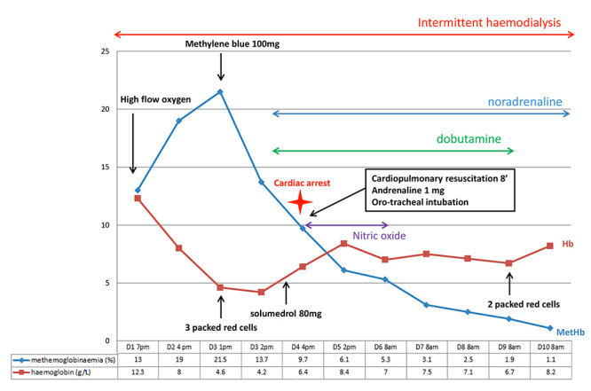
On arrival, she was conscious and haemodynamically stable. She was fitted with a urinary catheter, which yielded only a few millilitres of “port-coloured” urine. 6 hours after arrival, O2 saturation on pulse oximetry dropped to 74% without breathlessness. Her pulmonary examination was normal. Arterial blood gas analysis at that time showed: pH 7.18, PO2 70 mmHg, pCO2 30 mmHg, HCO3- 11.2 mmol/L and SpO2 99% (Fig. 1). Methemoglobinaemia level was 13% and explained the saturation gap. Otherwise, the laboratory tests gave the following findings: creatinine 612 μmol/L, urea 22.7 mmol/L, Na+ 128 mmol/L, WBC 47G/L (neutrophil count 37G/L), Hb 12.3g/dL, CPK 3427 UI/L, bilirubin 73 μmol/L, ultra-sensitive troponine 2,380 μg/L, CRP 186 mg/L, PCT 14 μg/L. The laboratory called us to warn us that the blood was black and viscous, and that they could not give us the results for potassium levels, hepatic enzymes, the balance of haemostasis and the detail of bilirubin.
Figure 1.

Main events and elements of medical care during the first ten days
The patient was admitted to the ICU. We began a treatment with high-flow nasal oxygen and intermittent haemodialysis. MetHb rose to 23% and we used one dose of methylene blue.
48 hours later, she developed brutal anaemia to 4.2g/dL due to massive haemolysis (hyperbilirubinaemia, high LDH level, normal haptoglobin, and positive Coombs test, IgG 1+). There was no glucose-6-phosphate dehydrogenase (G6PD) deficiency. The troponin level also continued to rise, without any echocardiographic abnormalities. She was treated with 8 packed red blood cell transfusions, and received an 80 mg dose of methylprednisolone due to suspicion of autoimmune haemolytic anaemia, but this hypothesis was abandoned after consultation with the regional expert centre in autoimmune cytopenia.
72 hours after admission, the patient suffered a 2-minute long cardiac arrest, recovering after 8 minutes of cardiac massage, 1 mg IV adrenaline and mechanical ventilation. Cardiac ultrasound revealed an enlarged right ventricle and paradoxical septum, but left ventricular function was preserved. A thoracic angioscan excluded a pulmonary embolism. The diagnosis reached was toxic myocarditis. Dobutamine infusion and nitric oxide therapy were necessary for 24 hours (Fig. 1).
About a week later, analysis of the powder she had taken revealed that it was pentahydrated copper sulphate.
The patient’s blood copper level at day 6 after ingestion was 25 mcmol/L (1.59 mg/L, N 11–20 mcmol/L).
She was extubated on day 15 of hospitalisation. She recovered diuresis of about 250 cc per day but has persistent chronic renal failure and requires haemodialysis three times a week.
DISCUSSION
Copper sulphate poisoning is a rare but often fatal intoxication, mainly related to suicide attempts. The substance is often taken orally. Our patient is uncommon because of the reason for taking it (trying to get pregnant) and the route of administration used (intra rectal).
Copper physiology and physiopathology in the human body
Copper is a trace element essential for human life. The total body content of copper is 150 mg. Copper is distributed in the tissues, with higher concentrations in the heart, muscles, brain, kidneys and liver[1]. It is the cofactor of many human enzymes, and copper deficiency or overload is the cause of many pathologies (Wilson’s disease, familial forms of Alzheimer’s disease, Creutzfeld-Jacob’s encephalitis)[2].
Ingested copper first has a hepatic passage, then binds in the blood to the ceruloplasmin at about 90%. The free form of copper, weakly bound to albumin, is the toxic form and increases during copper intoxication. It is responsible for cell damage. Indeed, copper has an oxidising effect with formation of free radicals, and it also inhibits certain enzymes including G6PD. It is eliminated mainly (>70%) by the liver through the bile, although a small part is eliminated by the kidneys (<5%)[1].
Copper sulphate has not always been known for its toxic properties: it has been used for a long time in the debridement of wounds because of its antiseptic properties, and as an emetic agent in intoxications. It is still contained in certain dental cements or intrauterine devices[3]. Many domestic and industrial products contain copper, but poisoning is rare, and fortunately often limited to gastrointestinal disease. Nevertheless, some cases may be fatal with mortality rates of up to 23% [4].
The most common route of intoxication is oral. Nevertheless, intravenous, cutaneous via wounds, and even intra-uterine (for abortion) routes have been reported[5, 6]. To our knowledge, our patient is the first case of rectal intoxication. Moreover, the reason for intoxication (in order to become pregnant) differs from the usual reasons reported in the literature (suicide attempts, accidental intoxication).
Clinical features
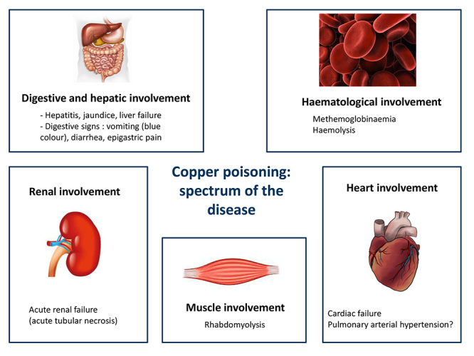
The clinical spectrum of copper poisoning is summarised in Figure 2.
Figure 2.

Clinical spectrum of copper sulphate poisoning
Digestive and hepatic disorders
The first and most frequent (80–100%) symptom of copper poisoning is digestive [7]. It begins with vomiting, epigastric pain or diarrhoea. A characteristic sign is bluish vomiting [8].
Copper, which is toxic to intestinal mucosal cells, can cause ulcerations responsible for haematemesis and melena, with haemorrhagic or hypovolaemic shock. In our case, rectal administration may explain the presence of diarrhoea only at admission.
Copper then passes to the liver where it is particularly toxic to hepatocytes. One study has shown a correlation between the level of hepatic cytolysis and mortality[4]. Copper hepatotoxicity causes acute hepatitis and jaundice, and can lead to death from liver failure.
Haemolysis
Haemolysis is also a frequent manifestation, related to the accumulation of copper in red blood cells. It is related to both the direct cellular toxicity of copper, and the inhibition of the enzymes responsible for protecting the red blood cells against oxidative stress (mainly G6PD and glutathione reductase)[9, 10]. The presentation of our case was quite uncommon: haemolysis occurred at day 4 following intake, whereas it is usually described within 12 to 24 hours after intoxication[1].
Not all the criteria for toxic haemolysis were met: haptoglobin was normal, and Coombs test was positive. However, we did not find any other aetiology for this brutal anaemia, in particular, no external evidence of gastrointestinal bleeding.
Rhabdomyolysis
Rhabdomyolysis is a rare complication of copper sulphate poisoning[1]. It has also been described in Wilson’s disease[11]. The mechanism is thought to be related to copper inactivation of the Na+ / K+ ATPase pump, resulting in increased cell permeability in human musculoskeletal cells[12].
Acute renal failure
About 40% of cases of acute copper sulphate poisoning are complicated by acute renal failure (ARF)[13].
The condition is caused by acute tubular necrosis, linked to several factors: myoglobinuria, haemoglobinuria, direct toxicity of cupper and hypovolaemia related to digestive symptoms. The advent of haemodialysis has reduced ARF-related mortality[4].
However, in our case, renal function was never recovered, which may suggest a more complex mechanism with definitive lesions in the kidney.
Methemoglobinaemia
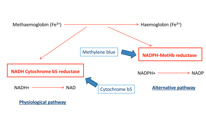
Methemoglobin is haemoglobin in which oxidised iron has been transformed from the ferrous state (Fe ++) to the ferric state (Fe +++), which renders it unfit for oxygen transportation. Two main erythrocyte enzymatic systems are involved in the reduction of physiologically produced methemoglobin (Fig. 3):
Figure 3.

Diagram of the different ways of reducing methemoglobin to haemoglobin
a physiological pathway via cytochrome b5 reductase, NADH-dependent
an alternative pathway via nicotinamide adenine dinucleotide phosphate hydrogen methemoglobin (NADPH-MetHb) reductase, which is involved in oxidative stress. It uses NADPH which is formed through G6PD in the hexose monophosphate shunt. Certain exogenous electron donor agents, such as methylene blue, are able to favour this pathway[14].
Methemoglobinaemia is asymptomatic below 10%. Above these levels, the following will gradually appear: cyanosis and brown blood staining, signs of hypoxia, neurological signs and death.
Copper sulphate poisoning is a rare cause of methemoglobinemia. Poisoning with dapsone or with amyl-nitrites through the use of poppers are more famous causes[15].
Cardiac disorders
Cardiac involvement is rarely reported in cases of copper poisoning, although it is known that copper also accumulates in myocardial cells. Several hypotheses may be formulated to explain a cardiac arrest during copper intoxication: direct toxicity of the metal on the myocytes, tissue hypoxia induced by methemoglobinaemia, or a metabolic disorder such as hyperkalaemia related to the renal failure. In our patient, cardiac ultrasound showed isolated right heart failure. An experimental study in sheep showed that IV administration of copper sulphate induces an increase in pulmonary resistance[16], although the underlying mechanism is not clear. It may be related to the implication of copper sulphate in the synthesis of catecholamines.
Diagnosis and management
Copper intoxication can be diagnosed through direct analysis of the substance, if available, as was the case in our patient. It can also be done by measuring blood copper level, although this decreases rapidly after ingestion [7], and is not related to the severity of the prognosis[4,17].
Once the diagnosis is made, the treatment is based on the administration of copper chelators, although these have not been proven to be effective, symptomatic treatment of malfunctions (including dialysis during renal failure), methylene blue and high-dose oxygen administration in cases of symptomatic methemoglobinemia. The available chelators are D-penicillamine, dimercaprol and edentate calcium diphosphore (EDTA). These treatments can chelate copper and promote its elimination through the urinary tract. We did not use them because our patient was anuric. Nevertheless, they have already been successfully used in some cases reported to be anuric[18]. The rationale for this use was that haemodialysis can filter copper-bound chelators. Haemodialysis alone has not proven to be effective in purifying copper sulphate[19], which first binds to plasma proteins in the blood, and is then rapidly stored in liver, muscle and erythrocyte cells[4].
The literature also reports cases successfully treated with plasmapheresis[20] and more recently the use of veno-venous extracorporeal membrane oxygenation (ECMO) for treatment of acute respiratory distress syndrome secondary to copper sulphate poisoning[21].
To conclude, copper poisoning remains rare, especially in Western countries, but it causes significant mortality and morbidity. It must be quickly recognised despite its polymorphic presentation, in order to quickly establish appropriate treatment and anticipate its numerous complications. This treatment is based on chelators and symptomatic treatment, and requires a suitable technical platform including haemodialysis, mechanical ventilation and even sometimes plasma exchanges or ECMO.
Acknowledgments
The authors would like to thank all the members of the medical and paramedical team who took care of this patient. We also wish to thank the toxicology laboratory for analysis of the copper powder.
Footnotes
Conflicts of Interests: The Authors declare that there are no competing interest
REFERENCES
- 1.Saravu K, Jose J, Bhat M, Jimmy B, Shastry B. Acute ingestion of copper sulphate: A review on its clinical manifestations and management. Indian J Crit Care Med. 2007;11:74–80. [Google Scholar]
- 2.Laliberté J, Labbé S. The molecular bases for copper uptake and distribution: lessons from yeast. Med Sci (Paris) 2008;24:277–283. doi: 10.1051/medsci/2008243277. [DOI] [PubMed] [Google Scholar]
- 3.Mortazavi F, Jahfari-Javid A. Acute renal failure due to copper sulfate poisoning; a case report. Iran J Pediatr. 2009;19:75–78. [Google Scholar]
- 4.Naha K, Saravu K, Shastry BA. Blue vitriol poisoning: a 10-year experience in a tertiary care hospital. Clin Toxicol (Phila) 2012;50:197–201. doi: 10.3109/15563650.2012.660572. [DOI] [PubMed] [Google Scholar]
- 5.Zhukov VF, Novoselov VP. [Fatal poisoning with blue vitriol]. Sud Med Ekspert. 1983;26:55–56. [PubMed] [Google Scholar]
- 6.Park KS, Kwon JH, Park SH, Ha W, Lee J, An HC, et al. Acute copper sulfate poisoning resulting from dermal absorption. Am J Ind Med. 2018;61:783–788. doi: 10.1002/ajim.22892. [DOI] [PubMed] [Google Scholar]
- 7.Chuttani HK, Gupta PS, Gulati S, Gupta DN. Acute copper sulfate poisoning. Am J Med. 1965;39:849–854. doi: 10.1016/0002-9343(65)90105-1. [DOI] [PubMed] [Google Scholar]
- 8.Higny J, Vanpee D, Boulouffe C. Bluish vomiting: a rare clinical presentation of poisoning. Acta Clin Belg. 2014;69:299–301. doi: 10.1179/2295333714Y.0000000033. [DOI] [PubMed] [Google Scholar]
- 9.Valsami S, Stamoulis K, Lydataki E, Fountoulaki-Paparizos L. Acute copper sulphate poisoning: a forgotten cause of severe intravascular haemolysis. Br J Haematol. 2012;156(3):294. doi: 10.1111/j.1365-2141.2011.08881.x. [DOI] [PubMed] [Google Scholar]
- 10.Sood N, Verma P. Life-threatening haemolysis in a patient with acute copper sulphate poisoning. Indian J Anaesth. 2011;55:204–205. doi: 10.4103/0019-5049.79878. [DOI] [PMC free article] [PubMed] [Google Scholar]
- 11.Propst A, Feichtinger H, Judmaier G, Willeit J, Vogel W. Gastroenterology. 1995;108:885–887. doi: 10.1016/0016-5085(95)90465-4. [DOI] [PubMed] [Google Scholar]
- 12.Benders AA, Li J, Lock RA, Bindels RJ, Bonga SE, Veerkamp JH. Copper toxicity in cultured human skeletal muscle cells: the involvement of Na+/K(+)-ATPase and the Na+/Ca(2+)-exchanger. Pflugers Arch. 1994;428:461–467. doi: 10.1007/BF00374566. [DOI] [PubMed] [Google Scholar]
- 13.Chugh KS, Sharma BK, Singhal PC, Das KC, Datta BN. Acute renal failure following copper sulphate intoxication. Postgrad Med J. 1977;53:18–23. doi: 10.1136/pgmj.53.615.18. [DOI] [PMC free article] [PubMed] [Google Scholar]
- 14.Ludlow JT, Wilkerson RG, Nappe TM. StatPearls. Treasure Island (FL): StatPearls Publishing; 2019. [Accessed 15 Aug 2019]. Methemoglobinemia. Available from: https://www.ncbi.nlm.nih.gov/books/NBK537317/ [Google Scholar]
- 15.Wilkerson RG. Getting the blues at a rock concert: a case of severe methaemoglobinaemia. Emerg Med Australas. 2010;22:466–469. doi: 10.1111/j.1742-6723.2010.01336.x. [DOI] [PubMed] [Google Scholar]
- 16.Ahmed T, Januszkiewicz A, Eyre P, Robinson MJ, Sackner MA. Acute pulmonary hemodynamic effects of intravenous copper sulfate: role of alpha-adrenergic system. J Appl Physiol. 1981;51:1204–1213. doi: 10.1152/jappl.1981.51.5.1204. [DOI] [PubMed] [Google Scholar]
- 17.Wahal PK, Mehrotra MP, Kishore B, Patney NL, Mital VP, Hazra DK, et al. Study of whole blood, red cell and plasma copper levels in acute copper sulphate poisoning and their relationship with complications and prognosis. J Assoc Physicians India. 1976;24:153–158. [PubMed] [Google Scholar]
- 18.Takeda T, Yukioka T, Shimazaki S. Cupric sulfate intoxication with rhabdomyolysis, treated with chelating agents and blood purification. Intern Med. 2000;39:253–255. doi: 10.2169/internalmedicine.39.253. [DOI] [PubMed] [Google Scholar]
- 19.Agarwal BN, Bray SH, Bercz P, Plotzker R, Labovitz E. Ineffectiveness of hemodialysis in copper sulphate poisoning. Nephron. 1975;15:74–77. doi: 10.1159/000180495. [DOI] [PubMed] [Google Scholar]
- 20.Du Y, Mou Y. The role of plasmapheresis in treating lethal cupric sulfate poisoning. Am J Med Sci. 2019;357:338–342. doi: 10.1016/j.amjms.2018.11.014. [DOI] [PubMed] [Google Scholar]
- 21.Cho YS, Moon JM, Jeong YH, Lee DH, Chun BJ. Successful extracorporeal life support in respiratory failure after copper sulphate ingestion. Natl Med J India. 2018;31:83–85. doi: 10.4103/0970-258X.253166. [DOI] [PubMed] [Google Scholar]


