Abstract
BACKGROUND AND PURPOSE: One of the dilemmas facing clinicians treating patients with thyroid cancer is the evaluation of postthyroidectomy patients with rising serum thyroglobulin levels and indeterminate or normal findings on neck sonography. In this study, we examine the role of MR imaging in this subgroup of patients.
MATERIALS AND METHODS: We retrospectively reviewed MR images of patients with thyroid cancer with abnormal lymph nodes in the retropharyngeal and parapharyngeal spaces and determined the size and signal-intensity characteristics of these nodes. We reviewed patient charts for the following history: 1) thyroidectomy, 2) rising thyroglobulin levels, 3) iodine-131 radiation therapy, 4) neck dissection, and 5) pathology on neck sonography and chest CT. We reviewed pathology findings to determine if thyroid cancer metastases were present in these lymph nodes.
RESULTS: Eight patients had abnormal retropharyngeal space nodes, and 1 patient had a parapharyngeal space mass. Lymph nodes ranged from 7 to 25 mm. On MR imaging, 1 patient had a cystic node, 2 had complex nodes, and 6 had solid nodes. Eight patients had rising serum thyroglobulin levels and a history of thyroidectomy, radioiodine therapy, and neck dissection. Two of these patients had no pathologic nodes on sonography and normal findings on chest CT. Six patients had tissue sampling of their skull base node, and metastatic thyroid cancer was present in 5.
CONCLUSIONS: MR imaging of the neck should be considered in thyroidectomy patients with rising serum thyroglobulin levels and a history of radioiodine therapy and neck dissection. Radiologists should carefully examine the retropharyngeal and parapharyngeal spaces in these patients because nodal metastases may occur there more commonly than realized.
The National Cancer Institute estimates that approximately 22,500 new cases of thyroid cancer are diagnosed each year.1 Papillary and follicular variants, collectively known as differentiated thyroid cancer, account for 90% of cases.2 Papillary thyroid cancer has a remarkable 40-year survival rate of 93%–94%.2,3 Mortality increases in the setting of recurrent papillary cancer, but the risk is dependent on the site of recurrence.3 Metastases are most common in levels III and IV but may occur in lymph nodes throughout the neck.4,5 The American Thyroid Association recommends total thyroidectomy for papillary carcinoma. If cervical nodal metastases are present, dissection of the anterior and lateral compartments of the neck is also recommended.6
Rising serum thyroglobulin levels in thyroidectomy patients with differentiated thyroid cancer is a marker for residual or recurrent cancer and is evaluated with clinical examination and ultrasonography of the neck to assess metastatic disease. In the setting of elevated thyroglobulin levels, sonography is 96% sensitive for locoregional metastases.7 However, sonography is limited in areas of the neck where bone and air restrict visualization, such as the retropharyngeal and parapharyngeal spaces at the base of skull. Although metastatic thyroid cancer in these areas is thought to be rare, the existing literature may not reflect the true incidence. Although several imaging techniques are available to the clinician in the assessment of occult metastases, the advantages of MR imaging early in the diagnostic algorithm are addressed in this study.
Materials and Methods
We identified and retrospectively reviewed patients with thyroid cancer with abnormal lymph nodes in the retropharyngeal and parapharyngeal spaces detected on MR imaging during a 6-year period (2003–2008). The institutional review board at our institution approved this study. All patients were imaged at our institution on a 1.5T unit by using the following pulse sequences: sagittal and axial T1-weighted, axial fat-suppressed T2-weighted, and gadolinium-enhanced fat-suppressed axial and coronal T1-weighted fast multiplanar spoiled gradient-recalled acquisition in the steady state. A board-certified neuroradiologist with expertise in head and neck disease reviewed all the images for the following: presence and size of lymph nodes, nodal contour, T1- and T2-weighted signal-intensity characteristics, and enhancement pattern. Additional imaging and reports were reviewed to determine the presence of these nodes on [18F]fluorodeoxyglucose positron-emission tomography (FDG-PET) and iodine-123 or I-131 scans.
We reviewed patient charts to determine the presence of the following clinical data: 1) prior thyroidectomy for differentiated thyroid cancer, 2) prior I-131 radiation therapy, 3) prior neck dissection, 4) rising serum thyroglobulin levels, 5) abnormal nodes on neck sonography, and 6) abnormalities on chest CT. We also recorded patient age, sex, primary tumor histology, and prior thyroid cancer treatments. Pathology results from CT-guided FNA and/or surgical resection were reviewed to determine the presence of metastatic thyroid cancer in these nodes.
Results
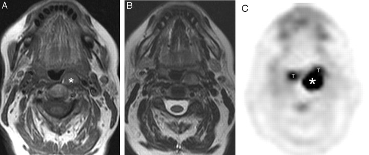
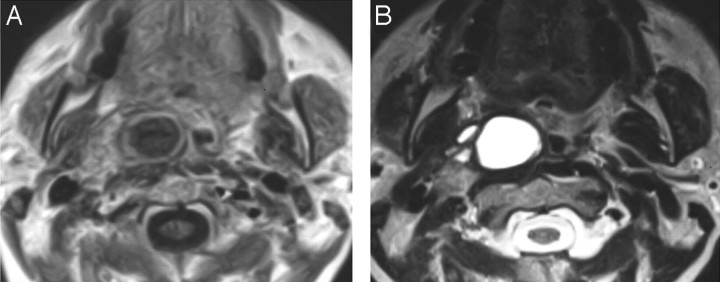
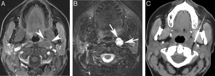
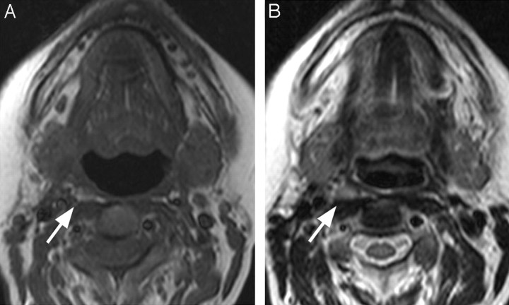
Our review identified 9 patients with thyroid cancer with abnormal lymph nodes in the retropharyngeal (n = 8) or parapharyngeal space (n = 1) on MR imaging (Table). All nodes were characterized as rounded and ranged from 7 to 25 mm (mean, 13 mm). Six patients had nodes that were solid on MR imaging (Fig 1), 1 patient had a cystic node (Fig 2), and 2 had complex nodal masses with solid and cystic components (Fig 3). The solid nodal metastases were hyperintense to muscle on T1-weighted imaging (Fig 4). Four patients had undergone [18F]FDG-PET imaging within the prior year, and 2 had [18F]FDG-avid lesions in the retropharyngeal space (Fig 2). Four patients underwent I-123 or I-131 scanning within the prior year, and none of these patients had abnormal radioiodine uptake in the retropharyngeal or parapharyngeal spaces.
Characteristics of patients with retropharyngeal or parapharyngeal space nodal metastases
| Age*/Sex | Prior Surgery | RP/PPS Node (Location/mm) | MR Signal | PET | I-123 | FNA Cytology | Surgical Pathology |
|---|---|---|---|---|---|---|---|
| 47/F | Thyroidectomy, b/l neck dissection | Left RP/10 | Complex | N/A | N/A | Papillary thyroid ca. | Papillary thyroid ca. |
| 68/F | Thyroidectomy, R neck dissection | Right RP/16 | Solid | Pos | Neg | Papillary thyroid ca. | Papillary thyroid ca. |
| 74/M | Thyroidectomy, L neck dissection | Left RP/20 | Complex | N/A | Neg | Anaplastic thyroid ca. | N/A |
| 58/F | Thyroidectomy, L neck dissection | Left RP/12 | Solid | Pos | Neg | No tumor | Papillary thyroid ca. |
| 49/F | Thyroidectomy, L neck dissection, b/l paratracheal dissection | Left RP/10 | Solid | Neg | N/A | No tumor | N/A |
| 52/F | None | Right PPS/25 | Cystic | N/A | N/A | N/A | Papillary thyroid ca. |
| 47/F | Thyroidectomy, R neck dissection | Right RP/7 | Solid | Neg | Neg | N/A | N/A |
| 47/F | Thyroidectomy, b/l neck dissection, posterior neck dissection | Left RP/7 | Solid | N/A | N/A | N/A | N/A |
| 56/F | Thyroidectomy, R neck dissection | Right RP/11 | Solid | N/A | N/A | N/A | N/A |
Note:—RP indicates retropharyngeal node; PPS, parapharyngeal space node; b/l, bilateral; L, left; R, right; Neg, negative; Pos, positive; N/A, not applicable; ca., cancer; PET, positron-emission tomography; FNA, fine needle aspiration.
Age at identification of pathologic lymph node on MR imaging.
Fig 1.
A 51-year-old woman with a history of thyroidectomy, left neck dissection, and rising thyroglobulin levels. A, Axial T1-weighted MR image shows a 12-mm solid left retropharyngeal node (asterisk) that is hyperintense relative to muscle. B, Axial T2-weighted MR image shows the left retropharyngeal lymph node. C, FDG-PET shows avid uptake in the left retropharyngeal node at the skull base, which did not concentrate radioiodine (not shown). Surgical resection confirmed metastatic papillary carcinoma. T indicates physiologic uptake in the palatine tonsils.
Fig 2.
A 52-year-old woman being evaluated with MR imaging for an unrelated indication. An incidental right parapharyngeal space mass was identified. A, Gadolinium-enhanced axial T1-weighted MR image shows a multiseptate cystic rim-enhancing mass in the right parapharyngeal space. B, Axial T2-weighted MR image shows the multiseptate parapharyngeal mass. The patient went directly to surgical resection, which identified metastatic papillary cancer in a high level II lymph node in the parapharyngeal space. Subsequent thyroidectomy identified a 4-mm papillary carcinoma in the thyroid gland. No other pathologic nodes in the neck were identified on MR imaging.
Fig 3.
A 47-year-old woman with a history of thyroidectomy, bilateral neck dissections, and rising thyroglobulin levels. A, Gadolinium-enhanced axial T1-weighted MR image shows a 1-cm complex solid and cystic lymph node metastasis in the left retropharynx (arrowheads). B, Corresponding axial T2-weighted MR image shows the complex retropharyngeal node (arrows). C, CT-guided FNA reveals metastatic papillary cancer confirmed at surgical resection.
Fig 4.
A 47-year-old woman treated with thyroidectomy and right neck dissection for papillary carcinoma presented with progressive rising thyroglobulin levels. A, Axial unenhanced T1-weighted MR image shows a 7-mm right retropharyngeal node (arrow) that is hyperintense relative to muscle. B, Axial T2-weighted MR image shows the node (arrow). Both FDG-PET and radioiodine showed increased uptake (not shown).
The patients in our study ranged from 47 to 74 years of age (mean, 55 years) and included 8 women and 1 man (Table). Eight of these patients had an initial diagnosis of papillary thyroid cancer and had undergone thyroidectomy, radioiodine treatment, and neck dissection. In these patients, the neck dissection was ipsilateral to the skull base metastasis that subsequently developed. All except 1 patient had rising serum thyroglobulin levels. Seven patients had neck sonography within the prior year, and 5 had pathologic nodes that were rounded, enlarged, and, in 1 patient, contained calcifications. Six patients had undergone chest CT within the prior year, and 3 had lung nodules suggestive of metastatic disease.
Six patients had tissue sampling of their skull base lymph nodes. Five patients had CT-guided FNA, of whom 2 had metastatic papillary carcinoma and 1 had anaplastic transformation in the nodal metastasis. Two had no cancer identified on FNA; however, 1 of these patients underwent surgical resection at which metastatic papillary carcinoma was identified in the node. The other patient with negative findings on CT-guided FNA is being followed clinically. Four patients went on to surgical resection of their retropharyngeal or parapharyngeal space nodal masses, where pathology confirmed metastatic papillary carcinoma. Three patients in our study did not have their retropharyngeal nodes histologically sampled. In 2 patients, this was due to small nodal size (7 mm) on MR imaging. The other patient has elected close clinical and radiologic observation.
Discussion
In the United States, approximately 367,000 people are living with the diagnosis of thyroid cancer. Women are 3 to 4 times more likely than men to be diagnosed with thyroid cancer, but men are more likely to die of this disease. European Americans are 17 times more likely to be diagnosed with thyroid cancer than are African or Hispanic Americans, but the mortality rate is similar among these populations (0.5–0.6 per 100,000).1 Differentiated thyroid cancer accounts for 90% of diagnoses and comprises the following subtypes: papillary (80%), follicular (11%), and Hürthle cell (3%).2
Papillary carcinoma generally has an excellent prognosis, though certain factors are associated with a higher risk for recurrent disease and cause-specific mortality, such as being younger than 15 years of age or older than 45 years of age, male gender, family history of thyroid cancer, and violation of the thyroid gland capsule at the time of treatment.8,9 Recurrence and mortality rates are higher in the setting of tumors >4.0 cm, the presence of bilateral or mediastinal lymph node metastases, extrathyroidal extension of the primary tumor, vascular invasion, poor radioiodine accumulation, and distant metastases.8,10,11 Certain histologic subtypes behave more aggressively. Hürthle cell, tall cell, and columnar cell variants carry a worse prognosis and frequently present with extrathyroidal extension, including vascular or laryngeal invasion. Papillary thyroid cancer metastasizes to lymph nodes in approximately 40%–60% of patients.8,9,12 Mortality increases by 12% in recurrent disease and 48% with distant metastases.3
Lymphatic spread is the most common mode of metastasis in differentiated thyroid cancer. The thyroid gland has an extensive anastomotic network of lymphatic drainage. The superomedial, superolateral, inferomedial, and inferolateral trunks provide the primary lymphatic drainage to the prelaryngeal nodes, upper deep cervical nodes, pretracheal nodes, and supraclavicular and subclavian nodes, respectively. A fifth lymphatic pathway, the posterosuperior trunk, exists in 20% of the population and drains into the lateral retropharyngeal nodes.13 Metastases may reach retropharyngeal nodes and high level II nodes in the parapharyngeal space by retrograde flow through the upper jugular lymphatics. Neck dissection removes most of the lymph nodes in the central and lateral compartments of the neck, altering lymphatic drainage and potentially redirecting metastases to the retropharyngeal nodes or high level II nodes in the parapharyngeal space. Large cervical nodal metastases may also play a role in redirecting lymphatic flow by obstructing normal lymphatic drainage.
Reports of metastatic papillary thyroid cancer to retropharyngeal and parapharyngeal nodes date back to 197014 and 1985,15 respectively. The English language literature reported 24 cases of retropharyngeal metastases14,16–20 and 22 cases of parapharyngeal metastases.15,21–36 Reports of metastatic medullary thyroid cancer in retropharyngeal and parapharyngeal space nodes also exist.20,31,37 Eight of our 9 patients had abnormal nodes in the retropharyngeal space, whereas 1 had an abnormal node in the level II nodes of the parapharyngeal space. All of our patients with metastases to the retropharyngeal space had prior neck dissection ipsilateral to the retropharyngeal metastasis. Where surgical data are reported in the literature, 10 of 12 patients with retropharyngeal metastases had prior neck dissection, whereas only 4 of 16 with parapharyngeal metastases had prior neck dissection. Only 1 of our patients had a nodal metastasis in the parapharyngeal space, and in this patient, this was the presenting finding of thyroid cancer in the absence of palpable nodules in the thyroid or lymph nodes in the neck. The literature describes 8 patients with parapharyngeal space nodal metastases as a presenting symptom in the initial diagnosis of thyroid cancer,15,21,25,27,29,32–34 but only 1 patient with a retropharyngeal metastasis presenting this way.16 These observations suggest that retropharyngeal nodal metastases may be more common in patients with treated thyroid cancer and prior neck dissection, whereas parapharyngeal space nodal metastasis may be more likely to occur in untreated patients.
Most cases (80%) of thyroid cancer metastases to the retropharyngeal and parapharyngeal space lymph nodes have been identified in the last 10 years by using CT and MR imaging. Although sonography is the preferred technique for detecting nodal metastases in the central and lateral neck, interference from bone and air limit the diagnostic capabilities of sonography in the retropharyngeal and parapharyngeal spaces. Although both MR imaging and CT provide excellent anatomic imaging of deep extramucosal spaces in the neck, MR imaging has several distinct advantages in patients with thyroid cancer. In these patients, MR imaging avoids the limitations inherent to iodinated contrast agents, which can interfere with radioiodine-based diagnostic or treatment scans for 6–8 weeks after administration.38
Because thyroid cancer metastases may contain high levels of thyroglobulin or colloid, they frequently appear hyperintense on T1-weighted imaging (Fig 4).39 FDG-PET and radioiodine imaging can be useful adjuncts in confirming nodal metastases, but neither has the sensitivity to be a reliable tool alone in detecting metastatic thyroid cancer. Although the specificity of radioiodine approaches 100%, it detects only half of recurrent disease in patients previously treated with thyroidectomy and I-131 radioablation.40 None of our 4 patients with radioiodine scans had uptake in the retropharyngeal or parapharyngeal spaces, whereas MR imaging identified abnormal nodes in all of these patients. FDG-PET has its highest sensitivity and specificity in the setting of treated patients with rising thyroglobulin levels and negative findings on radioiodine imaging.41 Three of our patients imaged with FDG-PET met these criteria, but only 2 of the 3 had [18F]FDG-avid metastases at the skull base.
All of our patients with retropharyngeal nodal metastases had evidence of extensive disease in their histories, raising their risk for recurrence and disease-specific morbidity and mortality. All were older than 45 years of age and had recurrences in the setting of prior thyroidectomy, radioiodine treatment, and neck dissection. Six of these 8 patients had a history of bilateral disease and evidence of metastasis to lymph nodes in the chest, and 4 had metastases with poor radioiodine accumulation, all poor prognostic indicators. Although it is not economically feasible to offer MR imaging to all patients with thyroid cancer as a screening for deep extramucosal metastases in the neck, certain patients may benefit from neck MR imaging in their diagnostic evaluation. Our review revealed that patients with retropharyngeal and parapharyngeal space nodal metastases fell into 3 broad categories. Six of our patients underwent MR imaging of the neck in the setting of rising thyroglobulin levels and equivocal neck nodes on sonography. Two patients had rising serum thyroglobulin levels and normal sonography of the neck, suggesting an occult metastasis requiring evaluation of the deeper neck spaces. In 1 patient the parapharyngeal space nodal metastasis was an incidental finding on MR imaging performed for an unrelated indication.
Our observations suggest that neck MR imaging is useful in evaluating thyroidectomy patients with rising thyroglobulin levels with equivocal or negative ultrasound of the neck. Patients with prior I-131 radiation therapy and neck dissection may be more likely to present with retropharyngeal metastases. When reviewing imaging from these patients, radiologists should carefully examine the retropharyngeal space and level II nodes in the parapharyngeal space for nodal metastases because these are likely to occur more commonly than recognized. Although not all pathologic nodes in the retropharyngeal or parapharyngeal spaces in patients with thyroid cancer will require immediate biopsy or resection, it is important to monitor these nodes for growth in subsequent MR imaging studies.
Conclusions
Thyroid cancer metastatic to retropharyngeal space lymph nodes should be considered in patients with thyroid cancer who have been treated with thyroidectomy, radioiodine, and neck dissection and present with rising serum thyroglobulin levels. We identified 9 patients at our institution in the past 6 years with metastases to retropharyngeal and parapharyngeal space lymph nodes, which may not be as rare as has been suggested in the literature. In the work-up of these patients, MR imaging provided excellent evaluation of retropharyngeal and parapharyngeal space metastases without the need for iodinated contrast.
Footnotes
Paper previously presented at: Annual Meeting of the American Society of Head and Neck Radiology, September 25–30, 2007; Seattle, Wash.
References
- 1.Ries LAG MD, Krapcho M, Mariotto A, et al. eds. SEER Cancer Statistics Review, 1975–2004. Bethesda, Md: National Cancer Institute;2007
- 2.Hundahl SA, Fleming ID, Fremgen AM, et al. A National Cancer Data Base report on 53,856 cases of thyroid carcinoma treated in the U.S., 1985–1995. Cancer 1998;83:2638–48 [DOI] [PubMed] [Google Scholar]
- 3.Mazzaferri EL, Kloos RT. Clinical review 128: current approaches to primary therapy for papillary and follicular thyroid cancer. J Clin Endocrinol Metab 2001;86:1447–63 [DOI] [PubMed] [Google Scholar]
- 4.Kupferman ME, Patterson M, Mandel SJ, et al. Patterns of lateral neck metastasis in papillary thyroid carcinoma. Arch Otolaryngol Head Neck Surg 2004;130:857–60 [DOI] [PubMed] [Google Scholar]
- 5.Sivanandan R, Soo KC. Pattern of cervical lymph node metastases from papillary carcinoma of the thyroid. Br J Surg 2001;88:1241–44 [DOI] [PubMed] [Google Scholar]
- 6.Cooper DS, Doherty GM, Haugen BR, et al. Management guidelines for patients with thyroid nodules and differentiated thyroid cancer. Thyroid 2006;16:109–42 [DOI] [PubMed] [Google Scholar]
- 7.Pacini F, Molinaro E, Castagna MG, et al. Recombinant human thyrotropin-stimulated serum thyroglobulin combined with neck ultrasonography has the highest sensitivity in monitoring differentiated thyroid carcinoma. J Clin Endocrinol Metab 2003;88:3668–73 [DOI] [PubMed] [Google Scholar]
- 8.Mazzaferri EL, Jhiang SM. Long-term impact of initial surgical and medical therapy on papillary and follicular thyroid cancer. Am J Med 1994;97:418–28 [DOI] [PubMed] [Google Scholar]
- 9.McConahey WM, Hay ID, Woolner LB, et al. Papillary thyroid cancer treated at the Mayo Clinic, 1946 through 1970: initial manifestations, pathologic findings, therapy, and outcome. Mayo Clin Proc 1986;61:978–96 [DOI] [PubMed] [Google Scholar]
- 10.Hawk WA, Hazard JB. The many appearances of papillary carcinoma of the thyroid. Cleve Clin Q 1976;43:207–15 [DOI] [PubMed] [Google Scholar]
- 11.Rossi RL, Nieroda C, Cady B, et al. Malignancies of the thyroid gland: The Lahey Clinic experience. Surg Clin North Am 1985;65:211–30 [DOI] [PubMed] [Google Scholar]
- 12.Mirallie E, Visset J, Sagan C, et al. Localization of cervical node metastasis of papillary thyroid carcinoma. World J Surg 1999;23:970–73, discussion. 973–74 [DOI] [PubMed] [Google Scholar]
- 13.Rouviere H. Anatomy of the Human Lymphatic System: A Compendium Translated from the Original and Rearranged for the Use of Students and Practitioners by MJ Tobias. Ann Arbor, Mich: Edwards Brothers;1938
- 14.McCormack KR, Sheline GE. Retropharyngeal spread of carcinoma of the thyroid. Cancer 1970;26:1366–69 [DOI] [PubMed] [Google Scholar]
- 15.Robbins KT, Woodson GE. Thyroid carcinoma presenting as a parapharyngeal mass. Head Neck Surg 1985;7:434–36 [DOI] [PubMed] [Google Scholar]
- 16.DiLeo MD, Baker KB, Deschler DG, et al. Metastatic papillary thyroid carcinoma presenting as a retropharyngeal mass. Am J Otolaryngol 1998;19:404–06 [DOI] [PubMed] [Google Scholar]
- 17.Le TD, Cohen JI. Transoral approach to removal of the retropharyngeal lymph nodes in well-differentiated thyroid cancer. Laryngoscope 2007;117:1155–58 [DOI] [PubMed] [Google Scholar]
- 18.Leger AF, Baillet G, Dagousset F, et al. Upper retropharyngeal node involvement in differentiated thyroid carcinoma demonstrated by 131I scintigraphy. Br J Radiol 2000;73:1260–64 [DOI] [PubMed] [Google Scholar]
- 19.Otsuki N, Nishikawa T, Iwae S, et al. Retropharyngeal node metastasis from papillary thyroid carcinoma. Head Neck 2007;29:508–11 [DOI] [PubMed] [Google Scholar]
- 20.Shellenberger T, Fornage B, Ginsberg L, et al. Transoral resection of thyroid cancer metastasis to lateral retropharyngeal nodes. Head Neck 2007;29:258–66 [DOI] [PubMed] [Google Scholar]
- 21.Aygenc E, Kaymakci M, Karaca C, et al. Papillary thyroid carcinoma metastasis to the parapharyngeal space. Eur Arch Otorhinolaryngol 2002;259:322–24 [DOI] [PubMed] [Google Scholar]
- 22.Cetik F, Yazici D, Uguz A. A thyroid tumor extending to the parapharyngeal space. BMC Ear Nose Throat Disord 2006;6:3. [DOI] [PMC free article] [PubMed] [Google Scholar]
- 23.Desuter G, Lonneux M, Plouin-Gaudon I, et al. Parapharyngeal metastases from thyroid cancer. Eur J Surg Oncol 2004;30:80–84 [DOI] [PubMed] [Google Scholar]
- 24.Ducci M, Bozza F, Pezzuto RW, et al. Papillary thyroid carcinoma metastatic to the parapharyngeal space. J Exp Clin Cancer Res 2001;20:439–41 [PubMed] [Google Scholar]
- 25.Erdem T, Miman MC, Oncel S, et al. Metastatic spread of occult papillary carcinoma of the thyroid to the parapharyngeal space: a case report. Kulak Burun Bogaz Ihtis Derg 2003;10:244–47 [PubMed] [Google Scholar]
- 26.Ferrario F, Roselli R, Macchi A. Occult thyroid carcinoma presenting as a parapharyngeal mass. J Laryngol Otol 1995;109:1204–06 [DOI] [PubMed] [Google Scholar]
- 27.Horvath M, Plas H, Termote JL, et al. Thyroid-related papillary carcinoma presenting as a cystic lesion in the parapharyngeal space. Rofo 1991;155:373–74 [DOI] [PubMed] [Google Scholar]
- 28.Imai T, Tanaka Y, Matsuura N, et al. Successful surgical treatment of a solitary parapharyngeal metastasis from thyroid cancer, using the mandibular swing-transcervical approach: report of a case. Surg Today 1999;29:378–81 [DOI] [PubMed] [Google Scholar]
- 29.Lombardi D, Nicolai P, Antonelli AR, et al. Parapharyngeal lymph node metastasis: an unusual presentation of papillary thyroid carcinoma. Head Neck 2004;26:190–96 [DOI] [PubMed] [Google Scholar]
- 30.Ortapamuk H, Arican P, Alp A. Parapharyngeal lymph node involvement in papillary thyroid carcinoma. Clin Nucl Med 2003;28:947–48 [DOI] [PubMed] [Google Scholar]
- 31.Pearlman SJ, Lawson W, Biller HF. Occult medullary carcinoma of the thyroid presenting as neck and parapharyngeal metastases. Otolaryngol Head Neck Surg 1988;99:509–12 [DOI] [PubMed] [Google Scholar]
- 32.Ruiz Clemente J, Ortiz Bish E, Gonzalez Garcia J, et al. Papillary carcinoma of the thyroid metastatic to the parapharyngeal space[in Spanish]. Acta Otorrinolaringol Esp 2005;56:135–37 [DOI] [PubMed] [Google Scholar]
- 33.Saydam L, Kalcioglu T, Demirkiran A, et al. Occult papillary thyroid carcinoma presenting as a parapharyngeal metastasis. Am J Otolaryngol 1999;20:166–68 [DOI] [PubMed] [Google Scholar]
- 34.Sirotnak JJ, Loree TR, Penetrante R. Papillary carcinoma of the thyroid metastatic to the parapharyngeal space. Ear Nose Throat J 1997;76:342–44 [PubMed] [Google Scholar]
- 35.Thomas G, Pandey M, Jayasree K, et al. Parapharyngeal metastasis from papillary microcarcinoma of thyroid: report of a case diagnosed by peroral fine needle aspiration. Br J Oral Maxillofac Surg 2002;40:229–31 [DOI] [PubMed] [Google Scholar]
- 36.Tomoda C, Matsuzuka F, Miyauchi A. Parapharyngeal metastasis from papillary thyroid carcinoma: a case diagnosed by thyroglobulin measurement in peroral fine-needle aspiration of a cystic metastatic lymph node. J Laryngol Otol 2005;119:155–57 [DOI] [PubMed] [Google Scholar]
- 37.Gourgiotis L, Sarlis NJ, Reynolds JC, et al. Localization of medullary thyroid carcinoma metastasis in a multiple endocrine neoplasia type 2A patient by 6-[18F]-fluorodopamine positron emission tomography. J Clin Endocrinol Metab 2003;88:637–41 [DOI] [PubMed] [Google Scholar]
- 38.van der Molen AJ, Thomsen HS, Morcos SK. Effect of iodinated contrast media on thyroid function in adults. Eur Radiol 2004;14:902–07 [DOI] [PubMed] [Google Scholar]
- 39.Som PM, Brandwein M, Lidov M, et al. The varied presentations of papillary thyroid carcinoma cervical nodal disease: CT and MR findings. AJNR Am J Neuroradiol 1994;15:1123–28 [PMC free article] [PubMed] [Google Scholar]
- 40.Grunwald F, Kalicke T, Feine U, et al. Fluorine-18 fluorodeoxyglucose positron emission tomography in thyroid cancer: results of a multicentre study. Eur J Nucl Med 1999;26:1547–52 [DOI] [PubMed] [Google Scholar]
- 41.Feine U, Lietzenmayer R, Hanke JP, et al. Fluorine-18-FDG and iodine-131-iodide uptake in thyroid cancer. J Nucl Med 1996;37:1468–72 [PubMed] [Google Scholar]


