Abstract
BACKGROUND AND PURPOSE: The CT grading system for otosclerosis was proposed by Symons and Fanning in 2005. The purpose of this study was to determine if this CT grading system has high interobserver and intraobserver agreement.
MATERIALS AND METHODS: All 997 petrous bone CTs performed between December 2000 and September 2007 were reviewed. A total of 81 subjects had CT evidence of otosclerosis on at least 1 side; 68 (84%) had bilateral disease. Because otosclerosis was clinically suspected in both ears of all 81 subjects even if CT evidence was only unilateral, both petrous bones (162 in total) were included. Two blinded neuroradiologists independently graded disease severity using the Symons/Fanning grading system: grade 1, solely fenestral; grade 2, patchy localized cochlear disease (with or without fenestral involvement) to either the basal cochlear turn (grade 2A), or the middle/apical turns (grade 2B), or both the basal turn and the middle/apical turns (grade 2C); and grade 3, diffuse confluent cochlear involvement (with or without fenestral involvement). One reviewer repeat-graded the petrous bone CTs to determine intraobserver agreement with a 7-month intervening delay to mitigate recall bias.
RESULTS: There were 154 agreements (95%) comparing the first grading of reviewer 1 with that of reviewer 2 (κ = 0.93). When the repeat 7-month delayed grading of reviewer 1 was compared with that of reviewer 2, there were 151 (93%) agreements (κ = 0.90). Therefore, mean interobserver agreement was excellent (mean κ = 0.92). There were 155 agreements (96%) comparing the original grading of reviewer 1 with the delayed grading (κ = 0.94), demonstrating excellent intraobserver agreement.
CONCLUSIONS: A recently published CT grading for otosclerosis on the basis of location of involvement yielded excellent interobserver and intraobserver agreement.
Otosclerosis is an idiopathic disease that can result in spongiosis or sclerosis of portions of the petrous bone leading to conductive, sensorineural, or mixed hearing loss. Hearing loss from otosclerosis is often bilateral, and the effect on quality of life can be profound.
Otosclerosis has traditionally been diagnosed by characteristic clinical findings, which include progressive conductive hearing loss, a normal tympanic membrane, and no evidence of middle ear inflammation. Seen through the tympanic membrane, the promontory may have a faint pink tinge reflecting the vascularity of the lesion, referred to as the Schwartze sign.1
Conductive hearing loss is usually secondary to impingement of abnormal bone on the stapes footplate. This involvement of the oval window forms the basis of the name fenestral otosclerosis. The most common location of involvement of otosclerosis is the bone just anterior to the oval window at a small cleft known as the fissula ante fenestram.2–5 The fissula is a thin fold of connective tissue extending through the endochondral layer, approximately between the oval window and the cochleariform process, where the tensor tympani tendon turns laterally toward the malleus.
Imaging is often not pursued in patients who present with uncomplicated conductive hearing loss and characteristic clinical findings. Patients with only conductive hearing loss are often treated medically or with surgery without imaging. The diagnosis of otosclerosis may be unclear clinically in cases of sensorineural or mixed hearing loss and may become apparent only on imaging. Therefore, imaging is often performed (and always at our institution) when the hearing loss is sensorineural or mixed. The mechanism of sensorineural hearing loss in otosclerosis is less well understood. It may result from direct injury to the cochlea and spiral ligament from the lytic process or from release of proteolytic enzymes into the cochlea.
High-resolution CT can show very subtle bone findings. It is the imaging technique of choice in the evaluation of osseous changes in the petrous bones and has been described by many authors with respect to otosclerosis.6, 7 This body of knowledge is evolving because of the advent of better and higher-resolution CT techniques. Previous publications of the prevalence of otosclerosis on CT are likely underestimations given the newer CT techniques.
It has been shown by some authors that the severity of cochlear disease on CT scanning correlates with the degree of sensorineural hearing loss.8, 9 This group of patients can benefit from cochlear implantation. However, others have not been able to significantly correlate the severity of disease with the severity of hearing loss, though sample sizes were small in some of these studies.10, 11
Various authors have used CT grading systems of otosclerosis in their studies. Valvassori initially proposed a grading system for cochlear otosclerosis on the basis of disease site and progression.2 Shin et al8 divided subjects into fenestral and pericochlear, with the pericochlear group subdivided into extended or not extended to the cochlear endosteum. Kiyomizu et al9 graded fenestral disease as group A, no pathologic CT findings; group B1, demineralization localized to the fissula ante fenestram; group B2, demineralization extending toward the cochleariform process from the anterior region of the oval window; group B3, extensive demineralization surrounding the cochlea; and group C, thick anterior and posterior calcified plaques.9 Rotteveel et al12 described a classification system on the basis of appearance of involvement of the otic capsule: type 1, solely fenestral involvement; type 2, cochlear (with or without fenestral) involvement and divided into types 2a (“double ring effect”), type 2b (narrowed basal turn), and type 2c (“double ring effect” and narrowed basal turn); and type 3, severe cochlear involvement (unrecognizable otic capsule). No system has gained wide acceptance.
Symons and Fanning13 have recently published a CT grading system for otosclerosis: grade 1, solely fenestral, either spongiotic or sclerotic lesions, evident as a thickened stapes footplate, and/or decalcified, narrowed or enlarged round or oval windows; grade 2, patchy localized cochlear disease (with or without fenestral involvement) to either the basal cochlear turn (grade 2A), or the middle/apical turns (grade 2B), or both the basal turn and the middle/apical turns (grade 2C); and grade 3, diffuse confluent cochlear involvement of the otic capsule (with or without fenestral involvement). Grade 3 is differentiated from grade 2C by the diffuse confluent involvement in grade 3 of the entire cochlea, where as grade 2C has patchy focal involvement of the entire cochlea.
The aim of this study was to identify all patients with otosclerosis from our PACS data base of a tertiary referral center for hearing loss, and assess the interobserver and intraobserver agreement of the grading system described by Symons and Fanning.13 The authors chose to evaluate this grading system because it categorizes the presence of otosclerosis on the basis of both location and appearance of disease (spongiosis and/or sclerosis). This grading system also allows for more precise localization of cochlear disease.
Materials and Methods
Patient Selection
We obtained approval of this study from our local research ethics board. Informed patient consent was waved for this retrospective study. All neuroradiologists involved in image analysis in this study were certified by the American Board of Radiology (ABR) in Diagnostic Radiology, had completed a 2-year neuroradiology fellowship, and were eligible for or certified with an ABR Neuroradiology Certificate of Advanced Qualification. All CT studies performed per our petrous protocol were identified on our PACS. We identified 997 studies from December 2000 to September 2007. The images were examined by 2 neuroradiologists, and a consensus was made that otosclerosis was present on at least 1 side. The CT evidence of otosclerosis included sclerosis or spongiosis at the cochlear promontory; sclerosis or spongiosis around the cochlea; thickened stapes footplate; or sclerotic, spongiotic, narrowed, or enlarged round or oval windows. A total of 81 subjects (9%) were identified as having imaging findings consistent with otosclerosis on at least 1 side. There were 68 (84%) who had bilateral CT evidence of otosclerosis. Because otosclerosis was clinically suspected in both ears of all subjects even if CT evidence was only unilateral, both petrous bones (162 total) of these 81 subjects were included in the analysis.
CT Studies
From 2000 through 2005, the CTs of the petrous bone were performed on a 4-section CT scanner (LightSpeed Plus; GE Healthcare, Milwaukee, Wis) with 0.625-mm section thickness and both axial and coronal imaging. From 2005 to 2007, a 64-section CT scanner (VCT; GE Healthcare) was used with 0.625-mm section thickness axial imaging with coronal re-formations at 0.6 mm. All studies were performed without contrast, and imaging included the entire petrous bone.
Image Review
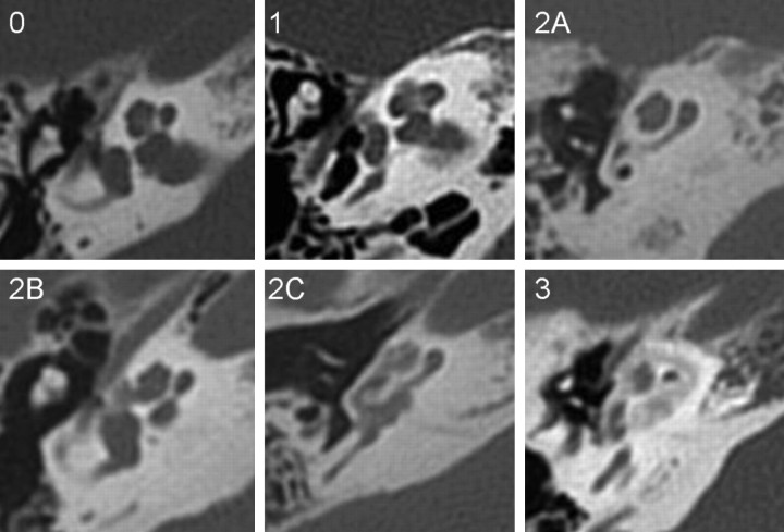
A total of 162 petrous temporal bones were graded independently and in a blinded fashion by 2 neuroradiologists. One neuroradiologist was senior (7 years of experience) and the other, junior (1 year of experience). The appearance of the otic capsule was graded as follows (Fig 1): grade 1, solely fenestral, either spongiotic or sclerotic lesions, evident as a thickened stapes footplate, and/or decalcified, narrowed, or enlarged round or oval windows; grade 2, patchy localized cochlear disease (with or without fenestral involvement) to either the basal cochlear turn (grade 2A), or the middle/apical turns (grade 2B), or both the basal turn and the middle/apical turns (grade 2C); and grade 3, diffuse confluent cochlear involvement of the otic capsule (with or without fenestral involvement). The senior neuroradiologist repeat-graded the same series of petrous bone CT scans with a 7-month gap between the first grading and the repeated grading to mitigate recall bias. The Symons/Fanning grading system is illustrated in Fig 1.
Fig 1.
Axial CT images of the petrous bone in patients with otosclerosis. Grade 0: normal. Grade 1: small lucent lesion at the fissula ante fenestram. Grade 2A: sclerosis and narrowing of the basal turn (also has spongiotic fenestral disease). Grade 2B: lucent lesion extending from the fissula ante fenestram to the middle turn of the cochlea. Grade 2C: patchy lucency around the lateral aspect of basal, middle, and apical turns of the cochlea, the medial aspect of the cochlea appears spared. Grade 3: severe, confluent lucency around the cochlea.
Results
There were 154 agreements (95%) among the 162 petrous bones graded when comparing the first grading of reviewer 1 with that of reviewer 2 (κ = 0.93). When the repeated 7-month delayed grading of reviewer 1 was compared with that of reviewer 2, there were 151 (93%) agreements (κ = 0.90). Mean interobserver agreement was therefore excellent (mean κ = 0.92).
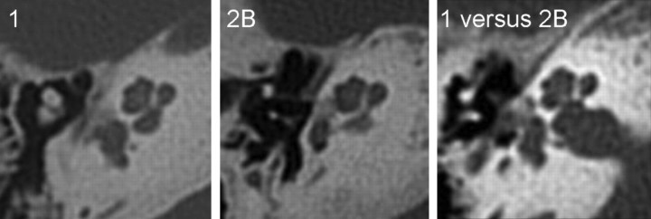
There were 8 disagreements when comparing the first grading of reviewer 1 with that of reviewer 2: five 1 vs 2B, one 2A vs 2C, and two 2B vs 2C. There were 11 disagreements when comparing the repeat 7-month delayed grading of reviewer 1 with that of reviewer 2: one 1 vs 2A, five 1 vs 2B, three 2B vs 2C, and one 2C vs 3. An example of one of the most common disagreements, 1 vs 2B, is shown in Fig 2.
Fig 2.
Disagreement between grade 1 vs grade 2B. Grade 1: there is a lucent lesion in the cochlear promontory with preservation of the adjacent middle turn otic capsule. Grade 2B: there is a lucent lesion in the cochlear promontory, with clear extension to the adjacent otic capsule of the middle turn. Grade 1 vs grade 2B: there is a lucent lesion in the cochlear promontory with debatable extension to the otic capsule of the middle turn and this was graded as 1 by the first neuroradiologist and 2B by the second neuroradiologist.
There were 155 agreements (96%) among the 162 petrous bones graded when comparing the original grading of reviewer 1 to the repeat 7-month delayed grading of reviewer 1 (κ = 0.94), demonstrating excellent intraobserver agreement. There were 7 disagreements: one 1 vs 2A, one 2A vs 2B, one 2A vs 2C, three 2B vs 2C, and one 2C vs 3.
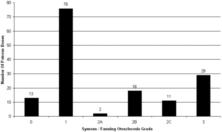
When the first grading of reviewer 1, the first grading of reviewer 2, and the repeat 7-month delayed grading of reviewer 1 were all compared; there were 149 agreements (92%). The distribution of these agreements are demonstrated in Fig 3. Of 81 patients, 68 (84%) had bilateral CT evidence of otosclerosis.
Fig 3.
Grades of otosclerosis. The distribution of grades 0 through 3 among the 149 agreements.
Discussion
There are surgical grading systems of otosclerosis as published by Portmann,14 Bellucci,15 and others. There are also histiologic grading systems as described by Lindsay16 and others. There is no universally accepted imaging grading system for otosclerosis, though many grading systems have been proposed. The CT grading of otosclerosis published by Symons and Fanning13 is based on anatomic localization of disease. Focal spongiotic areas, sclerotic areas, and “double ring effect” are included, but classification is based on the presence of these at a particular location around the cochlea. The “double ring effect” is because of confluence of spongiotic foci within the thickness of the capsule and may be limited to a segment of the capsule or follow the entire cochlear contour.17 The classification proposed by Rotteveel et al12 is partially based on location (solely fenestral, basal turn, or complete cochlea), and on “double ring effect” or basal turn narrowing. There are similarities between the 2 systems of classification. Both Symons/Fanning grade 1 and Rotteveel type 1 are solely fenestral disease. Both Symons/Fanning grade 3 and Rotteveel type 3 are compatible with a complete cochlear “double ring effect.” The difference is in classification of nonconfluent cochlear disease, Symons/Fanning grade 2 and Rotteveel type 2. One advantage of the Symons/Fanning classification is that it provides for more precise localization of disease around the cochlea. The Symons/Fanning classification divides the cochlea into 2 segments: basal turn and middle/apical turns. Symons/Fanning grade 2 is patchy cochlear disease, divided into grades 2A, 2B, and 2C. Grade 2A involves only the basal turn; grade 2B, the middle and/or apical turns; and grade 2C, both the basal turn and the middle and/or apical turns. Grade 3 involves the entire cochlea in a confluent manner and is differentiated from grade 2C, which involves segments of the entire cochlea (basal turn and apical and/or midturn) but in a patchy, rather than confluent, manner. With recent improvements in CT resolution that allow section thicknesses in the half-millimeter range or less, more precise localization of disease is now possible. Future advances in CT technology will continue to make precise localization even easier. The Symons/Fanning classification allows for more precise localization of disease afforded by these more advanced CT scanners. The Symons/Fanning classification also encompasses all types of cochlear disease changes: spongiotic, sclerotic, and the “double ring effect.” One disadvantage of the Rotteveel classification is that grade 2 is limited to either a “double ring effect” and/or narrowing of the basal turn. However, many cases of cochlear disease have neither of these findings but do have focal erosions or sclerosis around the cochlea. It does not seem possible to classify these cases in the Rotteveel grading.
A classification must have excellent interobserver and intraobserver agreement to be clinically useful. The interobserver agreement of the Symons/Fanning classification was excellent (mean κ = 0.92). This excellent agreement was determined by a senior neuroradiologist (7 years of experience) and a junior neuroradiologist (1 year of experience), who demonstrated that the classification system is robust even for level of experience. Intraobsever agreement of the Symons/Fanning classification was excellent (κ = 0.94). Rotteveel et al12 obtained good interobserver agreement (κ = 0.77), but they did not discuss intraobserver agreement in their study. Our agreement measurements were based on 162 petrous bones, compared with 36 in the study by Rotteveel et al.12 Shin et al8 and Kiyomizu et al9 did not assess the reliability of their grading systems.
A small focus of demineralization anterior to the oval window has been classically described as early fenestral otosclerosis. Long-term follow-up suggests that approximately 10% of petrous bones with fenestral otosclerosis develop cochlear involvement.18, 19 In several cases of the current study where a patient had a follow-up study several years later, there was no obvious progression. This may suggest that the nature of progression is so indolent that follow-up of decades is required, or perhaps that after an initial brief period of progression, the sequelae become relatively stable. In rare circumstances, the lesion has been described as attenuated, approaching the same attenuation as the otic capsule.1 This increase in attenuation was rarely observed in our study and may be a reflection of less qualitative difference with increased attenuation as opposed to lysis.
Otosclerosis has been described in the literature as bilateral in 80% of cases.20–23 This figure corresponds well with findings in our study in which, of 81 patients, 68 (84%) had bilateral CT evidence of otosclerosis.
In the cochlear implant in otosclerosis series by Marshall et al13 and Rotteveel et al,12 most patients had cochlear disease, 75% and 77%, respectively. The percentage of purely fenestral disease was slightly more in the Marshall et al series (17% vs 7%). Both of these findings are in contrast to Shin et al,8 who studied CT scans of 437 cases of otosclerosis, of which 91% had positive findings on CT; however, only 12% showed cochlear disease. This is probably attributable to the fact that Shin et al8 were studying the CT scans of patients with much milder hearing loss undergoing stapedotomy, rather than candidates for cochlear implant. In our current series, 40% had cochlear disease (60/149 agreements between the first grading of reviewer 1, the grading of reviewer 2, and the repeat 7-month delayed grading of reviewer 1).
Because patients were identified primarily by imaging findings in our study, one might argue that some of the identified patients may have had imaging consistent with otosclerosis but had an alternate diagnosis. Fenestral otosclerosis has a specific appearance, but cochlear otosclerosis can be mimicked by various diseases that demineralize the cochlear capsule. Such diseases include osteogenesis imperfecta, Paget disease, ankylosing rheumatoid arthritis, and syphilis.1, 4 However, unlike otosclerosis, these diseases have widespread manifestations that are usually apparent on imaging. None of our patients had any of these manifestations. Labyrinthitis ossificans is a different entity occurring as a result of a previous inflammatory process such as meningitis, middle ear infection, trauma, or surgery24 and, again, has associated imaging findings or clinical history. None of our patients had the typical combined imaging findings and history to suggest labyrinthitis ossificans rather than otosclerosis.
It has been shown that this grading system is clinically relevant. Marshall et al13 demonstrated that patients with grade 3 otosclerosis have a higher risk for facial nerve stimulation after cochlear implantation, particularly with nonmodiolar hugging electrodes. Therefore, implant surgeons should be cognizant of the type of implant used when a grade 3 petrous bone is implanted. Grading systems of otosclerosis are also clinically relevant as many authors have shown that as the severity of cochlear otosclerosis increases, the severity of sensorineural hearing loss increases.8, 9
Cochlear otosclerosis presents with combined conductive and sensorineural hearing loss. Specific portions of the cochlea are attuned to certain frequencies of sound. The low-frequency tones have maximal effective amplitude at the apical turn, whereas high-frequency tones have maximal effective amplitude at the basilar turn, where the basilar membrane is relatively thin. Some authors have successfully correlated the frequency of hearing loss with the location of involvement of the cochlear capsule.25, 26 This suggests a local effect by the otosclerotic foci on that part of the cochlea. However, other authors have postulated that the sensorineural hearing loss is a result of cytotoxic enzymes causing hyalinization of the spiral ligament,27, 28 in which case there may be no correlation between the exact location of a lytic focus on the cochlea and the frequency of hearing loss. Additional study of whether the grade of otosclerosis correlates with frequency of hearing loss would be interesting. Audiometric findings may also help resolve which of the disagreement cases are more appropriately graded as solely fenestral vs cochlear otosclerosis.
Conclusions
The CT classification system of otosclerosis proposed by Symons and Fanning has high interobserver and intraobserver agreement. Future correlation with audiometric data may help clarify disagreements in classification.
Footnotes
Paper previously presented in part at: Annual Meeting of the American Society of Neuroradiology, June 2, 2008, New Orleans, La; and Annual Meeting of the American Society of Head and Neck Radiology, September 11, 2008, Toronto, Ontario, Canada.
References
- 1.Sakai O, Curtin HD, Hasso AN, et al. Otosclerosis and dysplasias of the temporal bone. In: Som PM, Curtin HD, eds. Head and Neck Imaging. 4th ed. St. Louis: Mosby;2003. :1245–73
- 2.Valvassori, GE. Imaging of otosclerosis. Otolaryngol Clin North Am 1993;26:359–71 [PubMed] [Google Scholar]
- 3.Rovsing H. Otosclerosis: fenestral and cochlear. Radiol Clin North Am 1974;12:505–15 [PubMed] [Google Scholar]
- 4.Swartz JD, Harnsberger HR. The otic capsule and otodystrophies In: Imaging of the Temporal Bone. 3rd ed. New York: Thieme;1998. :240–317
- 5.Bretlau P. Relation of the otosclerotic focus to the fissula ante-fenestram. J Laryngol Otol 1969;83:1185–93 [DOI] [PubMed] [Google Scholar]
- 6.Palacios E, Valvassori G. Cochlear otosclerosis. Ear Nose Throat J 2000;79:494. [PubMed] [Google Scholar]
- 7.Marx SV, Langman AW. Cochlear otosclerosis. Am J Otol 1997;18:404. [PubMed] [Google Scholar]
- 8.Shin YJ, Fraysse B, Deguine O, et al. Sensorineural hearing loss and otosclerosis: a clinical and radiological survey of 437 cases. Acta Otolaryngol 2001;121:200–04 [DOI] [PubMed] [Google Scholar]
- 9.Kiyomizu K, Tono T, Yang D, et al. Correlation of CT analysis and audiometry in Japanese otosclerosis. Auris Nasus Larynx 2004;31:125–29 [DOI] [PubMed] [Google Scholar]
- 10.Grayeli AB, Saint Yrieix C, Imauchi Y, et al. Temporal bone density measurements using CT in otosclerosis. Acta Otolaryngol 2004;124:1136–40 [DOI] [PubMed] [Google Scholar]
- 11.Naumann IC, Porcellini B, Fisch U. Otosclerosis: incidence of positive findings on high-resolution computed tomography and their correlation to audiological test data. Ann Otol Rhinol Laryngol 2005;114:709–16 [DOI] [PubMed] [Google Scholar]
- 12.Rotteveel LJ, Proops DW, Ramsden RT, et al. Cochlear implantation in 53 patients with otosclerosis: demographics, computed tomographic scanning, surgery, and complications. Otol Neurotol 2004;25:943–52 [DOI] [PubMed] [Google Scholar]
- 13.Marshall AH, Fanning N, Symons S, et al. Cochlear implantation in cochlear otosclerosis. Laryngoscope 2005;115:1728–33 [DOI] [PubMed] [Google Scholar]
- 14.Portmann M. Classifications des lesions stapedo-vestibulaires en cas d'otosclerose chirurgicale. In: Traite de technique chirurgicale O.R.L. et cervico-faciale, vol 1. Oreille et os temporal, Paris: Masson;1986. :119–20
- 15.Bellucci RJ. A guide for stapes surgery based on a new surgical classification of otosclerosis. Laryngoscope 1958;68:741–59 [DOI] [PubMed] [Google Scholar]
- 16.Lindsay JR. Histopathology of otosclerosis. Arch Otalaryngol 1973;97:24–29 [DOI] [PubMed] [Google Scholar]
- 17.Mafee MF, Valvassori GE, Becker M. Imaging of the temporal bone: otosclerosis and bone dystrophies. In Valvassori's Imaging of the Head and Neck, 2nd ed. New York: Thieme;2005. :122–29
- 18.Browning GG, Gatehouse S. Sensorineural hearing loss in stapedial otosclerosis. Ann Otol Rhinol Laryngol 1984;93:13–16 [DOI] [PubMed] [Google Scholar]
- 19.Ramsay HA, Linthicum FH Jr. Mixed hearing loss in otosclerosis: indication for long-term follow-up. Am J Otol 1994;15:536–39 [PubMed] [Google Scholar]
- 20.Schuknecht HF. Disorders of bone. In: Schuknecht HF. Pathology of the Ear, 2nd ed. Malvern, Pa: Lea & Febiger;1993. :365–414
- 21.Nager GT. Otosclerosis. In: Nager GT. Pathology of the Ear and Temporal Bone. Baltimore: Williams & Wilkins;1993. :943–1010
- 22.Lindsay JR. Otosclerosis. In: Paparella MM, Shumrick DA, eds. Otolaryngology. Vol 2, The Ear, 2nd ed. Philadelphia: WB Saunders;1980. :1617–44
- 23.Ruedi L. Pathogenesis of otosclerosis. Arch Otolaryngol 1963;78:469–77 [DOI] [PubMed] [Google Scholar]
- 24.Swartz JD, Mandell DW, Faerber EN, et al. Labyrinthine ossification: CT appearance and possible etiology. Radiology 1985;157:395–98 [DOI] [PubMed] [Google Scholar]
- 25.Swartz JD, Mandell DW, Berman SE, et al. Cochlear otosclerosis (otospongiosis): CT analysis with audiometric correlation. Radiology 1985;155–147-50 [DOI] [PubMed]
- 26.Swartz JD, Mandell DW, Wolfson RJ, et al. Fenestral and cochlear otosclerosis: CT evaluation. Am J Otol 1985;6:476–81 [PubMed] [Google Scholar]
- 27.Antoli-Candela F Jr, McGill T, Peron D. Histopathological observations on the cochlear changes in otosclerosis. Ann Otol Rhinol Laryngol 1977;86:813–20 [DOI] [PubMed] [Google Scholar]
- 28.Parahy C, Linthicum FH. Otosclerosis: relationship of spiral ligament hyalinization to sensorineural hearing loss. Laryngoscope 1983;93:717–20 [DOI] [PubMed] [Google Scholar]


