Abstract
BACKGROUND AND PURPOSE: Recent advances in flat panel detector angiographic equipment have provided the opportunity to obtain physiologic and anatomic information from angiographic examinations. To exploit this possibility, one must understand the factors that affect the bolus geometry of an intra-arterial injection of contrast medium. It was our purpose to examine these factors in a canine model.
MATERIALS AND METHODS: Under an institutionally approved protocol conforming to Guide for the Care and Use of Laboratory Animals of the National Institutes of Health, 7 canines were placed under general anesthesia with isoflurane and propofol. Through a 5F catheter placed into the right common carotid artery, a series of biplane angiographic acquisitions was obtained to examine the effects caused by variation in the volume of injection, the rate of injection, the duration of injection, the concentration of contrast medium, and the catheter position on arterial, capillary, and venous opacification. The results of each injection protocol were determined from analysis of a time-contrast concentration curve derived from locations over an artery, in brain parenchyma, and over a vein. The curve was generated from 2D digital subtraction angiography acquisitions by using prototype software. The area under the curve, the amplitude of the curve, and the time to peak (TTP) were analyzed separately for each injection parameter.
RESULTS: Changes in the injection protocols resulted in predictable changes in the time-concentration curves. The injection parameter that contributed most to maximum opacification was the volume of contrast medium injected. When the injection rate was fixed and the volume was varied, there was an increase in opacification (maximal) proportional to the injected volume. The injected volume also had an indirect (secondary) impact on the temporal characteristics of the opacification. The time-concentration curve became wider, and the peak was shifted to the right as the injection duration increased. The impact of injected volume on maximal opacification was significant (P < .0001), regardless of the site of measurement (artery, tissue, and vein); however, the impact on the temporal characteristics of the time-concentration curve reached statistical significance only in measurements made in the artery and the vein (P < .05), but not in the tissue (P > .1). The impact of injected volume on maximal opacification became nonproportional in the tissue and vein when the volume was very large (>12 mL). Increasing the concentration of contrast medium resulted in a nonproportional increase in the height of the time-concentration curves (P < .05). Injection rate had an impact on both maximal opacification and TTP. The impact on TTP occurred only when the injection rate was very slow (1 mL/s). Changes of concentration had a similar impact on the time-concentration curve. Catheter position did not cause significant alterations in the shape of the curves.
CONCLUSIONS: There were predictable effects from modification of injection parameters on the contrast bolus geometry and on time-concentration curves as measured in an artery, brain parenchyma, or a vein. The amplitude, TTP, and area under the time-concentration curve depend mainly and proportionally on the amount of iodine traversing the vasculature per second. Other injection parameters were of less importance in defining bolus geometry. These findings mimic those observed in studies of parameters affecting bolus geometry following an intravenous injection.
The availability of flat panel detector angiographic equipment has made it possible to perform high-spatial-resolution soft-tissue imaging in the angiographic suite. Recent reports describe the utility of these C-arm CT techniques for the detection of central nervous system hemorrhages, for visualization of endoluminal devices and their relationship to the arterial wall and lumen, and for pretreatment planning and post-treatment evaluations.1–3 Because these applications, along with others that will likely emerge (eg, functional imaging), are often used in association with the intra-arterial administration of iodinated contrast medium, it is necessary to understand the influence of injection parameters that may affect arterial, tissue, and venous opacification.
A variety of parameters may be adjusted when one injects contrast medium. Among others, these include the volume of the injection, the rate of the injection, the duration of the injection, the concentration of the contrast medium that is injected, and the site of injection. Most previous work looking at the effects of variations of these parameters on bolus geometry has been done to try to optimize arterial opacification following an intravenous injection of contrast medium so as to improve the image quality of intravenous digital subtraction angiography (DSA), MR angiography, and CT angiography (CTA).4–7 Although there is very likely a close relationship between the effects of these manipulations on an intra-arterial injection and an intravenous injection in the same subject, to our knowledge, this has not been previously studied.
The purpose of our study was to examine the impact on contrast-bolus geometry after an intra-arterial injection (Table 1) of variation in: 1) the volume of an injection, 2) the rate of an injection, 3) the duration of an injection, 4) the concentration of the contrast injected, and 5) the catheter position.
Table 1:
Injection protocol
| Group | Volume (mL) | Rate (mL/s) | Duration (s) | Concentration | Catheter Position |
|---|---|---|---|---|---|
| 1 | Constant, 8 mL | Variable, 1, 2, 4, 8 mL/s | Variable, 1, 2, 4, 8 seconds | Constant, 100% | Constant, prox ICA |
| 2 | Variable, 4, 8, 12, 16 mL | Constant, 4 mL/s | Variable, 1–4 seconds | Constant, 100% | Constant, prox ICA |
| 3 | Variable, 4, 8, 12, 16 mL | Variable, 2, 4, 6, 8 mL/s | Constant, 2 seconds | Constant, 100% | Constant, prox ICA |
| 4 | Constant, 8 mL | Constant, 4 mL/s | Constant, 2 seconds | Variable, 25, 50, 75, 100% | Constant, prox ICA |
| 5 | Constant, 8 mL | Constant, 4 mL/s | Constant, 2 seconds | Constant, 100% | Variable, prox, middle, distal ICA |
Note:—Prox ICA indicates proximal internal carotid artery.
Materials and Methods
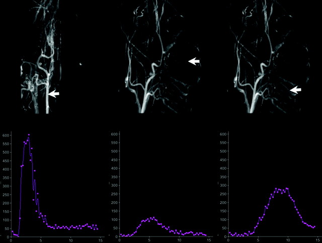
Under an institutionally approved protocol, 7 canines were placed under general anesthesia with isoflurane and propofol. A 5F sheath was placed percutaneously into the right common femoral artery, and through this, a 5F catheter was advanced over a guidewire into the right common carotid artery. There was constant monitoring of heart rate, oxygen saturation, and end-tidal carbon dioxide. Sequential biplane DSA acquisitions were obtained in conjunction with a variety of injection protocols (described below) by using a biplane flat panel detector angiographic system (Artis dBA; Siemens, Erlangen, Germany). Nonionic contrast (300 mg/mL) was power-injected in all studies (Accutron HP-D; Medtron, Saarbrücken, Germany). The angiographic acquisitions were loaded onto a workstation running prototype software (Siemens Healthcare, Forchheim, Germany), which allowed creation of a time-concentration curve of a contrast medium bolus at a selected position on the DSA image. Using these, we could measure specific characteristics and display them on a parametric map. The result from each injection protocol was evaluated by analyzing the time-concentration curves at points in the carotid artery, the torcula, and/or the transverse sinuses and the brain parenchyma. To avoid superimposition of other vascular structures, we made the arterial measurement on an anteroposterior projection, whereas the capillary (tissue) and venous measurements were made on a lateral projection. Care was taken so that the points selected for analysis were such that there was no overlap with other vascular structures (Fig 1). Time-to-peak (TTP) and the area under the curve (AUC) were extracted for the artery, vein, and tissue and were compared. Because there was no movement of the animal or the angiographic table during the acquisitions, the points were the same on all images from each injection protocol. This allowed image-to-image comparison of the curves.
Fig 1.
Three views from the 3D reconstruction used to determine time concentration curves for arterial, parenchyma and venous structures (A, B, and C). The corresponding time-concentration curves are shown in D, E, and F.
We systematically examined 5 variables: 1) volume, 2) rate, 3) duration, 4) concentration, and 5) catheter position, by using the injection protocols summarized in Table 1. Volume, rate, and duration are interrelated variables such that maintaining 1 variable constant requires changing the other 2. The effect of changes in contrast medium concentration was evaluated by using 8 mL of contrast injected at 4 mL/s for 2 seconds. Concentrations of 100%, 75%, 50%, and 25% were tested. This same injection protocol was used to evaluate the effect of varying catheter positions (proximal, middle, and distal) in the right common carotid artery.
TTP, peak opacification, and AUC results from each injection protocol were then evaluated by analyzing the time-concentration curves for the artery (common carotid artery at the bifurcation), vein (torcula), and capillary (brain parenchyma) (Fig 1).
All outcome variables listed in Tables 2–5 were analyzed by using 1-way analysis of variance (ANOVA) after transformation to the log scale to achieve constant variance in the residuals. If the P value for the 1-way ANOVA was significant (P < .05), then pair-wise comparisons of the various levels of the predictor variables were performed by using t tests.
Table 2:
P values from the assessment of TTP, peak opacification, and AUC differences when comparing injection protocols: variable rate and volume with constant duration (2 seconds)*
| Rate | TTP Capillary | TTP Arterial | TTP Venous | Peak Capillary | Peak Arterial | Peak Venous | AUC Capillary | AUC Arterial | AUC Venous |
|---|---|---|---|---|---|---|---|---|---|
| Overall | .0334 | .0102 | .0363 | .0009 | .0057 | .0003 | .0004 | .0003 | <.0001 |
| 2–4 mL/s | .9598 | .4923 | .4592 | .1147 | .0179 | .0020 | .1111 | .0152 | .0002 |
| 2–6 mL/s | .1024 | .0110 | .0302 | .0022 | .0038 | .0002 | .0007 | .0003 | <.0001 |
| 2–8 mL/s | .0126 | .0041 | .0114 | .0002 | .0011 | <.0001 | <.0001 | <.0001 | <.0001 |
| 4–6 mL/s | .1117 | .0404 | .1165 | .0503 | .4158 | .2065 | .0171 | .0512 | .0584 |
| 4–8 mL/s | .0139 | .0153 | .0465 | .0034 | .1592 | .0534 | .0018 | .0062 | .0020 |
| 6–8 mL/s | .2680 | .6066 | .6069 | .1678 | .5229 | .4355 | .2423 | .2739 | .0918 |
Note:—TTP indicates time-to-peak; AUC, area under curve.
P values for paired contrasts were not calculated if no overall significant difference of the group was observed.
Table 5:
P values from the assessment of TTP, peak opacification, and AUC differences when comparing injection protocols: variable rate and duration with constant volume*
| TTP Capillary | TTP Arterial | TTP Venous | Peak Capillary | Peak Arterial | Peak Venous | AUC Capillary | AUC Arterial | AUC Venous | |
|---|---|---|---|---|---|---|---|---|---|
| Overall | .0496 | .1288 | .0444 | .0128 | .0282 | .0805 | .0418 | .2567 | .0956 |
| 1–2 mL/s | .0483 | .0075 | .9195 | .2043 | .1035 | ||||
| 1–4 mL/s | .0291 | .0644 | .0171 | .0057 | .2754 | ||||
| 1–8 mL/s | .0108 | .0371 | .0174 | .0229 | .3659 | ||||
| 2–4 mL/s | .8036 | .3116 | .0137 | .0854 | .0109 | ||||
| 2–8 mL/s | .4775 | .4571 | .0140 | .2575 | .0165 | ||||
| 4–8 mL/s | .6419 | .7819 | .9934 | .5236 | .8458 |
P values for paired contrasts were not calculated if no overall significant difference of the group was observed.
Results
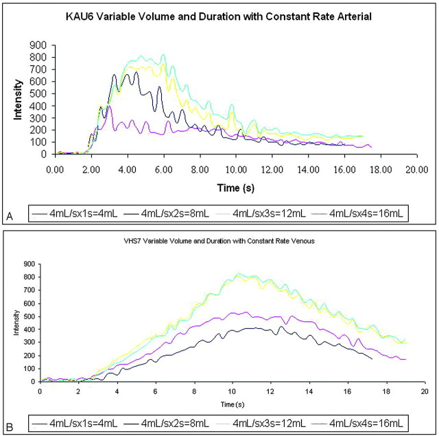
Variations in the injection protocols resulted in predictable changes in the time-concentration curves. The injection parameter that contributed the most to maximum opacification was the injected volume of contrast medium. The impact of this variable on maximal opacification was significant (P < .0001), regardless of the site of measurement (artery, tissue, and vein); however, the impact on the TTP reached statistical significance only with arterial and venous measurements (P < .05). The effect of the contrast volume on the maximal opacification could be observed as well when the injection duration was fixed and both the volume and injection rate varied (Table 2). When the injection rate was fixed, the maximal opacification increased proportionally with the injected volume (P < .01), as long as the injected volume was lower than 12 mL (Table 3). The injected volume had an indirect (secondary) impact on the temporal characteristics (TTP) as well. However, this only occurred in the artery and vein when the injection rate was constant (Table 3 and Fig 2).
Table 3:
P values from the assessment of TTP, peak opacification, and AUC differences when comparing injection protocols: variable volume and duration with constant rate*
| TTP Capillary | TTP Arterial | TTP Venous | Peak Capillary | Peak Arterial | Peak Venous | AUC Capillary | AUC Arterial | AUC Venous | |
|---|---|---|---|---|---|---|---|---|---|
| Overall | 0.9897 | <.0001 | .0161 | <.0001 | <.0001 | <.0001 | <.0001 | <.0001 | <.0001 |
| 4–8 mL | .7927 | .0111 | .0031 | <.0001 | .0014 | .0033 | .0016 | .0015 | |
| 4–12 mL | .0005 | .6923 | <.0001 | <.0001 | <.0001 | <.0001 | <.0001 | <.0001 | |
| 4–16 mL | .0001 | .9761 | <.0001 | <.0001 | <.0001 | <.0001 | <.0001 | <.0001 | |
| 8–12 mL | .0003 | .0046 | .0004 | .0027 | .0003 | .0004 | .0001 | .0002 | |
| 8–16 mL | <.0001 | .0104 | <.0001 | .0001 | .0001 | <.0001 | <.0001 | <.0001 | |
| 12–16 mL | .5841 | .7144 | .1945 | .1677 | .6919 | .2334 | .0495 | .3619 |
Pvalues for paired contrasts were not calculated if no overall significant difference of the group was observed.
Fig 2.
Time-concentration curves observed over an artery (A) and vein (B) at 4 different volumes while the rate of injection remained constant.
Alterations to the contrast concentration yielded results similar to those of altering the injection volume, with higher concentrations paralleling the relationship of higher injected contrast volumes. This effect was most significant in the arterial and venous TTP and in all 3 measurements for peak opacification when comparing full strength (100%) with dilute (25%) contrast medium (Table 4).
Table 4:
P values from the assessment of TTP, peak opacification, and AUC differences when comparing injection protocols: variable concentration*
| TTP Capillary | TTP Arterial | TTP Venous | Peak Capillary | Peak Arterial | Peak Venous | AUC Capillary | AUC Arterial | AUC Venous | |
|---|---|---|---|---|---|---|---|---|---|
| Overall | .2235 | <.0001 | .0072 | .0432 | .0040 | .0035 | .0789 | .0009 | .1901 |
| 100%–25% | <.0001 | .0086 | .0186 | .0023 | .0008 | .0004 | |||
| 100%–50% | .0977 | .0132 | .0122 | .0015 | .0038 | .0064 | |||
| 100%–75% | .2582 | .9593 | .1971 | .1609 | .1356 | .6618 | |||
| 25%–50% | .0004 | .8427 | .8466 | .8578 | .4980 | .2259 | |||
| 25%–75% | <.0001 | .0077 | .2277 | .0517 | .0245 | .0011 | |||
| 50%–75% | .0093 | .0118 | .1656 | .0360 | .0949 | .0166 |
P values for paired contrasts were not calculated if no overall significant difference of the group was observed.
Injection rate had an impact on both maximal opacification and TTP. The greatest impact on TTP occurred when comparing the slowest (1 mL/s) injection rate with the other rates (Table 5). On the other hand, when the injected volume was constant, the TTP was the most stable (Table 5).
Discussion
Studies of injection protocols on bolus geometry have previously analyzed the effects of injection parameters on arterial opacification following an intravenous injection.8,9 Our study assessed the impact of injection parameters on an intra-arterial bolus of contrast medium, looking not only at the effects on arterial opacification but also at opacification of parenchyma and venous structures.
The bolus geometry measured in the arterial capillary and venous phases represents the effects of the injection parameters modulated by the subject's physiology (eg, patient function). Because of this, measurements made near the site of injection (artery), where the subject's physiology has not yet exerted a full effect on the bolus, result in intensity time curves that are more striking than those in the capillaries (tissue) or veins.
Interpretation of independent variables such as contrast concentration and catheter position could be assessed individually. For the inter-related variables of volume, rate, and duration, analysis of all 3 led to an understanding of the effect of each variable. Many of the results were predictable. The more contrast that is given in a shorter time period, the greater and earlier is the peak opacification. This is more easily seen in the artery, with a greater change in the parameters being required to observe a difference in the capillaries and vein. Similar curves can be obtained by changing either the volume of the contrast injected or by altering the contrast concentration so that the total number of iodine molecules is similar.
Our results parallel previously published results of an intravenous-injection-parameter effect on intra-arterial opacification: The volume of the contrast injected causes the greatest effect on arterial, capillary, and venous opacification.1 Cademartiri et al5 summarized the effects of varying injection parameters in a review of intravenous injections for CTA. In our study using intra-arterial injections, we observed similar effects, especially when measurements were made in the artery.
As functional imaging methods are developed to measure perfusion parameters by using C-arm CT, it will be imperative to have an understanding of the effect of altering injection parameters on the bolus geometry. Due to the poor temporal resolution of C-arm CT, a bolus-tracking methodology, as used in perfusion CT, is currently not feasible. Instead, a relatively constant (steady) level of contrast medium must be maintained in the tissue for perfusion values to be valid.
Our observations, thus, have clinical significance in that they confirm the close relationships between the effects of manipulations of injection parameters on an intra-arterial injection and an intravenous injection in the same subject. As developments in perfusion measurements using C-arm CT evolve, it seems likely that serial measurements of perfusion in local areas of a particular tissue or organs will be facilitated through the use of an intra-arterial injection. This would further increase the value of understanding the interactions that we describe. Choosing a set of optimal parameters will depend on the goal of a particular imaging acquisition and on the subject's individual physiology.4
Conclusions
Contrast volume was the parameter that had the strongest impact on the time-concentration curve. It had more impact on the maximal opacification than on the temporal characteristics. The impact on maximal opacification diminished when the volume was very large (>12 mL). The injection rate impacted both maximal opacification and temporal characteristics. When the volume was fixed, the impact on TTP occurred only when the injection rate was very slow (1 mL). With regard to the variation of the time-concentration curve, the injection protocol with constant volume seemed to be the most stable.
References
- 1.Heran HS, Song JK, Namba K, et al.The utility of DynaCT in neuroendovascular procedures. AJNR Am J Neuroradiol 2006;27:330–32 [PMC free article] [PubMed] [Google Scholar]
- 2.Benndorf G, Strother CM, Claus B, et al. Angiographic CT in cerebrovascular stenting. AJNR Am J Neuroradiol 2005;26:1813–18 [PMC free article] [PubMed] [Google Scholar]
- 3.Wallace MJ, Kuo MD, Glaiberman C, et al., Three-dimensional C-arm cone-beam CT: applications in the interventional suite. J Vasc Interv Radiol 2008;19:799–813 [DOI] [PubMed] [Google Scholar]
- 4.Burbank FH, Brody WR, Bradley BR. Effect of volume and rate of contrast medium injection on intravenous digital subtraction angiographic contrast medium curves. J Am Coll Cardiol 1984;4:308–15 [DOI] [PubMed] [Google Scholar]
- 5.Cademartiri F, van der Lugt A, Luccichenti G, et al. Parameters affecting bolus geometry in CTA: a review. J Comput Assist Tomogr 2002;26:598–607 [DOI] [PubMed] [Google Scholar]
- 6.Bae KT. Peak contrast enhancement in CT and MR angiography: When does it occur and why? Pharmacokinetic study in a porcine model. Radiology 2003;227:809–16 [DOI] [PubMed] [Google Scholar]
- 7.Vrachliotis TG, Kostaki GB, Ahmad H, et al. Atypical chest pain: coronary, aortic and pulmonary vasculature enhancement at biphasic single-injection 64-section CT angiography. Radiology 2007;243:368–76 [DOI] [PubMed] [Google Scholar]
- 8.Claussen CD, Banzer D, Pfretzschner C, et al. Bolus geometry and dynamics after intravenous contrast medium injection. Radiology 1984;153:365. [DOI] [PubMed] [Google Scholar]
- 9.Reiser UJ. Study of bolus geometry after intravenous contrast medium injection: dynamic and quantitative measurements (Chronogram) using an x-ray CT device. J Comput Assist Tomogr 1984. :8:251–62 [PubMed] [Google Scholar]


