The following figures are corrected versions of the ones that were published in the article “The MR Imaging Appearance of the Vascular Pedicle Nasoseptal Flap” (Kang MD, Escott E, Thomas AJ, Carrau RL, Snyderman CH, Kassam AB, and Rothfus W. AJNR Am J Neuroradiol 2009;30:781–86; Published ahead of print on February 12, 2009 doi: 10.3174/ajnr.A1453).
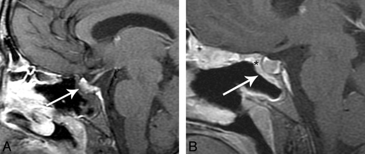
Fig 6.
A, Immediate postoperative sagittal T1-weighted postcontrast MR image with fat suppression shows no significant enhancement of the flap (white arrow). There is “C” shaped soft tissue in the defect that is presumably the flap (white arrow). B, Follow-up postoperative sagittal T1-weighted MR image postcontrast with fat suppression now shows an enhancing “C” nasoseptal flap (white arrow) in the surgical defect and increased enhancement to the tuberculum.
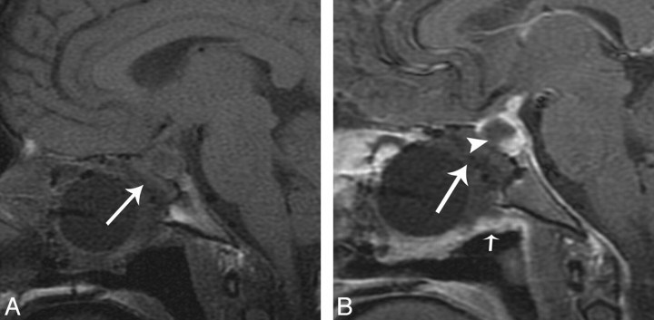
Fig 7.
This example is from another data group but illustrates how a flap may be displaced. A, Immediate postoperative sagittal T1-weighted MR image precontrast shows no enhancing nasoseptal flap in the expected region (white arrow). B, Immediate postoperative sagittal T1-weighted MR image postcontrast with fat suppression shows no enhancing C-shaped flap underlying the surgical defect (white arrowhead). There is linear soft tissue along the undersurface of the Foley balloon (small white arrow). This is presumed to represent a displaced enhancing flap. A CSF leak developed in this patient.


