Abstract
BACKGROUND AND PURPOSE:
Anterior communicating artery (AcomA) aneurysm is the most frequent form of aneurysm. Stent placement is particularly difficult and of limited use for AcomA aneurysms. We report our experience with stent-assisted embolization for wide-neck AcomA aneurysms in 21 patients. Particular attention is given to the morphologic characteristics and strategy of stent deployment.
MATERIALS AND METHODS:
Between January 2005 and February 2008, stent-assisted coiling was performed in 21 patients with wide-neck AcomA aneurysms. Patient demographics, aneurysm morphology, procedures, and clinical and angiographic outcomes were retrospectively reviewed.
RESULTS:
Successful deployment of the stent in the targeted artery was achieved in all patients. Nineteen Neuroform 2 or Neuroform 3 stents and 2 LEO stents were used. The distal segment of the stent was positioned in the ipsilateral A2 in 12 patients, in the contralateral A2 across the AcomA in 5 patients, and into the aneurysm sac in 4 patients. Complete occlusion was achieved in 18 patients; near-complete occlusion, in 2 patients; and partial occlusion, in 1 patient. Intraoperative perforation of the aneurysm developed in 1 patient, which was secured by subsequent coiling. Angiographic follow-up in 12 patients for 6.9 months showed 1 recanalization and no in-stent stenosis.
CONCLUSIONS:
Our preliminary results suggest that stent-assisted embolization for wide-neck AcomA aneurysms is technically feasible and safe. Further follow-up is needed for long-term efficacy of stent placement.
The anterior communicating artery (AcomA) is the most common location for intracranial aneurysms, as reported in some large surgical or endovascular series.1,2 The microsurgical clipping of AcomA aneurysms is complex.3 Although several articles have reported successful embolization of AcomA aneurysms with minimal complications, endovascular treatment for wide-neck AcomA aneurysms remains technically challenging.4–6 Various adjunctive techniques have been used to facilitate coil embolization of these lesions, including neck remodeling by using balloons or microguidewires, and simultaneous deployment of coils.7,8 Stent-assisted embolization has also been proposed as an alternative to achieve endovascular reconstruction of complex aneurysms.9,10 However, its use for wide-neck AcomA aneurysms is limited. In this article, we describe our experience in 21 patients with wide-neck AcomA aneurysms treated by endovascular stent-assisted embolization.
Materials and Methods
Patient Population
We performed a review of the clinical and radiologic records of all patients during a 37-month period from January 2005 to February 2008. The detailed information, including the patient's age and sex, clinical manifestations, aneurysm morphology, and endovascular treatment strategy, was carefully reviewed. The study comprised 21 patients with AcomA wide-neck aneurysms who were treated with self-expandable nitinol Neuroform microstents (Boston Scientific/Target Therapeutics, Fremont, Calif) or LEO stents (Balt Extrusion, Montmorency, France) at Changhai Hospital. Among these patients, 14 were women and 7 were men. The ages ranged from 39 to 75 years (mean age, 57 years). According to the Hunt and Hess (HH) scale, 8 patients were classified as grade I; 5 patients, as grade II; 6 patients, as grade III; and 2 patients, as grade IV. There was no unruptured AcomA aneurysm in our series. All the interventional procedures were performed by extensively experienced interventional vascular neurosurgeons (J.L., Y.X., B.H., Q.H.).
Aneurysm Morphology
There were 3 patients with multiple aneurysms. The maximal diameter of the aneurysm sac ranged from 2.5 to 9.3 mm, with a mean of 4.6 mm. The wide-neck aneurysms were defined as a having a large neck (>4 mm) and/or a dome-to-neck ratio ≤1.5. A bilobular sac was noticed in 2 patients with tiny aneurysms and in 1 patient with a small aneurysm. No partial intrasaccular thrombosis was demonstrated on digital subtraction angiography (DSA) or MR imaging. The most common anomaly associated with AcomA aneurysms was hypoplasia/aplasia of 1 A1 segment (12 patients).
The aneurysm neck was located at the A1-A2 junction in 12 patients and based directly on the AcomA in the other 9 aneurysms. The projection of the aneurysm and the direction of the fundus were clarified on the basis of the system used by Gonzalez et al.6 There were 6 AcomA aneurysms with superior projection, 8 aneurysms with anterior projection, 3 with inferior projection, and 4 with complex projections. The diameter of the parent vessel was <1 mm in only 1 patient, who was treated with the “waffle-cone” technique which means deployment of the distal segment of stent into the aneurysm sac. However, the size was thought to be related to severe vasospasm. In the other 20 patients treated with stent placement from A1 into A2, the diameters of afferent vessels ranged from 2.0 to 2.9 mm, with a mean of 2.4 mm as measured on DSA images, and the diameters of efferent vessels ranged from 1.7 to 2.8 mm (mean, 2.2 mm).
Endovascular Treatment
All procedures were performed with the patient under general anesthesia. Both conventional and rotational intra-arterial DSA were performed for 3D reconstruction in all patients. After systemic heparinization, a guiding catheter (Envoy; Cordis, Miami Lakes, Fla) was placed in the distal internal carotid artery to obtain a stable position. For Neuroform stent placement, a 300-cm 0.014-inch microguidewire was introduced distally in the A2 segment, with the help of a suitable microcatheter. The Neuroform stents were advanced over the microguidewire and deployed when the precisely targeted location was confirmed. For LEO stent placement, the stents were pushed when Vasco microcatheters (Balt Extrusion) had been positioned at the distal segments of parent vessels. After stent placement, the aneurysm was catheterized via stent mesh and subsequently coiled in the same session. At the end of coiling, an image was acquired to confirm adequate coiling.
Timing of Endovascular Treatment
All ruptured aneurysms were treated within 15 days of rupture, except for 1 patient treated 5 months after initial bleeding. Concerning the timing of endovascular treatment, 5 patients were treated within 24 hours; 10 patients, within 24–72 hours; 5 patients, within 72 hours to 2 weeks; and 1 patient, at 5 months.
Pre- and Postanticoagulation and Antiplatelet Management
All patients received systemic heparinization throughout the procedure, and activated clotting time was maintained at 2–3 times the baseline. After stent placement, low-molecular-weight heparin was routinely subcutaneously injected for 3 days.
For aneurysms ruptured longer than 7 days before endovascular treatment, dual antiplatelet drugs (75-mg clopidogrel and 300-mg aspirin daily) were given for >3 days. However, in patients experiencing subarachnoid hemorrhage within 72 hours, 300-mg clopidogrel and 300-mg aspirin were administered either by mouth or rectally at 2 hours before stent placement. All the patients were maintained on aspirin and clopidogrel for 6 weeks, followed by aspirin alone, which was continued indefinitely. In the event of major gastrointestinal bleeding or craniectomy, the dual antiplatelet aggregation medication was temporarily suspended until a stable condition was achieved. However, the antiplatelet treatment was continued even in the case of extraventricular drainage.
Clinical and Angiographic Follow-Up
There was no strictly defined follow-up protocol. Patients were advised to come for follow-up visits at the end of 1, 3, 6, and 12 months and yearly thereafter; angiography was advised after 6 months to assess the status of the treated aneurysm. However, for aneurysms with partial or near-complete occlusion, the first angiographic follow-up was performed 1–3 months after treatment. Recurrence was defined as aneurysm progression between the initial postprocedural angiographic control and the subsequent angiographic control with change in the aneurysm category.
Results
Clinical Outcomes
Clinical evaluation was performed in all patients before their discharge from the hospital. One patient with a poor preprocedural condition (HH IV) died as a consequence of severe vasospasm within 12 days after the embolization procedure. Eighteen patients improved or remained neurologically unchanged; 2 patients experienced clinical deterioration. All these patients were followed up for 6–36 months (median, 18.5 months). No rebleeding occurred during the follow-up period. The clinical evaluation performed at 6 months in 20 patients showed improvement in 4 patients, no change in 16 patients, and no clinical deterioration.
Stent Deployment and Angiographic Results
Successful embolization for AcomA was performed in all 21 patients. Overall, Neuroform 2 or Neuroform 3 stents were used in 19 patients; and LEO stents, in 2 patients. There were only 2 cases in which stent deployment was performed after coiling. In the other 19 patients, we first deployed the stent across the aneurysm neck, followed by subsequent coiling via the mesh of stents. In 4 patients treated with the waffle-cone technique, the diameter of the Neuroform stent was 4.0 mm. In the other patients, the diameter of the stents was 2.5 mm in 12 patients, 3.0 mm in 3 patients, and 3.5 mm in 2 patients.
When attempts to deliver the stent distally failed because of sharp or short-radius curves, we switched to deploying and releasing the distal aspect of the device into the aneurysm sac. With regard to the position of the distal segments of stents, there were 4 cases with the distal segment of the stent in the aneurysm sac, 12 cases in the ipsilateral A2 segments, and 5 cases in the contralateral A2 segments. At the time of the procedure, complete occlusion (>95%) was observed in 18 patients (82.9%) and near-complete occlusion (90%–95%), in 2 patients. Partial embolization (<90%) was observed in 1 patient.
Angiographic Follow-Up
Of the 21 embolized aneurysms, 12 were followed up angiographically for a median of 5.6 months (range, 1–12 months). Sustained stable aneurysm occlusion was achieved for 11 patients. One aneurysm was recanalized at 7-month follow-up; this patient was a candidate for a second treatment. However, the patient refused further treatment. No obvious intimal hyperplasia or in-stent stenosis was confirmed in these patients. Among the other 9 patients unavailable for DSA follow-up, 4 patients had contrast-enhanced MR angiography follow-up with no obvious recanalization.
Complications
One patient had aneurysm perforation during intrasaccular coiling following stent placement. The maximal diameter of this aneurysm was 4 mm. Unfortunately, aneurysm perforation occurred during endosaccular packing with a complex coil (4 mm × 10 cm (Microplex; MicroVention, Aliso Viejo, Calif), and the bleeding was controlled by additional coil packing. The patient recovered well without any persistent neurologic consequence. There was no procedure-related thromboembolic event.
Discussion
Although microsurgical clipping for AcomA aneurysms has been an effective method, the major obstacles, including complex arterial relationships with perforators and the higher potential of cognitive dysfunction, make surgical clipping complicated. With the development of technology and devices, endovascular treatment has become an important technique in the management of intracranial aneurysms.11 However, compared with other aneurysm locations, the higher frequency of small aneurysms, the high proportion of complex aneurysms, and the adjacent anomalies of the AcomA pose much difficulty in endovascular embolization. Nevertheless, some experienced authors reported their successful endovascular treatments of AcomA aneurysms with minimal complication rates.4–6
The effect of endovascular embolization is closely related to the aneurysm neck size. Aneurysms with a small neck were more likely to be successfully coiled. However, endovascular embolization of wide-neck aneurysm imposes technical challenges. To address the problems, a variety of techniques and devices emerged for embolization of wide-neck aneurysms, including TriSpan (Boston Scientific), remodeling techniques using balloon or microguidewire, the simultaneous deployment of 2 coils at the beginning of the framing of the aneurysm, or the use of stents.7–9 The use of intravascular stents and balloons has greatly expanded the application of endovascular treatment for intracranial aneurysms. However, because of the small length of the AcomA in conjunction with the acute angles of adjacent vessels, the primary limitation of the balloon- or stent-assisted technique is the inability to access the parent vessel at the level of the aneurysm neck and the loops in internal carotid arteries may prohibit placement of stent or balloon navigation. Furthermore, for extremely wide-neck aneurysms, the balloon alone is not enough to prevent coil protrusion into parent arteries.
Currently, the self-expanding nature, microdelivery property, and low profile of Neuroform and LEO stents make then more flexible and easier to navigate to target lesions farther in the smaller and distal cerebral vessels. The efficacy and safety of intracranial stents for endovascular treatment of wide-neck intracranial aneurysms have been evaluated in several studies.9,12 Their findings are consistent with our results, with high success rates and low procedure-related mortality-morbidity rates. Turk et al13 described 8 patents who had safely and successfully undergone stent treatment in vessel diameters <2 mm with good short- and intermediate-term results, including 5 AcomA aneurysms.
The location of aneurysm neck affects the strategy of stent deployment. For aneurysms mainly involving the ipsilateral A2 segment, it is very important to preserve the patency of A2. We tried to deploy the stent from the ipsilateral A2 to A1 and cover the ostia of the AcomA. The anatomic variations should be taken into account, in the case of the aneurysm neck located at the junction of the A2 and AcomA. When bilateral anterior cerebral arteries (ACAs) are symmetric, the AcomA can be sacrificed without any major clinical consequence. On the basis of this anatomic characteristic, we prefer stent placement from A2 to A1 in the ipsilateral ACA. However, in patients with aneurysm necks located at the AcomA and asymmetric A1 segments, stent placement across the AcomA to the contralateral A2 segments becomes the first choice to preserve the patency of parent vessels.
Selection of the stent is very important for successful stent placement. In the acute or subacute phase of a ruptured AcomA aneurysm, there will be moderate or severe vasospasm. The stent should not be undersized due to an inadequate measurement of the vessel, which leads to the incorrect stent choice. From this viewpoint, we usually oversize the diameters of stents as in the cases presented here. The minimal diameter of stents used in our patients was 2.5 mm, even in vessels <1 mm. Actually, the parent artery restored the original diameter after deployment of oversized stents. However, the use of stents as adjuncts to coiling has been reported to increase the risk of complications.
Aneurysm perforation is an uncommon but potentially fatal complication, especially for tiny aneurysms.14 Compared with closed-cell stents (such as LEO or the Enterprise self-expanding stent [Cordis]), segmented stent design conforms better to vascular tortuosity.15 However, the Neuroform stents with open-cell design show increased opening of cells and outward prolapse of struts into an aneurysm neck.16 This may lead to reduction of the largest diameter of the aneurysm sac, which makes coil sizing more difficult. Taking this into consideration, we prefer to choose undersized coils for endosaccular packing after stent placement. In addition, open-cell stents were used in parent vessels with sharp or short-radius curves. Use of closed-cell stents in this situation will result in underexpansion of stent.
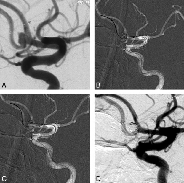
There are different strategies regarding the timing of stent deployment in relation to coiling.12 As demonstrated by cases presented here (Fig 2), the “jailed technique,” which means the microcatheter is caged between the vessel wall and the stent, permits stabilization of the microcatheter during coil delivery. However, it is unsuitable for the treatment of most AcomA aneurysms due to the small diameter of the ACA and vasospasm resulting from aneurysmal subarachnoid hemorrhage. So, in most of our patients, we first deployed the stents. The aneurysm sacs were packed with coils via catheterization from the mesh of stents. Only 1 patient was treated by coiling before stent deployment at the end of the session. We attempted treatment by coil packing alone, which resulted in coil protrusion into the parent vessel. For fear of subsequent thromboembolic events, we have adopted stent implantation to resolve the coil protrusion, as reported by Luo et al.17
Fig 2.
Images of a 58-year-old woman with an AcomA aneurysm. A, Angiogram shows a wide-neck AcomA aneurysm. B, Endosaccular coiling before stent placement. C, Stent placement after the first coil packing, D, Complete occlusion with further coiling after Neuroform stent placement.
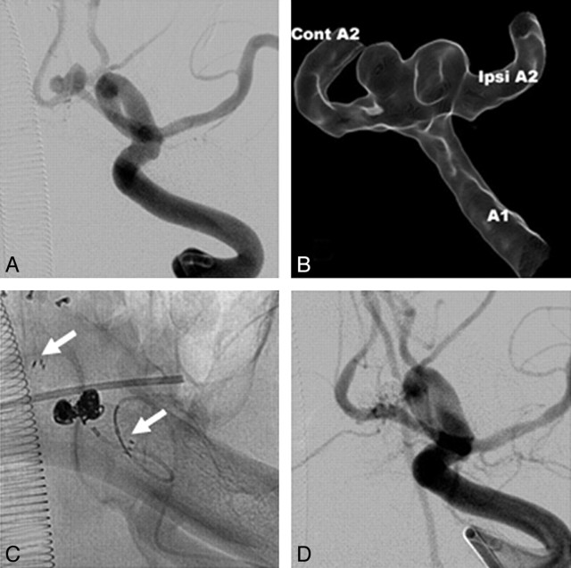
Fig 1.
Images of a 62-year-old man with an AcomA aneurysm. A and B, Conventional and 3D angiograms show a wide-neck AcomA aneurysm. C, Endosaccular coiling after successful stent placement across the AcomA (white arrow). D, Postembolization angiogram shows complete occlusion with patency of the parent vessels. Cont A2 indicates contralateral A2; Ipsi A2, ipsilateral A2.
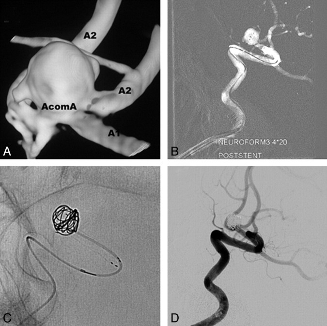
The angles between the A1, A2, and AcomA may be acute, which makes it difficult to catheterize the bilateral A2 segments and navigate stent systems. Microguidewires and microcatheters end up inside the aneurysm during attempts to deploy the stent across the neck. Moreover, stent navigation over the endosaccular microguidewire carries significant risk of a ruptured aneurysm. For aneurysms involving bilateral A2 segments and the AcomA, stent placement in any side of A2 is not enough for retaining the coils in the aneurysm sac and preservation of the patency of parent arteries. In both situations, the waffle-cone technique becomes a feasible method (Fig 3). In this series, we used this technique for wide-neck AcomA aneurysms in 4 patients. The immediate angiograms showed total occlusion in 3 patients and near-complete occlusion in 1 patient. However, there were disadvantages of this technique. First, placement of the stent into the aneurysm exposes the patient to potential risk of perforation, which is especially true with freshly ruptured and smaller aneurysms. In this light, we suggest that the technique should be used only for aneurysms larger than 4 mm. Second, unlike stent placement in parent vessels, flow direction into the aneurysm via the waffle-cone technique may predispose to more rapid rates of recanalization. In our patients, we had only 1 who experienced recanalization, which was treated by the waffle-cone technique. Regarding this latter point, it is not recommended as a standard treatment technique but could be an alternative choice for complicated conditions.
Fig 3.
Images of a 63-year-old man with an AcomA aneurysm. A, 3D angiogram shows a wide-neck AcomA aneurysm involving both A2 segments and the AcomA. B, A 4.0 × 20 Neuroform 3 stent is deployed into aneurysm sac. C, Endosaccular coil packing is protected by the Neuroform stent. D, Postembolization angiogram shows complete occlusion with patency of the parent vessels.
Some limitations of the present study include the retrospective design, patient selection bias, and the inadequate follow-up. In-stent stenosis often occurs in small vessels, which is a major problem that influences the treatment with stent-assisted coil embolization.18 Moreover, the hemodynamic changes after stent placement with the waffle-cone technique may increase the probability of coil compaction. Both of these need to be elucidated through longer follow-up.
Conclusions
Our preliminary results suggest that stent-assisted embolization for wide-neck AcomA aneurysms is technically feasible and safe. Detailed anatomic features of the aneurysms should be considered when attempting endovascular treatment.
Footnotes
This work was supported in part by the Key Project from the Shanghai Committee of Science and Technology (074119506) and the Shanghai Municipal Natural Science Foundation (08ZR1404200).
References
- 1. Molyneux A, Kerr R, Stratton I, et al. International Subarachnoid Aneurysm Trial (ISAT) of neurosurgical clipping versus endovascular coiling in 2143 patients with ruptured intracranial aneurysms: a randomised trial. Lancet 2002; 360: 1267– 74 [DOI] [PubMed] [Google Scholar]
- 2. Kassell NF, Torner JC, Haley EC, Jr, et al. , The international cooperative study on the timing of aneurysm surgery. Part 1: Overall management results. J Neurosurg 1990; 73: 18– 36 [DOI] [PubMed] [Google Scholar]
- 3. Agrawal A, Kato Y, Chen L, et al. Anterior communicating artery aneurysms: an overview. Minim Invasive Neurosurg 2008; 51: 131– 35 [DOI] [PubMed] [Google Scholar]
- 4. Andaluz N, Zuccarello M. Blister-like aneurysms of the anterior communicating artery: a retrospective review of diagnosis and treatment in five patients. Neurosurgery 2008; 62: 807– 11 [DOI] [PubMed] [Google Scholar]
- 5. Birknes J, Hwang S, Pandey A, et al. Feasibility and limitations of endovascular coil embolization of anterior communicating artery aneurysms: morphological considerations. Neurosurgery 2006; 59: 43– 52 [DOI] [PubMed] [Google Scholar]
- 6. Gonzalez N, Sedrak M, Martin N, et al. Impact of anatomic features in the endovascular embolization of 181 anterior communicating artery aneurysms. Stroke 2008; 39: 2276– 82 [DOI] [PubMed] [Google Scholar]
- 7. Kwon OK, Kim SH, Oh CW, et al. Embolization of wide-necked aneurysms with using three or more microcatheters. Acta Neurochir (Wien) 2006; 148: 1139– 45, discussion 1145. Epub 2006 Sep 29 [DOI] [PubMed] [Google Scholar]
- 8. Levy DI. Embolization of wide-necked anterior communicating artery aneurysm: technical note. Neurosurgery 1997; 41: 979– 82 [DOI] [PubMed] [Google Scholar]
- 9. Lubicz B, Leclerc X, Levivier M, et al. Retractable self-expandable stent for endovascular treatment of wide-necked intracranial aneurysms: preliminary experience. Neurosurgery 2006; 58: 451– 57 [DOI] [PubMed] [Google Scholar]
- 10. Lylyk P, Ferrario A, Pasbon B, et al. Buenos Aires experience with the Neuroform self-expanding stent for the treatment of intracranial aneurysms. J Neurosurg 2005; 102: 235– 41 [DOI] [PubMed] [Google Scholar]
- 11. Koebbe CJ, Veznedaroglu E, Jabbour P, et al. Endovascular management of intracranial aneurysms: current experience and future advances. Neurosurgery 2006; 59: S 93– 102 [DOI] [PubMed] [Google Scholar]
- 12. Biondi A, Janardhan V, Katz JM, et al. Neuroform stent-assisted coil embolization of wide-neck intracranial aneurysms: strategies in stent deployment and midterm follow-up. Neurosurgery 2007; 61: 460– 69 [DOI] [PubMed] [Google Scholar]
- 13. Turk AS, Niemann DB, Ahmed A, et al. Use of self-expanding stents in distal small cerebral vessels. AJNR Am J Neuroradiol 2007; 28: 533– 36 [PMC free article] [PubMed] [Google Scholar]
- 14. Tsutsumi M, Aikawa H, Onizuka M, et al. Endovascular treatment of tiny ruptured anterior communicating artery aneurysms. Neuroradiology 2008; 50: 509– 95 [DOI] [PMC free article] [PubMed] [Google Scholar]
- 15. Ebrahimi N, Claus B, Lee CY, et al. Stent conformity in curved vascular models with simulated aneurysm necks using flat-panel CT: an in vitro study. AJNR Am J Neuroradiol 2007; 28: 823– 29 [PMC free article] [PubMed] [Google Scholar]
- 16. Hsu SW, Chaloupka JC, Feekes JA, et al. In vitro studies of the Neuroform microstent using transparent human intracranial arteries. AJNR Am J Neuroradiol 2006; 27: 1135– 39 [PMC free article] [PubMed] [Google Scholar]
- 17. Luo CB, Chang FC, Teng MM, et al. Stent management of coil herniation in embolization of internal carotid aneurysms. AJNR Am J Neuroradiol 2008; 29: 1951– 55 [DOI] [PMC free article] [PubMed] [Google Scholar]
- 18. Fiorella D, Albuquerque FC, Woo H, et al. Neuroform in-stent stenosis: incidence, natural history, and treatment strategies. Neurosurgery 2006; 59: 34– 42 [DOI] [PubMed] [Google Scholar]


