Abstract
BACKGROUND AND PURPOSE: Dome-to-neck ratio of intracranial aneurysms is an important predictor of outcomes of endovascular coiling. 3D imaging techniques are increasingly used in evaluating the dome-to-neck ratio of aneurysms for intervention. The purpose of this study was to determine whether 3D rotational angiography (3DRA) can be used to determine accurately the dome-to-neck ratio of intracranial aneurysms when compared with conventional 2D digital subtraction angiography (2D DSA).
MATERIALS AND METHODS: A retrospective analysis of 180 patients with 205 intracranial aneurysms who underwent both 2D DSA and 3DRA for evaluation of previously untreated aneurysms was conducted. Dome-to-neck ratios were compared between 2D DSA and 3DRA images. The mean difference in dome-to-neck ratios between 2D DSA and 3DRA was calculated. The proportions of “wide-neck” aneurysms seen on 2D DSA and 3DRA were compared by using 2 different definitions of “wide-neck,” including <1.5 and <2.0.
RESULTS: The average dome-to-neck ratio was 1.81 ± 0.55 and 1.55 ± 0.48 for 2D DSA and 3DRA, respectively (P < .0001). When we defined “wide-neck” as a dome-to-neck ratio <1.5, sixty-nine (33.7%) aneurysms were wide-neck on 2D DSA compared with 119 (58%) on 3DRA (P < .0001). When we defined “wide-neck” as dome-to-neck ratio <2.0, one hundred forty-two (69.3%) aneurysms were wide-neck on 2D DSA compared with 173 (84.4%) on 3DRA (P = .0004).
CONCLUSIONS: In this retrospective study, 3DRA measurements resulted in significantly lower dome-to-neck ratios and significantly larger proportions of aneurysms defined as “wide-neck” compared with 2D DSA. Scrutiny of 2D DSA may offer substantial benefit over 3D techniques when triaging patients to or from endovascular therapy.
Aneurysm geometry represents one of the most important predictors of outcomes of endovascular coiling of intracranial aneurysms. Aspects of aneurysm geometry such as shape, size, dome-to-neck ratio, location, and relationship to the parent vessels all may impact decisions about how to treat individual aneurysms. Among these geometric features, dome-to-neck ratio is often the dominant factor in determining whether an endovascular approach is likely to be feasible for a given aneurysm.1-8
Common methods for determining the dome-to-neck ratios of aneurysms include standard digital subtraction angiography (DSA), 3D rotational DSA (3DRA), CT angiography (CTA), and MR angiography (MRA). 3DRA, CTA, and MRA all represent reconstructed images based on either multiple views of 2D angiography, in the case of 3DRA, or axial source images, in the case of CTA and MRA. Standard, or what we term 2D, DSA offers high spatial and contrast resolution with minimal postprocessing, unlike the 3D techniques listed above. Few studies to date critically evaluate the potential differences in apparent dome-to-neck ratios on various imaging techniques.9 Especially with the expanding application of CTA for triage of aneurysms to or from endovascular therapy without intervening DSA, it remains relevant to understand whether 3D techniques offer different apparent dome-to-neck ratios compared with 2D DSA. The purpose of this study was to determine whether systematic differences in dome-to-neck ratio are seen when comparing 2D DSA and 3DRA.
Materials and Methods
Patients
Following institutional review board approval, a retrospective analysis of 195 consecutive patients who underwent both 2D DSA and 3DRA for evaluation of intracranial aneurysms between January 2005 and November 2007 at our institution was conducted. Cases were excluded because of excessive artifacts distortion in 2D DSA or 3DRA images or no clear dome or neck identification on 2D DSA or 3DRA images. Of the 195 patients, 13 were excluded due to excessive artifacts distortion of 3DRA images and 2 were excluded due to a lack of a clear neck on 2D DSA images. In all, 205 intracranial aneurysms were studied in 180 patients. Sixty (29%) of these aneurysms were ruptured.
Angiographic Technique
2D DSA.
Typically, 5F or 6F catheters were placed into the internal carotid arteries or vertebral arteries. Biplane DSA images of the entire circulation were usually obtained, followed by “working-projection” DSA based on views identified on the 3DRA (see below). “Working-projection” images were those images that offered ideal separation between the aneurysm neck and parent artery. Small FOVs, usually 12.7 × 17.78 cm (5 × 7 inch), were used for these working-projection views.
3DRA.
All the 3DRA examinations were performed by using a biplane C-arm digital angiography suite (Integris; Philips Medical Systems, Best, the Netherlands) with an FOV of 17.78 cm (7 inches) and a frame rate 30 f/s. Images were acquired with a head-end propeller C-arm orientation at a rotational speed of 55°/s covering +120° to −120°. A volume of 16 mL of nonionic contrast medium was injected through a 5F-6F catheter by use of an injector with a velocity of 4 mL/s. Image acquisition was started 1–3 seconds after contrast material injection. The acquisition time of images was 4.4 seconds. Volume-rendered 3D images were reconstructed with a 100% magnification (a field of 37.56 cm2) and a matrix of 256 pixels3 by using the 3DRA volumetric measurement of the system software (Philips Medical Systems). The threshold for the volume-rendered image was fixed as the default value provided by the software.
Analysis of Images
Aneurysm location, maximum dimension, and aneurysm shape were recorded. Aneurysm dome-to-neck ratios in 2D DSA and 3DRA images were measured. Aneurysms were classified as those with simple-versus-complex shapes as previously described.6 Simple shapes were round, circular, or oval. Aneurysms with small blebs were included in this category. Aneurysms with complex shapes were classified as those with multiple lobes or domes or that were mushroom-shaped.
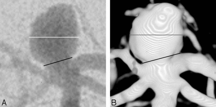
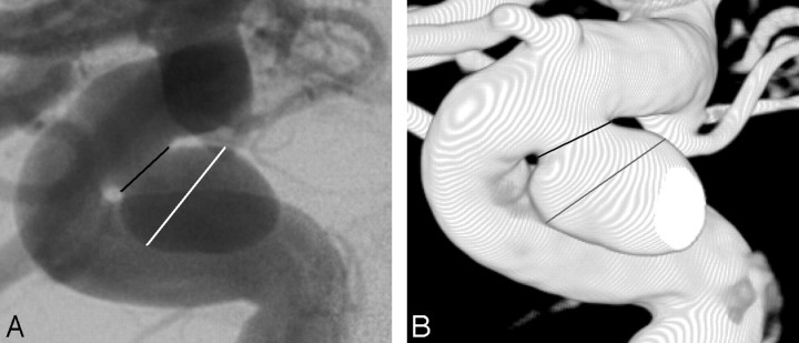
Measurement of dome-to-neck ratios was performed on PACS.10 A single reader selected an early or midarterial phase from the 2D DSA for measurement. The same working projection was used for both 2D DSA and 3DRA measurements. For aneurysms with simple shapes, the dome size was obtained by measuring the diameter of the dome. Blebs on simple-shaped aneurysms were not included in the dome measurement. For aneurysms with complex shapes, the diameter of the proximal dome was measured as described previously.6 The reader used clinical images, without manipulation of window or level settings for either the 2D DSA or 3DRA images, in an attempt to simulate the clinical environment. An electronic caliper was used to measure both the dome diameter and neck width (Figs 1 and 2). Measurements on 2D DSA images were performed on separate reading sessions from those using 3DRA images, to diminish bias from recall of one measurement or the other. Two weeks following the initial reading, the same reader repeated all measurements to assess intraobserver variability. Dome-to-neck ratios were calculated as the dome width to neck diameter. Mean dome-to-neck ratio was calculated for 2D DSA and 3DRA images. In addition, the mean difference in dome-to-neck ratio for each aneurysm on 2D DSA and 3DRA images was calculated. We also calculated the proportion of wide-neck aneurysms on the basis of 2D DSA and 3DRA images, by using a definition for “wide-neck” as dome-to-neck ratio <1.511 and <2.0.2,3
Fig 1.
Images from a patient with a basilar tip aneurysm. A, Anteroposterior (AP) 2D DSA shows the measurement of dome diameter (white line) and neck width (black line). Dome-to-neck ratio for this 2D DSA image is 1.7. B, AP 3DRA from the same patient as in A shows dome diameter (gray line) and neck width (black line). Dome-to-neck ratio for this 3DRA image is 1.3.
Fig 2.
Images from a patient with a superior hypophyseal aneurysm. A, Anteroposterior (AP) 2D DSA shows the measurement of dome diameter (white line) and neck width (black line). Dome-to-neck ratio for this 2D DSA image is 2.0. B, AP 3DRA from the same patient as in A shows dome diameter (gray line) and neck width (black line). Dome-to-neck ratio for this 3DRA image is 1.7.
Statistical Analysis
Statistical analysis was performed by using the software JMP (www.jmp.com). For analysis of the correlation between the 2 variables, a simple linear correlation (Pearson r) test was performed. For determining the impact of aneurysm shape on the 2D DSA and 3D measurements, a 2-sample t test was performed. For quantifying agreement between the 2 measurements of 2D DSA dome-to-neck ratios and the 2 measurements of 3DRA dome-to-neck ratios, a κ statistic was computed. For analysis of the difference between 2D DSA and 3DRA dome-to-neck ratios, a paired t test was performed. A Wilcoxon signed ranked test was used to correct for the non-normal distribution of the dome-to-neck ratios when computing the paired t test. For analysis of categoric data, a Pearson χ2 test was performed.
Results
The patient population consisted of 136 women and 44 men for a total of 180 patients. The average age of the patients was 59.0 ± 13.0 years (range, 21–88 years). One-hundred thirty-one (63.9%) aneurysms were in the anterior circulation, and 74 aneurysms (36.1%) were in the posterior circulation. The average maximum dimension for the 205 aneurysms included in this study was 6.5 ± 3.2 mm (range, 2–18 mm). The average 2D DSA dome-to-neck ratio was 1.81 ± 0.55, and the average 3DRA dome-to-neck ratios was 1.55 ± 0.48 (P < .0001). The mean difference in dome-to-neck ratios between 2D DSA and 3DRA images was 0.26. On average, 2D DSA dome-to-neck ratios were 16% greater than 3DRA dome-to-neck ratios.
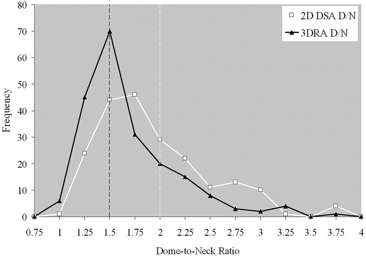
When we defined “wide-neck” as dome-to-neck ratio <1.5, 69 (33.7%) aneurysms were wide-neck on 2D DSA images compared with 119 (58.0%) on 3D images (P < .0001). With this same definition of wide-neck, 3DRA identified 24% more aneurysms as wide-neck than 2D DSA. When defining “wide-neck” as dome-to-neck ratio <2.0, 142 (69.3%) aneurysms were wide-neck on 2D DSA images compared with 173 (84.4%) on 3DRA images (P = .0004). With this same definition of wide-neck, 3DRA identified 15% more aneurysms as wide-neck than 2D DSA. Between the 2 sets of measurements, there was excellent agreement (κ = 1.0). All of the above data are summarized in the Table. Comparison of the distribution of dome-to-neck ratios in 2D DSA versus 3DRA images is shown in Fig 3.
Dome-to-neck ratios in 2D DSA versus 3DRA
| 2D DSA | 3DRA | P Value | |
|---|---|---|---|
| Mean D/N (SD) | 1.81 (0.55) | 1.56 (0.48) | <.0001* |
| D/N <1.5, No. (%) | 69 (33.7) | 119 (58.0) | <.0001† |
| D/N <2.0, No. (%) | 142 (69.3) | 173 (84.4) | .0004† |
Note:—D/N indicates dome-to-neck ratio; DSA, digital subtraction angiography; 3DRA, 3D rotational angiography.
Paired t test, Wilcoxon signed ranked test.
Pearson χ2 test.
Fig 3.
Comparison of the distribution of dome-to-neck ratios in 2D DSA and 3DRA. Histogram shows the frequency of aneurysms with various dome-to-neck ratios. Frequencies are calculated for dome-to-neck intervals of 0.25; for example, ratios from 2.0 to 2.25 are grouped into a single frequency number. The black line denotes 3DRA measurements, whereas the white line denotes 2D DSA measurements. Vertical dashed lines indicate previously published thresholds for wide-neck aneurysms (black line, 1.5; white line, 2.0). Thus, aneurysms to the left of a vertical line would be considered wide-neck.
There was a positive correlation between maximum size of the aneurysm and dome-to-neck ratio for both 2D DSA (r = 0.32, P < .0001) and 3DRA (r = 0.33, P < .0001). However, maximum aneurysm dimension did not correlate with the difference in dome-to-neck ratios between 2D DSA and 3DRA images (r = 0.03, P = .67).
Of the 205 aneurysms studied, 56 (27%) had complex shapes and 149 (73%) had simple shapes. On 2D DSA images, simple-shaped aneurysms had an average dome-to-neck ratio of 1.81 ± 0.59, and complex-shaped aneurysms had an average dome-to-neck ratio on 2D DSA of 1.80 ± 0.41 (P = .91). On 3D DSA images, simple-shaped aneurysms had an average dome-to-neck ratio of 1.57 ± 0.52, and complex-shaped aneurysms had an average dome-to-neck ratio of 1.53 ± 0.34 (P = .59). There was no difference in the dome-to-neck ratios between complex and simple-shaped aneurysms in either 2D DSA or 3DRA measurements.
Discussion
In this study, we found a systematic difference between the measurements of dome-to-neck ratios on 2D DSA-versus-3DRA images. Not only did 3DRA images yield a higher mean dome-to-neck ratio, but 3DRA images also identified substantially greater fractions of aneurysms as wide-neck than the 2D DSA images did. Because the 2D DSA images offer high spatial and contrast resolution without substantial postprocessing, we consider these 2D DSA images to be a relative “standard of reference” compared with 3DRA images.9,12,13 As such, it is likely that 3DRA imaging underestimates the true dome-to-neck ratio. Thus, careful scrutiny of 2D DSA images is warranted when performing DSA for aneurysm detection and characterization. Further, reliance on 3D images alone, as may be the case for CTA, might tend to increase the fraction of patients not considered good candidates for endovascular treatment on the basis of apparent unfavorable dome-to-neck ratios.
Previous authors have compared 2D DSA and 3DRA imaging and have reported substantial benefit from the latter technique.6,14-16 These previous comparisons have focused on determination of aneurysm detection rates as well as depiction of aneurysm shape, neck location, and relationship to the parent artery. We agree that 3DRA is superior to 2D DSA when evaluating these features. However, we have now demonstrated that 2D DSA probably is superior to 3DRA in determining one of the most clinically important geometric factors of an aneurysm, dome-to-neck ratio. We know of only 1 other study that compared measured dome-to-neck ratios of 2 different imaging techniques. Yoon et al9 reported that dome-to-neck ratio measurements for 2D DSA were 11% greater than those on multidetector row CTA, but this difference was not statistically significant. Our study not only describes a patient population nearly twice as large as that prior study but also offers a more-detailed analysis than that prior study regarding factors such as aneurysm size and shape on the difference between 2D DSA and 3DRA dome-to-neck ratios.
The current study has several limitations. First, we used the clinical images in the analysis of the 2D DSA and 3DRA images. We did not vary the window/level settings on 2D DSA or 3D images, which can substantially impact apparent diameter. Adjustment of threshold values to 3DRA reconstructions has been shown to be important in providing optimal visibility of vascular structures, and it has been suggested that they be adjusted individually for individual cases.17 However, the effects of changing threshold values on aneurysm treatment decisions have yet to be investigated.
A second limitation of our study is that the default threshold values on the equipment used in this study may not be identical to those on other types of equipment from other vendors. A third limitation is the use of only a single observer. However, the observer was blinded to all previous data obtained while conducting the measurements, and measurement of dome-to-neck ratios was held strictly to the methods described in previous protocols.6 A fourth limitation of this study is the lack of a proved gold standard in the assessment of dome-to-neck ratios of aneurysms. In our study, we deemed 2D DSA images likely to be more reliable than 3DRA images due to the high spatial and contrast resolution and minimal postprocessing with the former technique. Finally, although we noted significant differences in dome-to-neck ratios between techniques, we did not show that patient management would have necessarily been different in specific cases based on 2D DSA or 3DRA images.
Conclusions
In this study, we showed statistically significant differences in dome-to-neck ratios between 2D DSA and 3DRA images, with substantial differences in the frequency of wide-neck aneurysms between imaging techniques; however, these results may not necessarily apply to 3D DSA images obtained by using different equipment or different threshold values. These results suggest that 3D imaging techniques may be inferior to 2D DSA for triage of aneurysms to or from endovascular therapy.
Acknowledgments
We thank Beth Scheuler, PhD, for technical assistance.
References
- 1.Cloft HJ, Joseph GJ, Tong FC, et al. Use of three-dimensional Guglielmi detachable coils in the treatment of wide-necked cerebral aneurysms. AJNR Am J Neuroradiol 2000;21:1312–14 [PMC free article] [PubMed] [Google Scholar]
- 2.Debrun GM, Aletich VA, Kehrli P, et al. Selection of cerebral aneurysms for treatment using Guglielmi detachable coils: the preliminary University of Illinois at Chicago experience Neurosurgery 1998;43:1281–95, discussion 1296–87 [DOI] [PubMed] [Google Scholar]
- 3.Debrun GM, Aletich VA, Kehrli P, et al. Aneurysm geometry: an important criterion in selecting patients for Guglielmi detachable coiling Neurol Med Chir (Tokyo) 1998;38 (suppl):1–20 [DOI] [PubMed] [Google Scholar]
- 4.Fernandez Zubillaga A, Guglielmi G, Vinuela F, et al. Endovascular occlusion of intracranial aneurysms with electrically detachable coils: correlation of aneurysm neck size and treatment results. AJNR Am J Neuroradiol 1994;15:815–20 [PMC free article] [PubMed] [Google Scholar]
- 5.Gonzalez N, Sedrak M, Martin N, et al. Impact of anatomic features in the endovascular embolization of 181 anterior communicating artery aneurysms Stroke 2008;39:2776–82. Epub 2008 Jul 10 [DOI] [PubMed] [Google Scholar]
- 6.Kiyosue H, Tanoue S, Okahara M, et al. Anatomic features predictive of complete aneurysm occlusion can be determined with three-dimensional digital subtraction angiography. AJNR Am J Neuroradiol 2002;23:1206–13 [PMC free article] [PubMed] [Google Scholar]
- 7.Murayama Y, Nien YL, Duckwiler G, et al. Guglielmi detachable coil embolization of cerebral aneurysms: 11 years' experience. J Neurosurg 2003;98:959–66 [DOI] [PubMed] [Google Scholar]
- 8.Parlea L, Fahrig R, Holdsworth DW, et al. An analysis of the geometry of saccular intracranial aneurysms. AJNR Am J Neuroradiol 1999;20:1079–89 [PMC free article] [PubMed] [Google Scholar]
- 9.Yoon DY, Lim KJ, Choi CS, et al. Detection and characterization of intracranial aneurysms with 16-channel multidetector row CT angiography: a prospective comparison of volume-rendered images and digital subtraction angiography. AJNR Am J Neuroradiol 2007;28:60–67 [PMC free article] [PubMed] [Google Scholar]
- 10.Erickson BJ, Ryan WJ, Gehring DG. Functional requirements of a desktop clinical image display application. J Digit Imaging 2001;14:149–52 [DOI] [PMC free article] [PubMed] [Google Scholar]
- 11.Regli L, Uske A, de Tribolet N. Endovascular coil placement compared with surgical clipping for the treatment of unruptured middle cerebral artery aneurysms: a consecutive series. J Neurosurg 1999;90:1025–30 [DOI] [PubMed] [Google Scholar]
- 12.Jayaraman MV, Mayo-Smith WW, Tung GA, et al. Detection of intracranial aneurysms: multi-detector row CT angiography compared with DSA. Radiology 2004;230:510–18 [DOI] [PubMed] [Google Scholar]
- 13.Kouskouras C, Charitanti A, Giavroglou C, et al. Intracranial aneurysms: evaluation using CTA and MRA—correlation with DSA and intraoperative findings. Neuroradiology 2004;46:842–50 [DOI] [PubMed] [Google Scholar]
- 14.Anxionnat R, Bracard S, Ducrocq X, et al. Intracranial aneurysms: clinical value of 3D digital subtraction angiography in the therapeutic decision and endovascular treatment. Radiology 2001;218:799–808 [DOI] [PubMed] [Google Scholar]
- 15.Sugahara T, Korogi Y, Nakashima K, et al. Comparison of 2D and 3D digital subtraction angiography in evaluation of intracranial aneurysms. AJNR Am J Neuroradiol 2002;23:1545–52 [PMC free article] [PubMed] [Google Scholar]
- 16.van Rooij WJ, Sprengers ME, de Gast AN, et al. 3D rotational angiography: the new gold standard in the detection of additional intracranial aneurysms. AJNR Am J Neuroradiol 2008;29:976–79 [DOI] [PMC free article] [PubMed] [Google Scholar]
- 17.Hagen G, Lindgren PG, Jangland L, et al. Artifacts in 3D rotational angiography: an experimental study. Acta Radiol 2005;46:32–36 [DOI] [PubMed] [Google Scholar]


