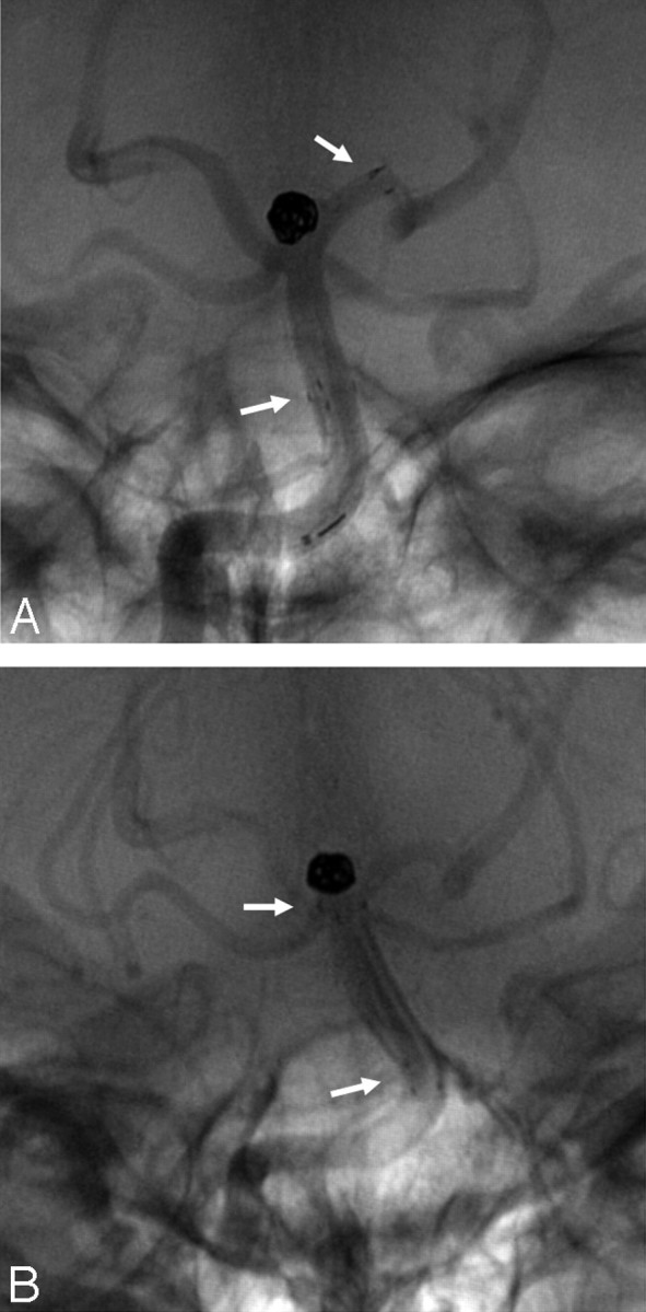
Kelly et al1 reported the first published case of a patient with delayed migration of an Enterprise stent (Cordis, Miami Lakes, Fla) used for assistance in endovascular coil embolization of a basilar apex aneurysm. The stent was placed, extending from the midbasilar artery to the P1 segment of the left posterior cerebral artery. It was proposed that a closed-cell design present in the Enterprise stent would have transmitted a force exerted on one end to the entire device. A smaller-vessel diameter in the distal aspect of the closed-cell stent might have then transferred a constant retrograde force that ultimately moved the stent to a more stable position. We have experienced a similar case of a patient with delayed migration of the Enterprise stent at this location.
A 54-year-old-woman was found to have an incidental broad-necked basilar apex aneurysm 4.0 × 3 mm on screening for family history of ruptured intracranial aneurysms in first-degree relatives. The patient elected to undergo endovascular aneurysmal treatment. She was given aspirin and clopidogrel 7 days before the endovascular stent-assisted coil embolization. A 4.5- × 14-mm Enterprise stent was used for assistance, extending from the midbasilar artery to the P1 segment of the left posterior cerebral artery (Fig 1A). At the end of the procedure, the patient reported a brief episode of visual disturbance described as “flashing lights” and, on physical examination, was noted to have nystagmus, which disappeared the next day. The patient was kept for observation overnight and was discharged the following day. Approximately 2 weeks later, she complained of another transient episode of visual disturbances. The patient was given aspirin and clopidogrel for 3 months, and then the clopidogrel was discontinued. Follow-up cerebral angiography 6 months later demonstrated almost 1 cm of proximal migration of the Enterprise stent (Fig 1B). She has remained asymptomatic.
Fig 1.

Basilar apex stent-assisted coil embolization in a 54-year-old woman with a broad-necked basilar apex aneurysm. Unsubtracted anteroposterior view of the right vertebral artery contrast injection. A, Final view after coil embolization. Note the arrows pointing to the distal and proximal ends of the stent. B, A 6-month follow-up of the same patient. Note that the stent has migrated into the basilar artery.
The relatively new Enterprise stent was designed for coil embolization assistance; it is highly flexible and, compared with its antecessors, can be repositioned and has a closed-cell design that gives a better scaffold and, therefore, protection against coil herniation.2 However, the closed-cell design may have its disadvantages when there is a differential in size between the distal and proximal ends, causing the stent to migrate into the larger vessel. We would also hypothesize that the presence of the distal stent in a curved segment, such as the P1 segment in both of our cases, might have contributed forces that squeezed the stent proximally. Our experience with the Enterprise stent in this location is limited to this single case.
References
- 1.Kelly ME, Turner RD 4th, Moskowitz SI, et al. Delayed migration of a self-expanding intracranial microstent. AJNR Am J Neuroradiol 2008;29:1959–60. Epub 2008 Aug 21 [DOI] [PMC free article] [PubMed] [Google Scholar]
- 2.Weber W, Bendszus M, Kis B, et al. A new self-expanding nitinol stent (Enterprise) for the treatment of wide-necked intracranial aneurysms: initial clinical and angiographic results in 31 aneurysms. Neuroradiology 2007;49:555–61 [DOI] [PubMed] [Google Scholar]


