Abstract
Objective
The purpose of the present study was to assess the symphyseal morphology and lower incisor angulation in different anteroposterior relationship and in different growth patterns and to investigate whether the symphyseal morphology had any correlation with dentofacial parameters.
Method
Random Sampling method and lateral cephalograms of 90 subjects, age group 16–30 years, were divided into 30 in each group, i.e. Class I, Class II & Class III after calculating the following parameters (ANB angle, wits appraisal). After that, groups were again divided into 10 in each subgroup i.e. Average, Horizontal and Vertical growers.
Results
Results showed the increase in actual symphysis width, inclination of the alveolar part, total height of symphysis and reduction in overall width along with retroclination of lower incisors in class III subjects as compared to class I and class II. Similarly actual and overall width of the symphysis were decreased and inclination of the alveolar part, symphyseal height and symphyseal ratio were increased in vertical growers.
Conclusion
The dimensions and configuration of Mandibular Symphysis in class III was found to be different than those in Class I and Class II relationships; the alveolar part of Mandibular Symphyseal compensated for the skeletal relationship in the Class III pattern. Mandibualr Symphysis dimensions were strongly correlated to anterior facial dimensions. Similarly the dimensions and configuration of Mandibular Symphysis was also different in vertical growers as compared to horizontal and average growers, moreover symphyseal morphology and lower incisor angulation had a correlation with dentofacial parameters.
Keywords: mandibular symphysis, morphology, lower incisor angulation
Introduction
The assessment of morphologic characteristics of the mandible is an important part for the diagnosis and treatment planning. The morphology of mandibular symphysis is important because it serves as the primary reference for the esthetics of the facial profile and is a determinant in planning the lower incisor position during orthodontic treatment by camouflage and orthognathic surgery.
There is some evidence that the morphology of the symphysis [1] and the antegonial notch depth may yield information about the growth of the mandible, although the later has been disputed by some investigators. Hence the study was undertaken. It is also of interest to discern whether the angulation of the lower incisors could be linked to a certain symphyseal morphology or other skeletal pattern.
Various studies have been carried out to study the symphyseal morphology in different growth patterns and malocclusions. There was a study which was conducted to assess the symphyseal morphology in different anteroposterior jaw relationships but they did not consider the different growth patterns [2]. On the other hand, some authors have studied symphyseal morphology in relation to growth patterns but in their study they did not consider the anteroposterior jaw relationships.
Similarly, lower incisor angulation has also been studied earlier in association with the morphology of the mandibular symphysis. In a study by Yu Q. et al [3] the position of the lower incisor and the morphology of the surrounding alveolar bone in males and females was investigated however they did not consider any growth patterns nor the antero-posterior jaw bases. In another study by Ishikawa H. et al [4] and Molina-Berlanga N. et al [5] studied the lower incisor angulation in skeletal Class I and skeletal Class III patients while they did not take into account the growth patterns neither did they involve Class II subjects.
Hence the purpose of the present study was to assess the symphyseal morphology and lower incisor angulation in different anteroposterior relations of skeletal bases and vertical, horizontal and average skeletal growth patterns and to investigate whether the Symphyseal morphology had any correlation with the dentofacial parameters.
Material and methods
Random Sampling method was used for the selection of subjects. Blinding was done so that while tracing lateral cephalogram the operator did not know about the group. Duly filled consent forms were signed by all the subjects. A total of 90 lateral cephalograms were traced of subjects ranging from 16–30 years of age and divided into 30 in each group i.e. Class I, Class II and Class III after calculating the following parameters (ANB angle, wits appraisal). These were further divided into 10 in each subgroup i.e. Average, Horizontal and Vertical based on the following parameters. Sn-Go-Gn, Sum of three angles, and Jarabak ratio.
The following were exclusion criteria:
1) Individuals with a history of orthodontic treatment. 2) Patients who have undergone orthognathic surgery. 3) Patients with craniofacial anomalies. 4) Patients’ with a history of trauma to the mandible. 5) Growing patients.
The following landmarks were used:
Point Me (Menton): The lowest point on the symphyseal shadow of the mandible seen on a lateral cephalogram
Point B (Supramentale): The deepest midline point on the mandible between infradentale and pogonion.
Pogonion (Pog): The most anterior point of the mandibular symphysis in the midline.
Li – Top most point of the incisal edge of the lower incisor.
The following were the Symphyseal Parameters used in the study:
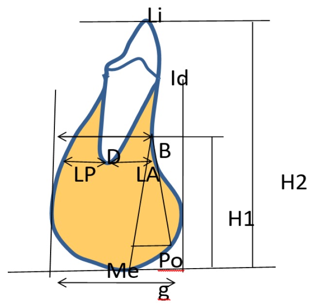
Linear parameters (Figure 1)
Figure 1.
Showing Linear symphyseal parameters.
Id–Me linear distance.
Perpendicular distance from Pog to B–Me line.
Perpendicular distance from point B to mandibular plane (H1).
Perpendicular distance from tip of the lower incisor to mandibular plane (H2).
Distance between anterior and posterior tangents to the symphysis perpendicular to the mandibular plane (W).
Bone posterior to the apex of the incisor (LP).
Bone anterior to the apex of the incisor (LA).
Actual symphysis: it is the portion of the mandible that lies anterior to a line perpendicular to the mandibular plane, passing through point B. This is measured parallel to the mandibular plane, from the most prominent point of the anterior border of bony symphysis.
Effective symphysis: it is the portion of the mandible which lies anterior to a continuation of line NB, measured perpendicular to line NB from the most prominent point of the anterior border of bony symphysis.
Ratio of Perpendicular distance from point B to mandibular plane (H1) to width. (H1/ W).
Ratio of Perpendicular distance from tip of the lower incisor to mandibular plane (H2) to width (H2/W).
Ratio depth to width (D/W).
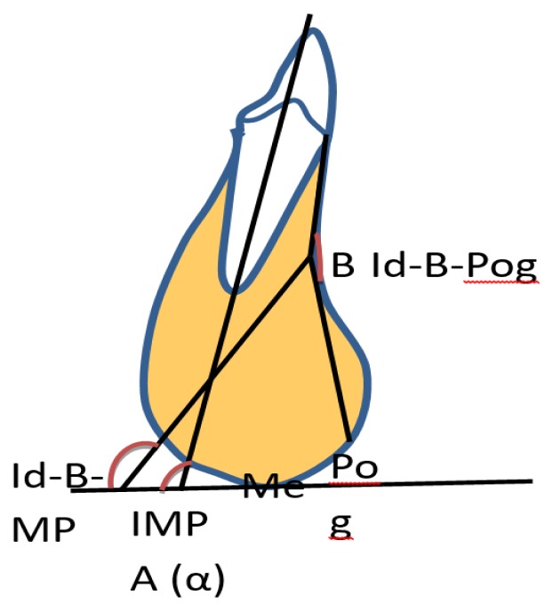
Angular parameters (Figure 2)
Figure 2.
Showing Angular symphyseal parameters.
Id–B–MP angle - The angle between a line connecting Id to Point B and the mandibular plane; it reflects the inclination of the alveolar part of the mandibular symphysis in relation to the mandibular plane.
Id–B-Pog angle - The angle between point Id, point B, and Pogonion; It reflects the concavity of the mandibular symphysis.
Angulation of lower incisor to mandibular plane (α).
Statistical Analysis
One way analysis of variance (ANOVA) test was used followed by post hoc tukeys HSD test to see if there was a statistically significant difference between symphyseal parameters and lower incisor angulation in different antero-posterior jaw relationships and skeletal growth patterns. Level of significance was set at p < 0.05; also unpaired t-test between male and female samples was applied for all the symphyseal parameters. intra-examiner and inter-examiner error was also checked. Spearman’s rank correlation coefficient was also used to see whether the Symphyseal parameters and lower incisor angulation had any correlation with the dentofacial parameters.
Results
Demographic details of the total sample including the mean age, standard deviation, standard error and p value of both the genders (male and female) have been illustrated in Table I.
Table I.
Demographic details of the total sample according to gender.
| Gender | N=90 | Age (years) | P value | ||
|---|---|---|---|---|---|
| Mean (mm) | SD (mm) | S.E. | |||
| Male | 40 | 19.886 | 3.847 | 3.847 | 0.6718 NS |
| Female | 50 | 20.108 | 3.446 | 3.446 | |
Comparison between MS parameters in different A-P skeletal relationships (Table II)
Table II.
Showing results of ANOVA with post hoc tukeys HSD test of symphyseal parameters in different A-P jaw relationships.
| Parameters | Class I (n=30) | Class II (n=30) | Class III (n=30) | p-value ANOVA |
Tukeys post hoc HSD test | |||||
|---|---|---|---|---|---|---|---|---|---|---|
| Mean | ±SD | Mean | ±SD | Mean | ±SD | I and II ‘p-value’ |
I and III ‘p-value’ |
II and III ‘p-value’ |
||
| Id-Me (mm) | 29.68 | 3.67 | 30.33 | 3.92 | 29.60 | 3.52 | 0.0067** | 0.631NS | 0.000** | 0.002** |
| Pog to B-Me (mm) | 4.78 | 0.86 | 4.55 | 0.93 | 4.92 | 1.03 | 0.0042** | 0.045* | 0.351NS | 0.004** |
| Width (w) (mm) | 14.08 | 1.68 | 14.53 | 2.18 | 13.02 | 1.90 | 0.009** | 0.62NS | 0.08 NS | 0.008** |
| Height (H1) (mm) | 20.30 | 4.77 | 20.35 | 2.78 | 20.90 | 3.39 | 0.831NS | 0.960NS | 0.576NS | 0.494NS |
| Height (H2) (mm) | 43.00 | 4.39 | 43.25 | 4.02 | 42.75 | 5.65 | 0.963Ns | 0.818NS | 0.848NS | 0.694NS |
| LA (mm) | 3.27 | 1.05 | 3.12 | 1.06 | 2.82 | 1.32 | 0.307Ns | 0.583NS | 0.148NS | 0.334NS |
| LP (mm) | 4.62 | 1.53 | 4.78 | 1.56 | 3.22 | 1.12 | 0.000** | 0.017* | 0.001** | 0.001** |
| H1/W (mm) | 1.50 | 0.28 | 1.43 | 0.28 | 1.61 | 0.29 | 0.049* | 0.570NS | 0.313NS | 0.040* |
| H2/W (mm) | 3.12 | 0.56 | 3.05 | 0.61 | 3.27 | 0.69 | 0.962NS | 0.656NS | 0.360NS | 0.201NS |
| D/W (mm) | 0.83 | 0.10 | 0.79 | 0.11 | 0.77 | 0.21 | 0.214NS | 0.130NS | 0.117NS | 0.575NS |
| Actual S. (mm) | 6.23 | 1.86 | 6.17 | 2.36 | 7.80 | 2.27 | 0.006** | 0.899NS | 0.017* | 0.012* |
| Eff. S.(mm) | 1.92 | 1.54 | 1.77 | 2.77 | 2.47 | 1.27 | 0.354Ns | 0.796NS | 0.137NS | 0.213NS |
| Id-B-Pog (deg) | 148.37 | 11.49 | 148.30 | 9.73 | 156.266 | 8.72 | 0.0011* | 0.8990NS | 0.003** | 0.003** |
| Id-B-MP (deg) | 91.70 | 7.93 | 92.20 | 7.85 | 81.23 | 8.72 | 0.000** | 0.807NS | 0.001** | 0.001** |
| α (deg) | 97.70 | 9.15 | 99.17 | 7.49 | 86.82 | 11.26 | 0.001** | 0.499NS | 0.001** | 0.001** |
p < 0.01,
p < 0.05,
NS-Non-significant
The results showed that, Id-Me (total length of mandibular symphysis) was statistically significantly larger in Class III group (32.60±3.52, p=0.0067) as compared to the other two groups. Perpendicular distance from Pogonion to B-Me line was statistically significantly larger in Class III group (p=0.042) as compared to Class I and Class II i.e. (Class III > Class I > Class II).
Symphyseal ratio (H1/W) was significantly different in Class III (1.61±0.29, p=0.049) subjects as compared to Class II being the highest amongst the class III subjects (Class III > Class I > Class II). LP was found to be highly statistically significantly smaller in the Class III (p=0.000) subjects as compared to Class I and Class II. The width of the mandibular symphysis was found to be highly statistically significantly lower in Class III subjects (13.02±1.90, p=0.009).
Actual symphysis was found to be statistically significantly larger in class III subjects as compared to Class I and II subjects (p≤0.01).
Id-B-Pog (anterior concavity of the mandibular symphysis) was found to be statistically significantly different in Class III as compared to Class I and II. Id-B-MP (inclination of the alveolar part of Mandibular Symphysis) was highly statistically significantly different in Class III subjects (p=0.000) as compared to Class I and II subjects (Class II > Class I > Class III). Inclination was more (indicated by a smaller angle) in the Class III subjects. Lower incisor mandibular plane angle (α) was found to be statistically highly significantly lower in Class III (p=0.000, 86.82±11.26) group as compared to Class I and II groups.
Comparison between MS parameters in different skeletal growth patterns (Table III)
Table III.
Showing results of ANOVA with post hoc tukeys HSD test of symphyseal parameters in different skeletal growth patterns.
| Parameters | Average (A) (N=30) | Horizontal (N=30) | Vertical (n=30) | P value ANOVA |
Tukeys post hoc HSD test | |||||
|---|---|---|---|---|---|---|---|---|---|---|
| Mean | ±SD | Mean | ±SD | Mean | ±SD | A and H ‘p value’ |
A and V ‘p value’ |
H and V ‘p value’ |
||
| Id-Me (mm) | 29.40 | 3.63 | 29.55 | 4.09 | 30.67 | 3.27 | 0.350NS | 0.881NS | 0.38NS | 0.472NS |
| Pog to B-Me (mm) | 4.90 | 0.84 | 4.78 | 1.01 | 4.57 | 0.96 | 0.3851NS | 0.630NS | 0.159NS | 0.399NS |
| Width (w) (mm) | 14.45 | 1.98 | 14.43 | 1.93 | 12.75 | 1.68 | 0.0005** | 0.899NS | 0.0019** | 0.0021** |
| Height (H1) (mm) | 20.37 | 4.23 | 20.17 | 3.18 | 21.02 | 3.71 | 0.655NS | 0.836NS | 0.529NS | 0.345NS |
| Height (H2) (mm) | 43.12 | 3.62 | 40.65 | 3.71 | 45.23 | 5.45 | 0.000** | 0.011* | 0.141NS | 0.000** |
| LA (mm) | 4.40 | 1.13 | 4.13 | 1.20 | 3.67 | 1.02 | 0.042* | 0.618NS | 0.030* | 0.246NS |
| LP (mm) | 3.97 | 1.30 | 4.88 | 1.49 | 3.77 | 1.71 | 0.011* | 0.053NS | 0.611NS | 0.014* |
| H1/W (mm) | 1.47 | 0.28 | 1.41 | 0.24 | 1.66 | 0.29 | 0.001** | 0.351NS | 0.023* | 0.001** |
| H2/W (mm) | 2.99 | 0.59 | 2.81 | 0.38 | 3.64 | 0.55 | 0.001** | 0.152NS | 0.001** | 0.001** |
| D/W (mm) | 0.80 | 0.21 | 0.81 | 0.11 | 0.78 | 0.10 | 0.698NS | 0.921NS | 0.542NS | 0.267NS |
| Actual S. (mm) | 7.08 | 2.23 | 5.82 | 1.66 | 7.30 | 2.63 | 0.023* | 0.073NS | 0.899NS | 0.011* |
| Eff. S. (mm) | 2.52 | 1.73 | 2.22 | 1.78 | 1.42 | 2.26 | 0.081NS | 0.511NS | 0.038* | 0.133NS |
| Id-B-pog (deg) | 148.7 | 13.70 | 150.70 | 7.55 | 150.70 | 8.60 | 0.704NS | 0.501NS | 0.515NS | 0.999NS |
| Id-B-MP (deg) | 89.70 | 10.49 | 90.93 | 8.00 | 84.50 | 9.05 | 0.020* | 0.848NS | 0.080NS | 0.022* |
| α (deg) | 97.03 | 9.87 | 97.75 | 10.14 | 88.90 | 10.51 | 0.0015** | 0.899NS | 0.007** | 0.003** |
p < 0.01,
p < 0.05,
NS-Non-significant.
The results showed that, the symphyseal width was highly statistically significantly smaller in vertical growers as compared to horizontal and average growers (p≤0.01) (Average > Horizontal > Vertical). LP was found to be significantly higher in the horizontal growers (p=0.012) whereas LA was found to be significantly higher in average growers (p=0.042). Symphyseal height (H2) was found to be highly statistically significantly different in horizontally growing subjects as compared to vertical and average growers (p=0.000, 40.65±3.71), (Vertical > Average > Horizontal) however, there was no statistically significant difference noted in vertical and average growers.
Symphyseal ratios (H1/W and H2/W) were found to be highly statistically significantly different in the vertical growers (p≤0.01) as compared to the horizontal and average growers (Vertical > Average > Horizontal), however there was no statistical difference observed in the horizontal and average growers (p≥0.05). The actual symphysis was found to be statistically significantly less in horizontally growing subjects (5.82±1.66, p=0.023) as compared to vertical and average growers, but when these two groups were compared there was no significant difference noted (p≥0.05).
Id-B-MP (Inclination of the alveolar part of Mandibular Symphysis) was significantly more in vertical growers (p=0.02) (indicated by a smaller angle) as compared to horizontal and average growers. Incisor mandibular plane angle (α) was found highly statistically significantly different in vertical growers (p=0.002) as compared to horizontal and average growers. It was observed to be lower in vertical growers (88.90±10.51), (Horizontal > Average > Vertical).
Table IV: Unpaired t test was applied between male and female subjects, however none of the symphyseal parameters or the lower incisor angulation were observed to have statistically significant differences (p≥0.05).
Table IV.
Showing mean standard deviation and t-test probability values between males and females for symphyseal parameters and IMPA.
| Parameters | Male group | Female group | Male and Female | |||
|---|---|---|---|---|---|---|
| Mean | ±SD | Mean | ±SD | Dif. | ‘p-value’ | |
| Id-Me (mm) | 29.59 | 3.65 | 30.17 | 3.73 | −0.58 | 0.455NS |
| Pog to B-Me (mm) | 4.68 | 1.00 | 4.82 | 0.88 | −0.13 | 0.505NS |
| Width (w) (mm) | 13.72 | 1.94 | 14.05 | 2.09 | −0.33 | 0.442NS |
| Height (H1) (mm) | 19.80 | 3.93 | 21.26 | 3.35 | −1.46 | 0.062NS |
| Height (H2) (mm) | 43.00 | 4.69 | 43.00 | 4.73 | 0.00 | 0.999NS |
| LA (mm) | 3.99 | 1.22 | 4.15 | 1.08 | −0.16 | 0.516NS |
| LP (mm) | 4.34 | 1.56 | 4.07 | 1.59 | 0.27 | 0.419NS |
| H1/W (mm) | 1.50 | 0.29 | 1.54 | 0.29 | −0.04 | 0.484NS |
| H2/W (mm) | 3.18 | 0.70 | 3.11 | 0.54 | 0.08 | 0.567NS |
| D/W (mm) | 0.79 | 0.11 | 0.80 | 0.18 | 0.00 | 0.910NS |
| Actual S. (mm) | 6.45 | 2.22 | 7.03 | 2.33 | −0.59 | 0.223NS |
| Eff. S. (mm) | 1.77 | 2.20 | 2.34 | 1.68 | −0.57 | 0.172NS |
| Id-B-pog (deg) | 150.20 | 12.04 | 149.91 | 8.05 | 0.29 | 0.894NS |
| Id-B-MP (deg) | 90.30 | 10.33 | 86.36 | 8.31 | 3.94 | 0.055NS |
| α (deg) | 95.16 | 11.40 | 93.93 | 10.32 | 1.23 | 0.593NS |
p<0.01,
p<0.05,
NS - Non-significant.
Correlation between Dentofacial parameters and MS parameters
Spearman’s rank correlation coefficient (Table V) showed significant correlations between symphyseal parameters and the dentofacial parameters, Total symphyseal height (H2) showed a strong positive significant correlation with upper anterior facial height (UAFH) and lower anterior facial height (LAFH) (r=0.628), however it showed a significant negative correlation with jaraback ratio (r=−0.486) and IMPA (α) (r=−0.282). Id-B-MP (inclination of alveolar part of Mandibular Symphysis) showed a strong positive significant correlation with ANB and IMPA (α) (r=0.797).
Table V.
Showing correlation between Symphyseal parameters and Dentofacial parameters.
| Parameters | ANB (deg) | jaraback (%) | UFH (mm) | LFH (mm) | α (deg) |
|---|---|---|---|---|---|
| Id-Me (mm) | −0.256* | −0.259* | 0.264* | 0.409** | −0.384** |
| Pog to B-Me (mm) | −0.148 | −0.077 | 0.353** | 0.262* | −0.165 |
| Width (w) (mm) | 0.260* | 0.028 | 0.144 | 0.116 | 0.272** |
| Height (H1) (mm) | −0.079 | −0.115 | 0.215* | 0.389** | −0.147 |
| Height (H2) (mm) | 0.030 | −0.486** | 0.355** | 0.628** | −0.282** |
| LA (mm) | 0.095 | −0.007 | −0.064 | −0.195 | 0.462** |
| LP (mm) | 0.230* | 0.184 | −0.224* | −0.218* | 0.341** |
| H1/W (mm) | −0.262* | −0.141 | 0.050 | 0.236* | −0.286** |
| H2/W (mm) | −0.142 | −0.320** | 0.042 | 0.269** | −0.368** |
| D/W(mm) | −0.135 | 0.093 | −0.013 | 0.028 | 0.296** |
| Actual S.(mm) | −0.257* | −0.187 | 0.306** | 0.168 | −0.576** |
| Eff. S. (mm) | −0.151 | 0.165 | 0.241* | −0.186 | −0.282** |
| Id-B-pog (deg) | −0.277** | 0.122 | −0.185 | −0.022 | −0.262* |
| Id-B-MP (deg) | 0.422** | 0.060 | 0.008 | −0.020 | 0.797** |
| α (deg) | 0.430** | 0.108 | −0.097 | −0.147 | 1.000 |
p<0.01,
p<0.05,
NS - Non-significant.
Discussion
The symphysis part of the mandible is thought to reflect growth behavior and future tendencies and is believed to be one of the predictors of the direction of growth rotation. Hence, it is of interest to know in details about the morphology of the symphysis in different Antero-Posterior jaw relationships and different skeletal growth patterns. Studying about the position and inclination of the lower incisor confined within the symphysis is necessary to achieve better stability and results. Moreover, it is also of interest to see whether the mandibular symphysis morphology and lower incisor angulation correlate to different dentofacial parameters.
The purpose of the present study was to assess the symphyseal morphology and lower incisor angulation in different anteroposterior relations of skeletal bases and skeletal growth patterns, and to investigate whether the Symphyseal morphology and lower incisor angulation have any correlation with the dentofacial parameters.
Class III subjects showed a greater inclination of the alveolar part of mandibular symphysis (Id-B-MP) which was indicated by a smaller angle as compared to the Class I and Class II subjects, also the anterior concavity of the mandibular symphysis (Id-B-Pog) was less pronounced in the Class III subjects as compared to the other two groups. These findings of ours were in agreement with the results of the studies conducted by Al – Khateeb et al. [2] and Yamada et al. [6] 2007 in which they also reported the similar findings with relation to inclination of the alveolar part and the anterior concavity of the mandibular symphysis.
Actual symphysis was found to be significantly greater in the skeletal Class III subjects as compared to the skeletal Class I and Class II. However, effective symphysis did not show any statistically significant difference in the three groups. This finding of ours could be due to the presence of a large mandible as a whole in the Class III skeletal relationship subjects. To the best of our knowledge no previous study [1,7–9] has been conducted to assess the actual and effective symphysis in all the three antero-posterior jaw relationships and skeletal growth patterns.
The lower incisor mandibular plane angle (α) was found to be highest in the Class II subjects in this study, indicating that the lower incisor is generally more proclined in Class II subjects as compared to Class I and Class III subjects. This finding also supported the results of Sangcharearn et al. [10] in which they reported that the skeletal Class II cases have proclined lower incisors. This kind of dentoalveolar compensation occurs in skeletal Class II cases to mask the skeletal discrepancy between maxilla and mandible and to achieve a near normal occlusion despite the presence of an underlying skeletal base discrepancy.
Handelman et al. [1] and others [2,11–12] have reported that the part of the alveolar bone present posterior to the apex of the lower incisor is markedly increased in Class III subjects as compared to the bone present anterior to the apex. This phenomenon could be due to the retroclination of the lower incisor in the Class III subjects.
Similarly in the present study we found the same, that the bone present posterior to the apex of the lower incisor i.e. LP was increased in Class III subjects as compared to LA, that is the bone present anterior to root apex, which caused the root apex to be closer to the labial cortical plate of the symphysis. Also, the inclination of the lower incisor (α) was lesser in Class III subjects, indicating that the lower incisor is retroclined in the Class III subjects. This, however, is a frequent finding in the Class III subjects to compensate for the skeletal discrepancy. It has been suggested that the outer surface of the dentoalveolar part of the mandibular symphysis undergoes surface remodelling due to this kind of retroclination of the lower incisor [3,6]. In these cases the lingual tipping of the incisor for orthodontic camouflage is not a suitable treatment option and these cases should be treated with appropriate surgical options.
Based on the above findings, we can conclude that the pre-treatment evaluation of the lower incisor angulation and its relationship to the underlying alveolar bone should be taken into consideration, to plan the limits of incisor movement, thus avoiding the associated root resorption and/or dehiscence.
Handelman et al. [1], 1996, also stated that the bone level lingual to the mandibular incisor apex (LP) and the total width (W) of the symphysis were narrower in Class III subjects as compared to Class I and class II. Our findings also were in agreement with his study that LP and the total width of the symphysis was lesser in class III as compared to Class I and Class II subjects.
Mandibular symphysis morphology in different skeletal growth patterns
The symphyseal width (W) was found to be significantly smaller in the vertical growers as compared to the horizontal and average growers. Also, the symphyseal heights (H1 and H2) and the symphyseal ratios (H1/W and H2/W) were found to be greater in vertical growers as compared to the other two groups. These findings observed in this study were in agreement with the studies conducted by Pintavirooj et al. [13], 2014, and others [14,15] they showed similar results. On the contrary, the studies conducted by Moshfeghi et al. [16], 2014, and Aki et al. [9], 1994, concluded from their studies that the symphyseal ratio was smaller in the vertical growers as compared to the horizontal growers. This was in contradiction to the findings observed in this study.
LP, i.e. bone present posterior to the apex of the incisor, was found to be significantly greater in the horizontal growers as compared to the vertical and average growers. Our results were in agreement with a few studies [5,17] in which they reported that a greater distance exists between the lower central incisor apices and the associated alveolar bone. They also stated that the LP cortex which was found significantly decreased in the vertical growers lies outside the dentoalveolar compensation mechanism of the lower incisor and responds to the more intense reformation process that takes place in the LP area.
Molina-Berlanga et al. [5], 2013, in their study reported a decreased IMPA (α) in the long faced subjects as compared to normal and short faced subjects. There results were in accordance with the findings the present study, that in the long faced patients the lower incisor was almost always found to be retroclined and there was a decrease in LP and LA and an overall increased length of the symphysis.
When assessing the morphology of the mandibular symphysis according to gender, we did not find any statistically significant difference between males and females in any of the symphyseal parameters or the lower incisor angulation, however, this result of the present study was in contrast with the results observed by Al–Khateeb et al. [2], 2014, and Aki et al. [9], 1994, in which the males had increased symphyseal length and total height of MS as compared to females. This finding could be due to the presence of increased number of vertical growers in female group than male group.
Correlation of mandibular symphyseal parameters with the dentofacial parameters
Al-Khateeb et al. [2] showed a weak but significant correlation between the lower incisor inclination (α) and inclination of the alveolar part of MS (Id-B-MP). However, our results were in contradiction to this in which we observed a strong positive correlation between the two (r = 0.797), this result of ours was supported by a few studies [6,18] that also showed a strong correlation between these two parameters, whereas they used different points and lines to study the inclination of Mandibular Symphysis.
Actual symphysis showed a strong negative correlation with the lower incisor inclination (α) (r=−0.576). This negative correlation could be because the B point might have receded with increase in lower incisor inclination.
Actual symphysis was found to be significantly decreased in the horizontal growers as compared to the average and vertical growers. This could be due to the fact that we found a positive correlation between the actual symphysis and upper anterior facial height (UAFH). However, we also found a negative correlation with IMPA and angle ANB. As we know that the anterior facial height is lesser in horizontal growers as compared to vertical growers and the lower incisor is more proclined in the horizontal growers which further decreases the ANB angle in them. Hence, we can say that a combined effect of these findings in the horizontal growers may lead to a decreased length of actual symphysis.
This study outlines and reflects the importance of carrying out a thorough analysis for each patient as a separate entity, taking into consideration his/her craniofacial composition and jaw and chin morphology for the purpose of proper diagnosis and treatment planning.
Further studies can be carried out to study the symphyseal morphology to see correlation of all the A-P skeletal jaw relationships and growth patterns separately with the dentofacial parameters by increasing the sample size of the study. Also with the advancement of CBCT, three dimensional imaging of the mandibular symphysis in different Antero-Posterior jaw relationships and growth patterns should be done and will be more conclusive.
Conclusions
The following conclusions were drawn from the study:
-
The Class III skeletal jaw relationship exhibited the following statistically significant differences as compared to Class II causing the symphysis to be narrower and elongated reflecting compensation for the skeletal pattern of the jaws :
Actual symphysis width was found to be increased.
Less concave anterior contour of mandibular symphysis.
Inclination of the alveolar part towards the mandibular plane increased.
-
The total height of the symphysis and the symphyseal ratio increased.
The overall width was reduced.
The lower incisors were retroclined.
-
The vertical growers exhibited the following statistically significant differences as compared to Horizontal growers.
Actual symphysis width was found to be decreased.
Inclination of the alveolar part towards the mandibular plane increased.
Increased symphyseal height and symphyseal ratio.
-
Decreased overall width of the symphysis.
The lower incisors were retroclined.
A strong positive correlation was found between the actual symphysis and the lower incisor angulation (α).
A strong positive correlation was also found between inclination of alveolar part of Mandibular Symphysis and IMPA (α).
References
- 1.Handelman CS. The anterior alveolus: its importance in limiting orthodontic treatment and its influence on the occurrence of iatrogenic sequelae. Angle Orthod. 1996;66:95–109. doi: 10.1043/0003-3219(1996)066<0095:TAAIII>2.3.CO;2. discussion 109–110. [DOI] [PubMed] [Google Scholar]
- 2.Al-Khateeb SN, Al Maaitah EF, Abu Alhaija ES, Badran SA. Mandibular symphysis morphology and dimensions in different anteroposterior jaw relationships. T Angle Orthod. 2014;84:304–309. doi: 10.2319/030513-185.1. [DOI] [PMC free article] [PubMed] [Google Scholar]
- 3.Yu Q, Pan XG, Ji GP, Shen G. The association between lower incisal inclination and morphology of the supporting alveolar bone--a cone-beam CT study. Int J Oral Sci. 2009;1:217–223. doi: 10.4248/IJOS09047. [DOI] [PMC free article] [PubMed] [Google Scholar]
- 4.Ishikawa H, Nakamura S, Iwasaki H, Kitazawa S, Tsukada H, Sato Y. Dentoalveolar compensation related to variations in sagittal jaw relationships. Angle Orthod. 1999;69:534–538. doi: 10.1043/0003-3219(1999)069<0534:DCRTVI>2.3.CO;2. [DOI] [PubMed] [Google Scholar]
- 5.Molina-Berlanga N, Llopis-Perez J, Flores-Mir C, Puigdollers A. Lower incisor dentoalveolar compensation and symphysis dimensions among Class I and III malocclusion patients with different facial vertical skeletal patterns. Angle Orthod. 2013;83:948–955. doi: 10.2319/011913-48.1. [DOI] [PMC free article] [PubMed] [Google Scholar]
- 6.Yamada C, Kitai N, Kakimoto N, Murakami S, Furukawa S, Takada K. Spatial relationships between the mandibular central incisor and associated alveolar bone in adults with mandibular prognathism. Angle Orthod. 2007;77:766–772. doi: 10.2319/072906-309. [DOI] [PubMed] [Google Scholar]
- 7.Chung CJ, Jung S, Baik HS. Morphological characteristics of the symphyseal region in adult skeletal Class III crossbite and openbite malocclusions. Angle Orthod. 2008;78:38–43. doi: 10.2319/101606-427.1. [DOI] [PubMed] [Google Scholar]
- 8.Gama A, Vedovello S, Vedovello Filho M, Lucato AS, Junior MS. Evaluation of the alveolar process of mandibular incisor in class I, II and III individuals with different facial patterns. J Health Sci. 2015;14:95–98. [Google Scholar]
- 9.Aki T, Nanda RS, Currier GF, Nanda SK. Assessment of symphysis morphology as a predictor of the direction of mandibular growth. Am J Orthod Dentofacial Orthop. 1994;106:60–69. doi: 10.1016/S0889-5406(94)70022-2. [DOI] [PubMed] [Google Scholar]
- 10.Sangcharearn Y, Ho C. Effect of incisor angulation on overjet and overbite in class II camouflage treatment: A typodont study. Angle Orthod. 2007;77:1011–1018. doi: 10.2319/111206-460.1. [DOI] [PubMed] [Google Scholar]
- 11.Singh GD, McNamara JA, Jr, Lozanoff S. Mandibular morphology in subjects with Class III malocclusions: finite-element morphometry. Angle Orthod. 1998;68:409–418. doi: 10.1043/0003-3219(1998)068<0409:MMISWC>2.3.CO;2. [DOI] [PubMed] [Google Scholar]
- 12.Gütermann C, Peltomäki T, Markic G, Hänggi M, Schätzle M, Signorelli L, et al. The inclination of mandibular incisors revisited. Angle Orthod. 2014;84:109–119. doi: 10.2319/040413-262.1. [DOI] [PMC free article] [PubMed] [Google Scholar]
- 13.Pintavirooj P, Sumetcherngpratya R, Chaiwat A, Changsiripun C. Relationship between mentalis muscle hyperactivity and mandibular symphysis morphology in skeletal Class I and II patients. Orthodontic Waves. 2014;73:130–135. [Google Scholar]
- 14.Gracco A, Luca L, Bongiorno MC, Siciliani G. Computed tomography evaluation of mandibular incisor bony support in untreated patients. Am J Orthod Dentofacial Orthop. 2010;138:179–187. doi: 10.1016/j.ajodo.2008.09.030. [DOI] [PubMed] [Google Scholar]
- 15.Mangla R, Singh N, Dua V, Padmanabhan P, Khanna M. Evaluation of mandibular morphology in different facial types. Contemp Clin Dent. 2011;2:200–206. doi: 10.4103/0976-237X.86458. [DOI] [PMC free article] [PubMed] [Google Scholar]
- 16.Moshfeghi M, Nouri M, Mirbeigi S, Baghban AA. Correlation between symphyseal morphology and mandibular growth. Dent Res J. 2014;11:375–379. doi: 10.4103/1735-3327.135915. [DOI] [PMC free article] [PubMed] [Google Scholar]
- 17.Lombardo L, Berveglieri C, Spena R, Siciliani G. Quantitative cone-beam computed tomography evaluation of premaxilla and symphysis in Class I and Class III malocclusions. Int Orthod. 2016;14:143–160. doi: 10.1016/j.ortho.2016.03.014. [DOI] [PubMed] [Google Scholar]
- 18.Guyer EC, Ellis EE, 3rd, McNamara JA, Jr, Behrents RG. Components of class III malocclusion in juveniles and adolescents. Angle Orthod. 1986;56:7–30. doi: 10.1043/0003-3219(1986)056<0007:COCIMI>2.0.CO;2. [DOI] [PubMed] [Google Scholar]


