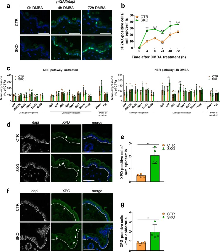
Fig. 2. Enhanced DNA-damage response and repair in SKOs.
a Immunostaining for γH2AX at the indicated time points after DMBA administration. Dapi was used for nuclear staining. Scale bars, 50 µm. b Quantification of γH2AX, as shown in panel a. The data are expressed as means ± SEM (n = 4). c qRT-PCR analysis of NER factors in the back skin of mice not treated (left panel) or treated for 4 h with DMBA (right panel). Hprt was used as a housekeeping gene for normalization. The data are expressed as means ± SD (n = 4). d Immunostaining for XPD in the back skin of mice treated for 24 h with DMBA. Arrows indicate positive stained cells. Dapi was used as a nuclear counterstain. Scale bars, 50 µm. e Quantification of the data in panel (d). The data are expressed as means ± SD (n = 4). f Immunostaining for XPG in the back skin mice treated for 24 h with DMBA. Arrows indicate positive stained cells. Dapi was used as a nuclear counterstain. Scale bars, 50 µm. g Quantification of the data in panel (f). The data are expressed as means ± SD (n = 4). *p < 0.05; **p < 0.01; ***p < 0.001 in unpaired Student’s t-test.

