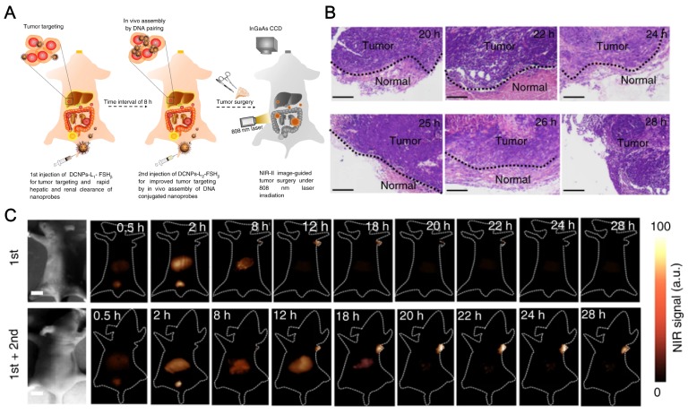
Figure 6.
NaGdF4 based NIR-II nanoprobes in-vivo assembly to improve IGS for metastatic ovarian cancer. (A) Schematic illustration of NIR-II nanoprobes fabrication for ovarian metastasis surgery under NIR-II bioimaging guidance. (B) Hematoxylin and eosin (H&E) staining results of the tumors resected in 20-28 h PI under NIR-II fluorescence bioimaging guidance. (C) NIR-II fluorescence bioimaging (1000 nm long-pass filter) of the nude mice with murine epidermal tumor by single caudal vein first injection and two-staged in sequence injection (first + second) (interval between two injection is 8 h) under 808 nm excitation (fluence rate = 40 mW cm-2). The concentration of DCNPs in single injection is same to the sum of that for two-staged injection. All scale bars: 1 cm. Representative images are for n = 5 per group. Adapted with permission from 150, Copyright 2017, Nature Springer.

