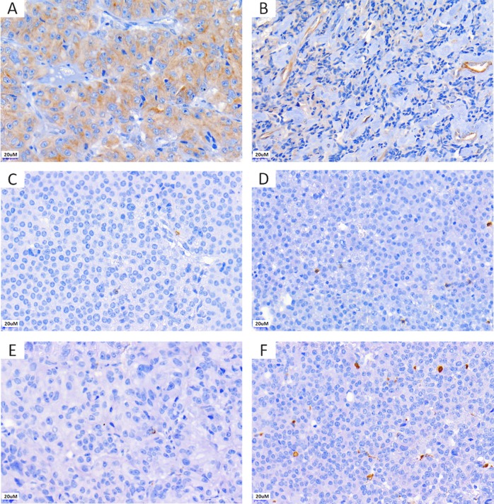
Fig. 1.
Representative pictures of the immunohistochemistry for endocan and the immune cell markers CD8 and CD68 in somatotroph pituitary neuroendocrine tumours. Immunohistochemistry for endocan showing moderate cytoplasmic expression in the tumour cells only (H-score 150) (a) and endothelial cell expression with weak tumour cell staining (H-score 70) (b). Immunohistochemistry for CD8 (c, d) and CD68 (e, f): tumours with sparse positive cells (c, e) and higher immune cell infiltrate are shown (d, f)

