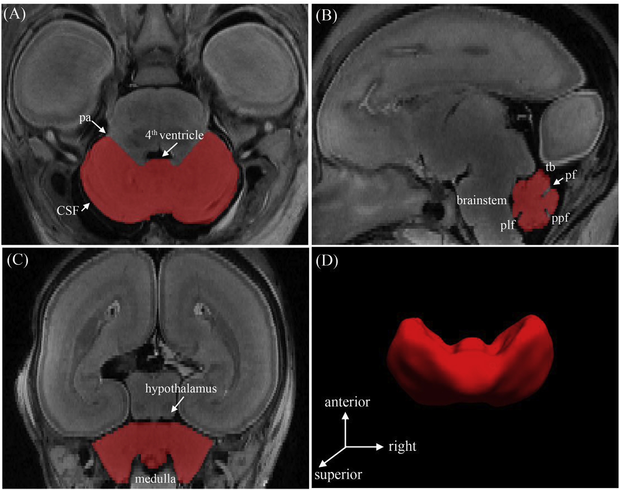
Fig. 1.

Segmentation and surface reconstruction of the cerebellum of a 20 GW subject. The boundaries of the cerebellum were delineated in the axial plane (a) and confirmed in the sagittal (b) and coronal (c) planes to verify the segmentation accuracy. (d) The 3D reconstructed cerebellar surface was displayed simultaneously. Abbreviations: (pa) pontocerebellar angle; (tb) tentorium cerebelli; (pf) primary fissure; (ppf) prepyramidal fissure; (plf) posterolateral fissure.
