Abstract
Photon upconversion in lanthanide-doped upconversion nanoparticles offers a wide variety of applications including deep-tissue biophotonics. However, the upconversion luminescence and efficiency, especially involving multiple photons, is still limited by the concentration quenching effect. Here, we demonstrate a multilayered core-shell-shell structure for lanthanide doped NaYF4, where Er3+ activators and Yb3+ sensitizers are spatially separated, which can enhance the multiphoton emission from Er3+ by 100-fold compared with the multiphoton emission from canonical core-shell nanocrystals. This difference is due to the excitation energy transfer at the interface between activator core and sensitizer shell being unexpectedly efficient, as revealed by the structural and temperature dependence of the multiphoton upconversion luminescence. Therefore, the concentration quenching is suppressed via alleviation of cross-relaxation between the activator and the sensitizer, resulting in a high quantum yield of up to 6.34% for this layered structure. These findings will enable versatile design of multiphoton upconverting nanoparticles overcoming the conventional limitation.
Subject terms: Nanoparticles, Nanoparticles, Optical physics
Photon upconversion in nanoparticles is often limited by concentration quenching. The authors present a multi-layered particle approach that enables increased upconversion efficiency by reducing cross-relaxation, through spatial separation of activator and sensitizer.
Introduction
The upconversion process involving multistep absorption of two or more low-energy photons to generate a high-energy photon in lanthanide-doped upconversion nanoparticles (UCNPs) enables promising applications in various fields, such as display1, microlaser2, solar cell3, deep-tissue biophotonics4–6 and super-resolution nanoscopy7. Typically, Yb3+ ions are doped into these nanoparticles as sensitizer ions with the purpose of transferring energy to activator ions (Er3+, Tm3+), thereby producing an efficient upconversion8–10. However, the brightness and upconverting efficiency of these UCNPs are still limited due to the relatively low doping concentrations of the sensitizer and activator ions. Thus, a direct way to improve their brightness is to increase the concentration of the dopants since the upconversion efficiency is largely dependent on the dopant concentration. Unfortunately, concentration quenching occurs in heavily doped UCNPs because nonradiative energy losses, including energy migration-induced surface quenching and cross-relaxation between neighboring dopant ions, will dominate in this case11,12.
Recently, substantial efforts have been made to overcome the above obstacles and to enhance the upconversion luminescence (UCL) for lanthanide-doped UCNPs. Inert shell passivation has been demonstrated to be successful in overcoming energy migration-induced surface quenching and enhancing emission intensity in UCNPs9,12–17. Energy back-transfer from the activator ions to sensitizer ions can be efficiently blocked in a sandwich structured UCNPs, yielding bright UCL18. Wang et al. revealed that a KYb2F7 host is favorable for multiphoton upconversion of Er3+ by minimizing the migration of excitation energy to the defects10. High-irradiance excitation is an alternative method that can be used to overcome concentration quenching by enriching the excitation energy4,19,20. In the meantime, both brightness enhancement and the promotion of upconverting efficiency has been achieved by construction of dye-UCNP hybrids21. Even though these existing approaches are effective for large emission enhancement and for the promotion of upconverting efficiencies, the intrinsic cross-relaxation energy loss in UCNPs has not yet been properly addressed, even though it is a key factor that contributes primarily to concentration quenching when surface quenching is negligible.
Here, we propose that cross-relaxation between dopant ions can be effectively suppressed in UCNPs by combining energy migration and interfacial energy transfer via multilayered structure design, as shown in Fig. 1a. In this structure, the separated location for the Er3+ and Yb3+ ions in the core and neighboring shell can alleviate Yb3+-Er3+ cross-relaxation (backward energy transfer) and assure efficient energy transfer at the core-shell interface due to the short Yb3+-Er3+ distance therein11,18,22,23. The inert shell herein can mitigate migration-induced surface quenching and promote energy transfer from Yb3+ to Er3+ ions. This multilayered structure enables us to overcome the concentration limitation and enrich excitation energy by thickening the sensitizer layer to enhance the UCL, especially at shorter wavelengths. The optimum α-NaYF4:10% Er@NaYbF4 @NaYF4 nanoparticles show an upconversion quantum yield (QY) of 6.34% at an excitation power density of 4.5 W cm−2, with a 100-fold enhancement for three-photon upconversion compared with canonical NaY0.78F4:Yb0.2Er0.02@NaYF4 core-shell nanocrystals.
Fig. 1. Schematic diagram for the UCNPs showing principle emissions.
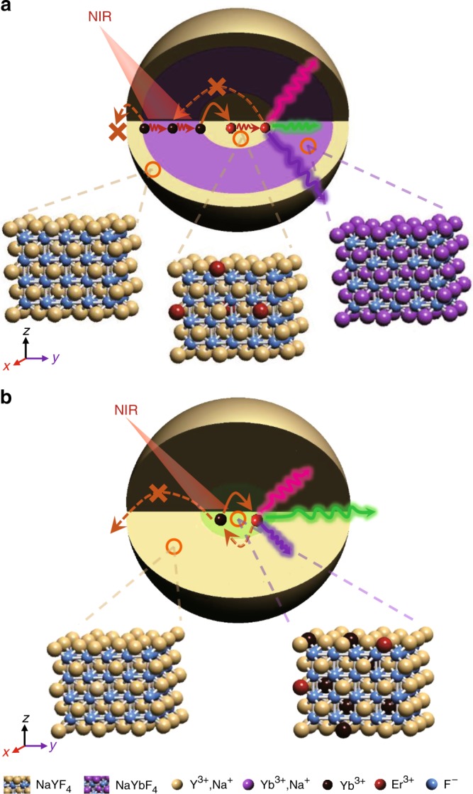
a α-NaYF4:Er@NaYbF4@NaYF4 core-shell-shell nanoparticles. b α-NaYF4:20% Yb, 2% Er@NaYF4 core-shell nanoparticles. Both backward energy transfer and surface quenching processes can be efficiently suppressed in a.
Results
Synthesis and brighter UCL of the trilayered UCNPs
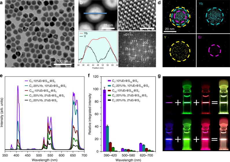
We synthesized both the canonical α-NaYF4:Yb,Er@NaYF4 (CY:20% Yb, 2% Er@SY) core-shell (Fig. 1b and Supplementary Fig. 1) and α-NaYF4:Er@NaYbF4@NaYF4 (CY:Er@SYb@SY) core-shell-shell nanoparticles by an epitaxial growth method (Fig. 1a and Supplementary Fig. 2). The crystal phase and high quality of these nanoparticles was confirmed by X-ray diffraction and transmission electron microscopy (Fig. 2a–c and Supplementary Fig. 3). Typically, the as-synthesized α-NaY0.78F4:Yb0.2Er0.02@NaYF4 nanoparticles, which are one of the most efficient ensembles for photon upconversion24, generate intense green emission (2H11/2, 4S3/2 → 4I15/2), competitive red emission (4F9/2 → 4I15/2) and weak violet emission (2H9/2 → 4I15/2) for Er3+ under a 980-nm excitation (Fig. 2e, Supplementary Fig. 4); these findings are consistent with previous reports24,25. The inert shell herein (~10.5 nm, Supplementary Fig. 3) is thick enough to suppress surface or solvent quenching of the UCL26,27. In stark contrast, once the activator and sensitizer were separately doped into the core and intermediate layer of the core-shell-shell nanoparticles at a higher doping level, these α-NaYF4:10% Er@NaYbF4 @NaYF4 (CY:10% Er@SYb@SY) nanoparticles exhibited a markedly increased UC emission, with an ~100-fold increment, especially for the multiphoton violet emission at ~407 nm (Fig. 2e, f). Both the violet and red emission are predominant in this multilayered structure, unlike the strong green emission in the canonical structure (Fig. 2g). Moreover, all of the multilayered structures with Yb3+ and Er3+ codoped into the core present a much lower UC luminescent intensity than our CY:10% Er@SYb@SY structure despite their particle sizes, sensitizer layers and inert layers all being nearly the same. This dynamic reveals that a much more efficient energy transfer upconversion occurs in this core-shell-shell structure, indicating the superiority of combining energy migration and interfacial energy transfer in the upconversion process. It should be pointed out that severe luminescence quenching will occur if the sensitizing layer is not encapsulated by an inert shell in this structure (Supplementary Fig. 5), indicating that the surface quenching is minimized by this means. It is also noted that the green emission in the CY:10% Er@SYb@SY structure increases by only fivefold compared with the canonical structure, consistent with the Er3+ content increasing from 2 to 10%. This finding may indicate population saturation for the 2H11/2 and 4S3/2 levels of Er3+, unlike those emitting violet and red light.
Fig. 2. Efficient multiphoton upconversion in trilayered nanoparticles.
a–d TEM (a) and high-resolution STEM images (b, c) revealing the single-crystalline nature of the α-NaYF4:Er@NaYbF4@NaYF4 nanocrystal (dSYb = 8.3 ± 0.7 nm, dSY = 2.3 ± 0.1 nm). The dSYb and dSY are used to designate the thickness of the intermediate NaYbF4 and the outmost NaYF4 shells, respectively. The EDX results (b, d) show distribution of Er3+, Yb3+ and Y3+ ions, consistent with the core-shell-shell structure. Nanoparticles with dSY = 5.6 ± 0.7 nm were selected for better contrast in b. e Upconversion emission spectra (collected with the mode of “emission correction off”) for different UCNPs upon 980-nm excitation (24.0 W cm−2): CY, CYb, SYb and SY are used to designate NaYF4 core, NaYbF4 core, NaYbF4 shell and NaYF4 shell, respectively. For CY:20%Yb, 2%Er@SY, dSY = 10.5 ± 0.8 nm. f, g The PL intensities (f) and luminescent photographs (g) for CY:20% Yb, 2% Er@SY (top) and CY:10% Er@SYb@SY nanoparticles (bottom). Error bars in f represent the standard deviation of three trials. Source data are provided as a Source Data file. The photographs were taken through violet, green and red color filters upon 980-nm excitation (15.2 W cm−2).
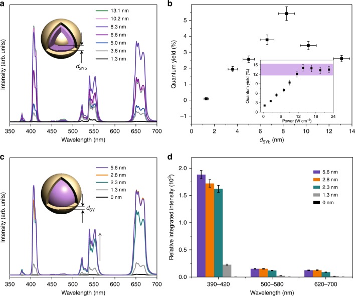
Enrichment of excitation energy can be realized not only by increasing the Yb3+ doping level to 100% but also via thickening of the sensitizing layer (dSYb). The UC emission for the CY:10% Er@SY:Yb@SY UCNPs increases continuously with Yb3+ concentration in the sensitizing layer, with no luminescence quenching observed (Supplementary Fig. 6). This phenomenon demonstrates that the contribution of cross-relaxation or self-quenching among Yb3+ ions to concentration quenching can be neglected, consistent with a previous report16. With increasing dSYb (Supplementary Fig. 7 and Supplementary Table 1), the overall UC emission and QY for the UCNPs also enhances continuously until dSYb = 8.3 nm (Fig. 3a, b). In particular, the violet and red emission increases much faster than the green emission, resulting in a constantly increased contribution from the violet and red emission to the overall emission. While the thickness of NaYbF4 shell further increases, the overall UC emission and QY for the UCNPs decreases instead. The QY of the optimum UCNPs firstly increases with increasing excitation power and then reaches saturation when the excitation power is relatively higher. Once dSY is thicker than 2 nm, the inert shell thickness (dSY, determined by the transmission electron microscopy (TEM) images in Supplementary Fig. 8 with details shown in Supplementary Table 1) seems to have little effect on the population of different Er3+ energy levels or the overall intensity of the UC emission (Fig. 3c, d). This dynamic is due to the luminescence from Yb3+ being more likely to be quenched by surface defects instead of solvent quenching and due to the relatively thin inert shell being sufficient to suppress the quenching26. In the meantime, the distance of the Er3+ from the surface is larger than 10 nm (dSYb + dSY) in this case, which is sufficient to prevent the solvent quenching of Er3+ emission26,27.
Fig. 3. Shell thickness-dependent upconversion luminescence.
a, c Upconversion emission spectra for the CY:10% Er@SYb@SY core-shell-shell nanoparticles with varying shell thickness: a NaYbF4 shell, dSY = 2.0–2.7 nm; c NaYF4 inert shell, dSYb = 8.3 ± 0.7 nm (collected with the mode of “emission correction off”). b The quantum yields (QY) as a function of NaYbF4 shell thickness (4.5 W cm−2). The inset: power-dependent quantum yields for NPs with dSYb of 8.3 ± 0.7 nm and dSY of 2.3 ± 0.1 nm. The error bars for QY represent the standard deviation of three trials. Source data are provided as a Source Data file. The violet shading in the inset is used to mark saturated QY. d Corresponding emission intensity for violet, green and red bands when the thickness of NaYF4 shell varies. The results indicate the key roles of excitation energy enrichment and inert shell protection in enhancement of multiphoton upconversion for the core–shell–shell nanoparticles. The standard deviation of three trials are shown as error bars. Source data are provided as a Source Data file.
Evidence of an efficient interfacial energy transfer
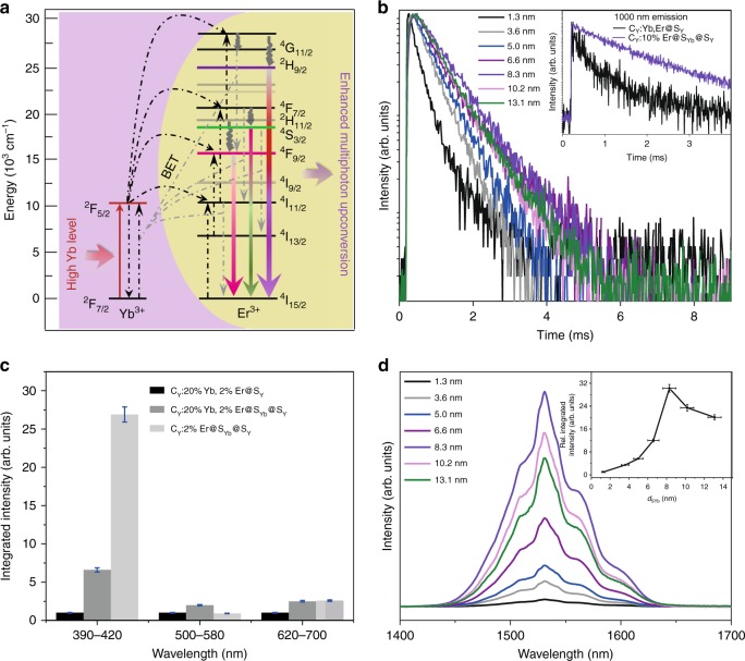
The power dependence for the UC emission from the CY:10% Er@SYb@SY multilayered nanoparticles (Supplementary Fig. 9) indicates that the 407-nm emission arises from the three-photon UC process, while the 540-nm emission is governed by the two-photon UC process. The slop factor calculated for the red emission is 2.3, suggesting contributions from both two- and three-photon processes (Fig. 4a)23,27–29. As we discussed above, the population of the green emitting levels for Er3+ may be saturated because two-photon UC is more efficient under low excitation power density19,30. On average, the amount of Yb3+ ions increases by ~22-fold when dSYb varies from 1.3 to 8.3 nm, with the green emission intensity increasing by ~20-fold correspondingly (Supplementary Fig. 10). This finding indicates that the green emission intensity increases linearly with the increment of Yb3+ sensitizer ions. In contrast, the violet and red emission intensities increase by 440 and 180 times, respectively, in the same condition, further validating the hypothesis that the 4F9/2 level may be populated primarily through a triphotonic transition accompanied by back energy transfer (Fig. 4a). The three-photonic population of the 2H9/2 and the 4F9/2 levels is greatly enhanced; thus, the violet and red emissions increase exponentially when dSYb is elevated. This finding explains why the contributions of the violet and red emissions for CY:10% Er@SYb@SY UCNPs become dominant at larger dSYb. Whereas once dSYb is larger than ~10.2 nm, the violet emission gradually reaches saturation while both the green and red emissions obviously decay. This phenomenon implies that the population of the 4F9/2 level through a triphotonic transition may be hindered when a much thicker NaYbF4 shell is coated.
Fig. 4. Efficient interfacial energy transfer in trilayered nanoparticles.
a Simplified energy-level diagram shows enhanced multiphoton upconversion from Yb3+ to Er3+ in the CY:10% Er@SYb@SY core-shell-shell nanoparticles (dSYb ≥ 3.2 nm, dSY ≥ 2.0 nm). b Luminescence decay curves of Er3+ emission at 407 nm for CY:10% Er@SYb@SY nanoparticles with dSYb increasing from 1.3 to 13.1 nm (dSY = 2.0–2.7 nm). Inset shows the decay curves of Yb3+ emission for the as-synthesized CY:10% Er@SYb@SY core-shell-shell (dSYb = 8.3 ± 0.7 nm) and CY:20% Yb, 2% Er@SY core-shell nanoparticles (dSY = 10.5 ± 0.8 nm). c Relative integrated UCL intensity of 2% Er3+-doped UCNPs (for trilayered structure, dSYb = 8.3 ± 0.7 nm, dSY = 2.3 ± 0.1 nm; for core-shell structure, dSY = 10.5 ± 0.8 nm). d NIR downshifting luminescence spectra and the relative integrated intensities (inset) for the CY:10% Er@SYb@SY nanoparticles with varying NaYbF4 shell thickness (dSY = 2.0–2.7 nm). The error bars in (c) and (d) (inset) represent the standard deviation of three trials. Source data are provided as a Source Data file.
To shed more light on the energy transfer between Yb3+ and Er3+ ions in the multilayered CY:10% Er@SYb@SY UCNPs, the decay kinetics of the Er3+ emission bands from these nanoparticles were measured and compared with those obtained from the canonical CY:20% Yb, 2% Er@SY samples. In comparison with the canonical structure, the separated distribution of Yb3+ and Er3+ ions in CY:10% Er@SYb@SY UCNPs undoubtedly slows the energy transfer from Yb3+ to Er3+ and prolongs the decay time for the Yb3+ 2F5/2 level (Fig. 4b). Consequently, the rise and decay times for the Er3+ emission are obviously prolonged in this core-shell-shell structure (Supplementary Fig. 11) since Yb3+ → Er3+ energy transfer occurs at the interface following Yb3+-Yb3+ energy migration in the sensitizer layer25. Moreover, with the increment of the sensitizers (dSYb), all the rise and decay times for the upconverted Er3+ emission at 407, 540, and 651 nm increase constantly until dSYb = 8.3 nm (Supplementary Fig. 12). This finding supports our conjecture that the excitation energy absorbed by the outmost Yb3+ ions can migrate a longer distance through the NaYbF4 layer and transfer to the Er3+ levels at the core-shell interface. However, luminescent quenching occurs if NaYbF4 layer is too thicker (larger than 10.2 nm), as demonstrated by the shortened lifetimes of the emission bands. It is also important to note that the UCL at 407 nm has single exponential decay kinetics for all the samples, except for the one with the thinnest sensitizing layer (~1.3 nm). In addition to the long decaying component resembling that of the others, thin protection layer (<4.0 nm) may lead to fast decay of 2H9/2 state from Er3+ ions neighboring sensitizing layer. In the meantime, the decay time for the red emission at 651 nm increases more strongly. This may be due to the increased contribution of the three-photon process with increasing amount of added NaYbF4. Similar to their UC emission intensities, the lifetimes of the emission bands at 407, 540 and 651 nm initially increase until dSY = 2.3 nm, and then, they reach an approximately constant maximum value (Supplementary Fig. 13), thereby verifying that a thin inert layer is enough for suppression of surface quenching in this structure.
To preclude the possibility that higher activator concentration (10 vs. 2%) plays a key role in enhancing energy transfer efficiency, we also synthesized CY:2% Er@SYb@SY UCNPs for comparison. The UCL intensity for the three 2% Er-doped UCNPs in Fig. 4c provides clear evidence for highly efficient interfacial energy transfer. The three-photo upconversion is enhanced by 6.6-fold if we coat an NaYbF4 layer onto the canonical core, while a 27-fold enhancement can be achieved if Yb3+ and Er3+ ions are separately located despite the total amount of Yb3+ ions being lower in the latter than that in the former. This finding also indicates that the cross-relaxation between the sensitizer Yb3+ and the activator Er3+ may contribute more to energy loss than that among the sensitizer Yb3+ ions. The fact that insertion of an inert layer between activator core and sensitizer layer will decrease UCL intensity of the multilayered UCNPs (Supplementary Fig. 14) also confirms a higher efficient energy transfer at activator@sensitizer interface. The near infrared (NIR) downshifting luminescence spectra in Fig. 4d further validate such a highly efficient energy transfer in this structure. As discussed above, the amount of Yb3+ ions increases by ~22-fold when dSYb varies from 1.3 to 8.3 nm, with the NIR emission intensity increasing by ~30-fold correspondingly. This phenomenon validates the fact that both upconversion and downconversion efficiencies increase when Yb3+ and Er3+ ions are spatially separated. Whereas the NIR emission will decay if NaYbF4 layer is further thickened, similar to that observed in UC emission. Furthermore, the increase in concentration of Er3+ from 10 to 50% in the core results in a slight increase in the overall upconversion emission (Supplementary Fig. 15), different from the recently reported nanostructures (Supplementary Fig. 16)16,18. Obvious concentration quenching occurs only after the concentration of Er3+ reaches 70%, suggesting successful suppression of concentration quenching. As anticipated, the energy transfer from Yb3+ to Er3+ accelerates with increasing Er3+ concentration (Supplementary Fig. 17). Surprisingly, the lifetimes for the upconverted Er3+ emission bands increase with increasing Er3+ concentration from 2 to 30% (Supplementary Fig. 18), unlike the lifetime shortening observed with increasing Er3+ content from 2 to 16% in the NaYF4@NaYbF4:Er@NaYF4 UCNPs16. The prolonged rise time for the Er3+ emission in 10 and 30% Er3+-doped samples may hinder Er3+-Er3+ interactions that produce nonradiative decay pathways, leading to fast decay of 2H9/2 state (Supplementary Table 2). This phenomenon further validates the fact that interfacial energy transfer enables an effective upconversion process with suppressed concentration quenching. In the meantime, the luminescent property of the larger CY:10% Er@SYb@SY UCNPs with a similar dSYb resembles that of the smaller samples (Fig. 3a and Supplementary Fig. 19), thus confirming the key role of dSYb in UCL for this multilayered structure.
The temperature-dependent photoluminescent (PL) properties of Er3+ in the trilayered CY:10% Er@SYb@SY UCNPs give further evidence for suppression of Yb3+-Er3+ cross-relaxation. Obviously, the violet emission band is greatly intensified at cryogenic temperatures, with an ~20-fold enhancement, whereas the red emission shows only an ~3.5-fold increase (Supplementary Figs. 20, 21), resulting in a dominant violet emission from the UCNPs. As a consequence, the CY:10% Er@SYb@SY UCNPs show temperature-dependent luminescence colors ranging from orange-red to violet when the temperature is decreased from 298 to 3.8 K. In contrast, the red emission increases by ~22-fold for the canonical NaYF4:20% Yb, 2% Er3+@ NaYF4 nanocrystals (Supplementary Fig. 22) at cryogenic temperatures, similar to that observed in a previous report31. Avoiding surface quenching by using a thick inert shell may result in higher temperature sensitivity for both UNCPs compared to previous reports32–34. It is reasonable that multiphonon-assisted relaxation will be greatly suppressed when temperature is decreased (Supplementary Fig. 23); therefore, three-photon upconversion will be enhanced in both nanostructures31. The cross-relaxation between Er3+ and Yb3+ ions in the canonical structure is favorable for the population of the Er3+ 4F9/2 level following the three-photon upconversion process. The segregation of Er3+ and Yb3+ ions in the core-shell-shell structure significantly suppresses the Er3+-Yb3+ cross-relaxation, thus enabling an increase in the population of the Er3+ 2H9/2 through the three-photon upconversion process.
Multiphoton upconversion enhancement in various structures
The construction strategy for such a sensitizer-activator separated multilayered core-shell-shell structure is an effective and general strategy for selective enhancement of multiphoton upconversion emission from different activators in various hosts (Supplementary Figs. 24–29). In comparison with the canonical nanoparticles, the UCL intensity for β-NaYF4:Er@NaYbF4@NaYF4 and LiYF4:Er@LiYbF4@LiYF4 UCNPs, especially their intensity for violet and red emission, is significantly increased (Supplementary Figs. 24, 25), consistent with that observed in α-NaYF4. Similarly, the UC emission bands attributed to the transition from the higher energy levels of Tm3+ (1D2) and Tb3+ (5D3) are remarkably intensified in these α-NaYF4:A@NaYbF4@NaYF4 UCNPs (Supplementary Figs. 26–29). In the canonical α-NaYF4: Yb/Tm@NaYF4 nanoparticles, the UCL intensity of Tm3+ in the range of 300–600 nm gradually decreases when the concentration is higher than 0.5 mol% (Supplementary Fig. 27), indicating characteristic concentration quenching19. Conversely, the integrated UCL intensity of Tm3+ (300–600 nm) in our trilayered structures continuously increases with increasing concentration of Tm3+ to 8 mol%, although the emission band at ~800 nm exhibits a complex variation trend. In addition, substantial luminescence reduction can also be observed when 10 mol% Tm3+ is doped into the core. The emissive enhancement of Tb3+ in core-shell-shell nanoparticles is also remarkable if we compare the two luminescent photographs (Supplementary Fig. 28). The core-shell UCNPs with Yb3+/Tb3+ codoped into the core emit weak green light, while core-shell-shell UCNPs emit bright blue light. All radiative transitions from 5D3 to 7F2,4,5,6 are significantly enhanced and predominate (Supplementary Fig. 29), suggesting an increased population of 5D3.
The highly efficient upconversion energy transfer in these trilayered core-shell-shell nanoparticles was also verified by QY measurements. Taking the UC emission of Er3+ as an example, the canonical nanoparticles with a thick inert shell present a QY of 0.61% at an excitation power density of 4.5 W cm−2, while all three trilayered UCNPs prepared in this work exhibit a QY of 5–6% despite having different sensitizer layer thicknesses and different lattice hosts (Table 1). These values are higher than most of the reported values measured at relatively higher excitation power density (Supplementary Table 3, Supplementary Fig. 30).
Table 1.
Upconversion quantum yields of Er3+-doped UCNPs synthesized in this work.
| Sample | Size (nm) | QY (%)a |
|---|---|---|
| α-NaYF4:20%Yb,2%Er@NaYF4 | 32.0 ± 2.5 | 0.61 ± 0.20 |
| α-NaYF4:10%Er@NaYbF4@NaYF4 (dSYb = 8.3 ± 0.7 nm, dSY = 2.3 ± 0.1 nm) | 32.2 ± 2.2 | 5.42 ± 0.43 |
| α-NaYF4:10%Er@NaYbF4@NaYF4 (dSYb = 8.3 ± 0.7 nm dSY = 5.6 ± 0.7 nm) | 38.8 ± 3.8 | 6.34 ± 0.48 |
| β-NaYF4:10%Er@NaYbF4@NaYF4 | 31.9 ± 1.3 | 6.82 ± 0.50 |
aExcitation power density: 4.5 W cm−2, QYs are means ± s.d. of three trials. Source data are provided as a Source Data file.
This concentration quenching suppression strategy enables versatile design of multilayered structures based on lanthanide-doped nanoparticles as bright and multicolor phosphors. As a proof-of-concept experiment, we synthesized four-layered α-NaYF4:A1@NaYbF4@NaYF4:A2@NaYF4 UCNPs with dual activators (Supplementary Table 4). Spatial separation of Yb3+, Er3+, and Tm3+ enables α-NaYF4:Tm@NaYbF4@NaYF4: Er@NaYF4 nanoparticles to emit bright light that appears cool white to the naked eye (Supplementary Fig. 31). The emission bands consist of those from both Er3+ and Tm3+ without deleterious cross-relaxation and cover the whole ultraviolet-visible range. In contrast, weaker and fewer emission bands are observed in α-NaYF4:Yb/Tm/Er@NaYF4 nanoparticles at the same doping level, which reveals substantial quenching and cross-relaxation between Er3+ and Tm3+. Similarly, intensified multiphoton emission from both Tm3+ and Tb3+ can be found in four-layered UCNPs doped with dual activators (Supplementary Fig. 32). The sensitizing layer here not only favors enriching excitation energy but also prohibits cross-relaxation between different activators, which assures bright and versatile upconversion emission from these multilayered UCNPs. This dynamic will benefit the design and construction of highly efficient UCL nanomaterials for various applications2,6,30,35,36.
Based on the above results, we hypothesize that separation location for the Yb3+ ions and Er3+ ions may not only alleviate Yb3+-Er3+ ion cross-relaxation, but also hinder nonradiative decay pathways induced by the cross-relaxation between Er3+ ions through prolonging the population process of the emitting levels. Consequently, concentration quenching will be suppressed in certain degree, leading to bright upconversion emission, especially stronger emission at shorter wavelengths, from these core-shell-shell nanoparticles. Although the energy transfer upconversion in Tm3+- and Tb3+-doped UCNPs is much more complex and needs further investigation, enhanced multiphoton upconversion can also be realized in this structure.
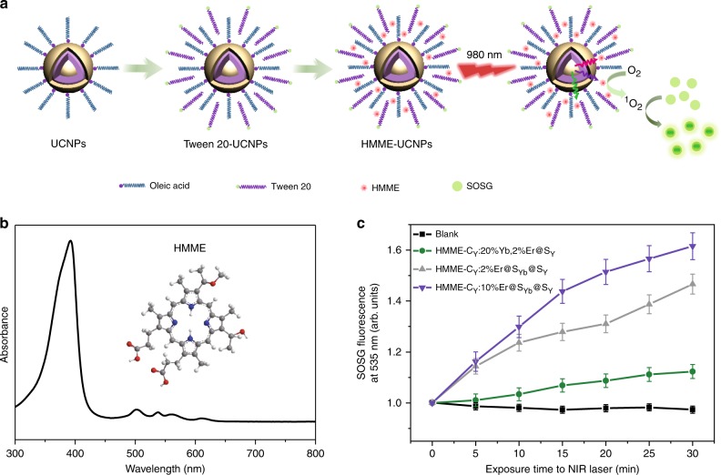
The intensified emission from high-energy levels may offer more opportunities for deep-tissue biophotonics, such as photodynamic therapy and optogenetics. Singlet oxygen (1O2) production activity of hematoporphyrin monomethyl ether (HMME), a commercial photosensitizer with strong absorption around 400 nm, upon exciting the UCNPs was tested here to validate our conjecture (Fig. 5a, b, Supplementary Fig. 33). Due to the weak absorption of HMME in the range of 500–650 nm, the augmentation of singlet oxygen sensor green reagent (SOSG) fluorescence in aqueous dispersion of CY:20% Yb, 2% Er@SY UCNPs is about 12.3% after 980 nm laser irradiation for about 30 min (Fig. 5c and Supplementary Fig. 34). In contrast, the SOSG fluorescence augmentation is about 46.4% and 61.5%, respectively, in aqueous dispersion of CY:2% Er@SYb@SY and CY:10% Er@SYb@SY UCNPs. While the fluorescence of the control sample decreases about 2.8%. As compared with CY:20% Yb, 2% Er@SY UCNPs, neglected enhancement in green and red emissions for CY:2% Er@SYb@SY UCNPs was observed (Fig. 4c); thus, the improved 1O2 production activity for the latter two samples should be attributed to their strong violet emission. The invariable lifetimes (Supplementary Fig. 35) for upconverted Er3+ emission before and after loading HMME exclude Förster resonance energy transfer (FRET) mechanism. In general, only a 1O2 QY of a photosensitizer can be detected by using Rose Bengal (RB) with a high 1O2 QY of 0.76 as a model photosensitizer37. Even though 1O2 QY of our UCNPs cannot be detected since these UCNPs are excited by infrared light, different from 488 nm for RB excitation. These results demonstrate CY:10% Er@SYb@SY UCNPs do emit much stronger violet light and initiate photoreactions more efficiently than the canonical UCNPs, indicating that they are superior to the latter in potential applications such as optogenetics.
Fig. 5. Singlet oxygen production activity upon exciting the UCNPs.
a Schematic diagram for loading hematoporphyrin monomethyl ether (HMME) and producing 1O2 on the UCNPs. b Structural formula and absorption spectrum of HMME. c Comparison of 1O2 production under 980 nm irradiation (24 W cm−2) as determined by the augmentation of singlet oxygen sensor green reagent (SOSG) fluorescence at 535 nm. For CY:20%Yb, 2%Er@SY, dSY = 10.5 ± 0.8 nm. For CY:Er@SYb@SY, dSYb = 8.3 ± 0.7 nm, dSY = 2.3 ± 0.1 nm. The error bars represent the standard deviation of three trials. Source data are provided as a Source Data file.
Discussion
We have demonstrated the critical role of cross-relaxation between sensitizers and activators in concentration quenching in lanthanide-doped UCNPs by designing and synthesizing different structures in which the codopants are distributed differently. We have revealed that spatially separated distribution of sensitizers and activators in neighboring layers can suppress their cross-relaxation and enable highly efficient energy transfer upconversion at the interface. As a result, the UCL from a multilayered structure NaYF4:Er@NaYbF4@NaYF4 is significantly enhanced at relatively high doping levels, especially for multiphoton upconversion emission. Therefore, a high QY of 6.34% is reached in optimum NaYF4:Er@NaYbF4@NaYF4 nanoparticles at a low excitation power density of 4.5 W cm−2. Moreover, the suppression of sensitizer-activator cross-relaxation enables the further enhancement of multiphoton upconversion emission at cryogenic temperatures. This segregated doping strategy provides an easy and efficient way to design and synthesize bright, versatile lanthanide-doped UCNPs to meet the demands of various applications.
Methods
Nanocrystal synthesis
We synthesized the NaYF4:Yb,Er@NaYF4 and NaYF4:Er@NaYbF4@NaYF4 nanoparticles following a procedure reported by Mai et al. with a slight modification24. Detailed experimental procedures are shown in Supplementary Methods.
Nanocrystal characterization
The phase and crystal structure of the samples were characterized by powder X-ray diffractometer (PANalytical X’pert PRO-DY2198). The size, shape and element distribution of the samples were observed by TEM (Jeol JEM 2100F) combined with energy dispersive X-ray spectrum operating at an acceleration voltage of 200 kV. Steady-state PL measurements were performed on an Edinburgh FLS 980 spectrofluorometer in conjunction with a continuous-wave (CW) 980-nm diode laser at an excitation power density of 24.0 W cm−2. Samples for temperature-dependent PL measurements were prepared using a drop-casting method on a quartz glass substrate. An Oxford Optistat DryBL4 cryostat and a Microstat HiRes2 with a temperature controller (MercuryiTC) were used for lowering (3.8–298 K) and mounting temperature (298–508 K), respectively. The samples were held at a certain temperature for at least 10 min to assure equilibration. Time-resolved PL spectra were collected by using an optical parametric oscillator (OPO) as the excitation source (197–2750 nm, 20 Hz repetition rate and ~3 ns pulse width). An excitation power density of 4.5 W cm−2 was used for all the measurements except for power-dependent QY measurements for CY: 10%Er@SYb@SY NPs (dSYb = 8.3 ± 0.7 nm, dSY = 2.3 ± 0.1 nm).
Detection of 1O2 production
The NaYF4:Yb,Er@NaYF4 and NaYF4:Er@NaYbF4@NaYF4 nanoparticles were loaded with HMME after modification by Tween 20, and then suspended in 2 mL of a SOSG aqueous solution. The mixture was injected into a quartz cuvette placed on a magnetic stirring apparatus and the solution was irradiated with an 980 nm laser at 24 W cm−2 for 5 min time intervals beginning from time (t) = 0–30 min, with the fluorescence emission of SOSG (excited by 380 nm) being measured between intervals using FLS980.
Supplementary information
Acknowledgements
The National Natural Science Foundation of China (No. 21673090), the National Key Research and Development Program of “Strategic Advanced Electronic Materials” (2016YFB0401100), Hubei Provincial Natural Science Foundation of China (2019CFA002), and the Fundamental Research Funds for the Central University (2019kfyXMBZ018) supported this work. Here, we thank Prof. X.Y. Chen (Fujian Institute of Research on the Structure of Matter, Chinese Academy of Sciences) for helpful discussion.
Source Data
Author contributions
The scientific concepts, ideas and experimental designs were the result of interactions and discussions between B.Z., Y.M., Z.G., J.Y., T.Z. B.Z. and C.Q. synthesized the nanoparticles and conducted the electron microscopy. B.Z. and B.T. conducted the spectroscopic measurements and the QY measurements. B.T. detected 1O2 production. B.Z., Y.M. and C.Z. wrote the paper, in coordination with all the authors.
Data availability
The data that support the findings of this study are available from the corresponding author upon reasonable request. The source data underlying Figs. 1a, 2f, 3b, d, 4c, d, 5c and Table 1 are provided as a Source Data file.
Competing interests
The authors declare no competing interests.
Footnotes
Peer review information Nature Communications thanks Timothy Thatt Yang Tan and the other, anonymous, reviewer(s) for their contribution to the peer review of this work. Peer reviewer reports are available
Publisher’s note Springer Nature remains neutral with regard to jurisdictional claims in published maps and institutional affiliations.
These authors contributed equally: Bin Zhou, Bing Tang
Supplementary information
Supplementary information is available for this paper at 10.1038/s41467-020-14879-9.
References
- 1.Sivakumar S, Veggel F, Raudsepp M. Bright white light through up-conversion of a single NIR source from sol-gel-derived thin film made with Ln3+-doped LaF3 nanoparticles. J. Am. Chem. Soc. 2005;127:12464–12465. doi: 10.1021/ja052583o. [DOI] [PubMed] [Google Scholar]
- 2.Fernandez-Bravo A, et al. Continuous-wave upconverting nanoparticle microlasers. Nat. Nanotechnol. 2018;13:572–577. doi: 10.1038/s41565-018-0161-8. [DOI] [PubMed] [Google Scholar]
- 3.Wang J, et al. Photon energy upconversion through thermal radiation with the power efficiency reaching 16% Nat. Commun. 2014;5:5669. doi: 10.1038/ncomms6669. [DOI] [PubMed] [Google Scholar]
- 4.Horton NG, et al. In vivo three-photon microscopy of subcortical structures within an intact mouse brain. Nat. Photon. 2013;7:205–209. doi: 10.1038/nphoton.2012.336. [DOI] [PMC free article] [PubMed] [Google Scholar]
- 5.Kachynski AV, et al. Photodynamic therapy by in situ nonlinear photon conversion. Nat. Photon. 2014;8:455–461. doi: 10.1038/nphoton.2014.90. [DOI] [Google Scholar]
- 6.Chen S, et al. Near-infrared deep brain stimulation via upconversion nanoparticle–mediated optogenetics. Science. 2018;359:679–684. doi: 10.1126/science.aaq1144. [DOI] [PubMed] [Google Scholar]
- 7.Liu Y, et al. Amplified stimulated emission in upconversion nanoparticles for super-resolution nanoscopy. Nature. 2017;543:229–233. doi: 10.1038/nature21366. [DOI] [PubMed] [Google Scholar]
- 8.Auzel F. Upconversion and anti-Stokes processes with f and d ions in solids. Chem. Rev. 2004;104:139–173. doi: 10.1021/cr020357g. [DOI] [PubMed] [Google Scholar]
- 9.Shen J, et al. Tunable near infrared to ultraviolet upconversion luminescence enhancement in (α-NaYF4:Yb,Tm)/CaF2 core/shell nanoparticles for in situ real-time recorded biocompatible photoactivation. Small. 2013;9:3213–3217. doi: 10.1002/smll.201370117. [DOI] [PubMed] [Google Scholar]
- 10.Wang J, et al. Enhancing multiphoton upconversion through energy clustering at sublattice level. Nat. Mater. 2014;13:157–162. doi: 10.1038/nmat3804. [DOI] [PubMed] [Google Scholar]
- 11.Wen S, et al. Advances in highly doped upconversion nanoparticles. Nat. Commun. 2014;9:2415. doi: 10.1038/s41467-018-04813-5. [DOI] [PMC free article] [PubMed] [Google Scholar]
- 12.Johnson NJJ, et al. Direct evidence for coupled surface and concentration quenching dynamics in lanthanide-doped nanocrystals. J. Am. Chem. Soc. 2017;139:3275–3282. doi: 10.1021/jacs.7b00223. [DOI] [PubMed] [Google Scholar]
- 13.Shen B, et al. Revisiting the optimized doping ratio in core/shell nanostructured upconversion particles. Nanoscale. 2017;9:1964–1971. doi: 10.1039/C6NR07687D. [DOI] [PubMed] [Google Scholar]
- 14.Ma C, et al. Optimal sensitizer concentration in single upconversion nanocrystals. Nano Lett. 2017;17:2858–2864. doi: 10.1021/acs.nanolett.6b05331. [DOI] [PubMed] [Google Scholar]
- 15.Chen X, et al. Confining energy migration in upconversion nanoparticles towards deep ultraviolet lasing. Nat. Commun. 2016;7:10304. doi: 10.1038/ncomms10304. [DOI] [PMC free article] [PubMed] [Google Scholar]
- 16.Liu Q, et al. Single upconversion nanoparticle imaging at sub-10 W cm−2 irradiance. Nat. Photon. 2018;12:548–553. doi: 10.1038/s41566-018-0217-1. [DOI] [PMC free article] [PubMed] [Google Scholar]
- 17.Tian B, et al. Low irradiance multiphoton imaging with alloyed lanthanide nanocrystals. Nat. Commun. 2018;9:3082. doi: 10.1038/s41467-018-05577-8. [DOI] [PMC free article] [PubMed] [Google Scholar]
- 18.Rabie H, et al. NIR biosensing of neurotransmitters in stem cell-derived neural interface using advanced core–shell upconversion nanoparticles. Adv. Mater. 2019;31:1806991. doi: 10.1002/adma.201806991. [DOI] [PMC free article] [PubMed] [Google Scholar]
- 19.Zhao J, et al. Single-nanocrystal sensitivity achieved by enhanced upconversion luminescence. Nat. Nanotechnol. 2013;8:729–734. doi: 10.1038/nnano.2013.171. [DOI] [PubMed] [Google Scholar]
- 20.Gargas DJ, et al. Engineering bright sub-10-nm upconverting nanocrystals for single-molecule imaging. Nat. Nanotechnol. 2014;9:300–305. doi: 10.1038/nnano.2014.29. [DOI] [PubMed] [Google Scholar]
- 21.Garfield DJ, et al. Enrichment of molecular antenna triplets amplifies upconverting nanoparticle emission. Nat. Photon. 2018;12:402–407. doi: 10.1038/s41566-018-0156-x. [DOI] [Google Scholar]
- 22.Nadort A, Zhao J, Goldys EM. Lanthanide upconversion luminescence at the nanoscale: fundamentals and optical properties. Nanoscale. 2016;8:13099–13130. doi: 10.1039/C5NR08477F. [DOI] [PubMed] [Google Scholar]
- 23.Hossan MY, et al. Explaining the nanoscale effect in the upconversion dynamics of β‑NaYF4:Yb3+, Er3+ core and core-shell nanocrystals. J. Phys. Chem. C. 2017;121:16592–16606. doi: 10.1021/acs.jpcc.7b04567. [DOI] [Google Scholar]
- 24.Mai H, Zhang Y, Sun L, Yan C. Highly efficient multicolor up-conversion emissions and their mechanisms of monodisperse NaYF4:Yb,Er core and core/shell-structured nanocrystals. J. Phys. Chem. C. 2007;111:13721–13729. doi: 10.1021/jp073920d. [DOI] [Google Scholar]
- 25.Zuo J, et al. Precisely tailoring upconversion dynamics via energy migration in core-shell nanostructures. Angew. Chem. Int. Ed. 2018;57:3054–3058. doi: 10.1002/anie.201711606. [DOI] [PMC free article] [PubMed] [Google Scholar]
- 26.Rabouw F, et al. Quenching pathways in NaYF4:Er3+,Yb3+ upconversion nanocrystals. ACS Nano. 2018;12:4812–4823. doi: 10.1021/acsnano.8b01545. [DOI] [PMC free article] [PubMed] [Google Scholar]
- 27.Würth C, et al. Quantum yields, surface quenching, and passivation efficiency for ultrasmall core/shell upconverting nanoparticles. J. Am. Chem. Soc. 2018;140:4922–4928. doi: 10.1021/jacs.8b01458. [DOI] [PubMed] [Google Scholar]
- 28.Würth C, et al. Excitation power dependent population pathways and absolute quantum yields of upconversion nanoparticles in different solvents. Nanoscale. 2017;9:4283–4294. doi: 10.1039/C7NR00092H. [DOI] [PubMed] [Google Scholar]
- 29.Fischer S, Bronstein ND, Swabeck JK, Chan EM, Alivisatos AP. Precise tuning of surface quenching for luminescence enhancement in core-shell lanthanide-doped nanocrystals. Nano Lett. 2016;16:7241–7247. doi: 10.1021/acs.nanolett.6b03683. [DOI] [PubMed] [Google Scholar]
- 30.Dong H, et al. Versatile spectral and lifetime multiplexing nanoplatform with excitation orthogonalized upconversion luminescence. ACS Nano. 2017;11:3289–3297. doi: 10.1021/acsnano.7b00559. [DOI] [PubMed] [Google Scholar]
- 31.Yang Y, Zhu Y, Zhou J, Wang F, Qiu J. Integrated strategy for high luminescence intensity of upconversion nanocrystals. ACS Photonics. 2017;4:1930–1936. doi: 10.1021/acsphotonics.7b00123. [DOI] [Google Scholar]
- 32.Wu K. Temperature dependent upconversion luminescence of Yb/Er codoped NaYF4 nanocrystals. J. Appl. Phys. 2011;110:053510. doi: 10.1063/1.3631822. [DOI] [Google Scholar]
- 33.Yu W, et al. Temperature-dependent upconversion luminescence and dynamics of NaYF4:Yb3+/Er3+ nanocrystals: influence of particle size and crystalline phase. Dalton Trans. 2014;43:6139–6147. doi: 10.1039/C3DT53562B. [DOI] [PubMed] [Google Scholar]
- 34.Wade S, Collins S, Baxter G. Fluorescence intensity ratio technique for optical fiber point temperature sensing. J. Appl. Phys. 2003;94:4743–4756. doi: 10.1063/1.1606526. [DOI] [Google Scholar]
- 35.Xu M, et al. Ratiometric nanothermometer in vivo based on triplet sensitized upconversion. Nat. Commun. 2018;9:2698. doi: 10.1038/s41467-018-05160-1. [DOI] [PMC free article] [PubMed] [Google Scholar]
- 36.Zuo J, et al. Near infrared light sensitive ultraviolet-blue nanophotoswitch for imaging-guided “off-on” therapy. ACS Nano. 2018;12:3217–3225. doi: 10.1021/acsnano.7b07393. [DOI] [PubMed] [Google Scholar]
- 37.Lin H, et al. Feasibility study on quantitative measurements of singlet oxygen generation using singlet oxygen sensor green. J. Fluoresc. 2013;23:41–47. doi: 10.1007/s10895-012-1114-5. [DOI] [PubMed] [Google Scholar]
Associated Data
This section collects any data citations, data availability statements, or supplementary materials included in this article.
Supplementary Materials
Data Availability Statement
The data that support the findings of this study are available from the corresponding author upon reasonable request. The source data underlying Figs. 1a, 2f, 3b, d, 4c, d, 5c and Table 1 are provided as a Source Data file.


