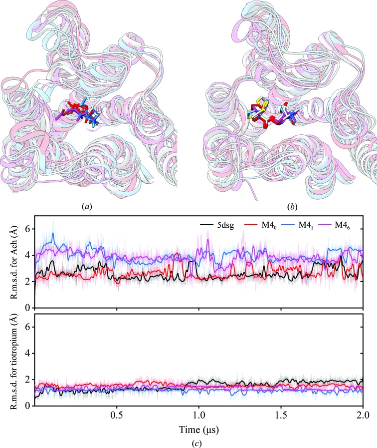
Figure 4.
Molecular-dynamics simulations of different forms [M4–tiotropium (PDB entry 5dsg), M40, M41 and M46] of M4. The last frames from the trajectories of the protein with Ach (a) and tiotropium (b) were aligned to show the locations of the ligands in M46 (purple), M41 (blue), M40 (red) and M4–tiotropium (PDB entry 5dsg; dark colour). (c) R.m.s.d. of the agonist Ach (top) and the antagonist tiotropium (bottom) with respect to the protein and its binding pocket during the simulations. Tiotropium is stable in the M46 and M41 templates when compared with Ach in the binding pocket.

