Abstract
Summary: We present a novel application of a transvascular rheolytic thrombectomy system in the treatment of symptomatic dural sinus thrombosis in a 54-year-old woman with somnolence and left-sided weakness. The diagnosis of bilateral transverse and superior sagittal sinus thrombosis was made and the patient was treated with anticoagulant therapy. After an initial period of improvement, she became comatose and hemiplegic 8 days after presentation. After excluding intracerebral hemorrhage by MR imaging, we performed angiography and transfemoral venous thrombolysis with a hydrodynamic thrombectomy catheter, followed by intrasinus urokinase thrombolytic therapy over the course of 2 days. This technique resulted in dramatic sinus thrombolysis and near total neurologic recovery. Six months after treatment, the patient showed mild cognitive impairment and no focal neurologic deficit. Our preliminary experience suggests that this technique may play a significant role in the endovascular treatment of this potentially devastating disease.
Cerebral venous sinus thrombosis is an unusual and potentially life-threatening condition (1). Standard therapy consists of anticoagulation (2). When this fails, intrasinus transcatheter thrombolytic therapy has proved beneficial in achieving thrombolysis and neurologic improvement. We describe the application of a rheolytic thrombectomy catheter, used in combination with urokinase thrombolysis, in the successful treatment of dural sinus thrombosis.
Patient and Technique
A 54-year-old right-handed woman presented with a 4-day history of progressive lethargy, diffuse myalgias, mild fever, and left-sided weakness. She suffered a generalized seizure and was admitted to an outside hospital. The diagnosis of dural sinus thrombosis involving the superior sagittal sinus, both transverse sinuses, the right sigmoid sinus, and the right jugular bulb was confirmed on MR imaging. The patient was also discovered to have an aspergillus left maxillary sinusitis, and a blood culture was positive for Streptococcus viridans, without sepsis. Anticoagulant therapy with heparin was instituted, and the patient's neurologic condition improved. However, 8 days after admission, she became comatose and hemiplegic on the left, despite adequate anticoagulation. She was transferred to our institution for the purpose of emergent transcatheter venous sinus thrombolysis.
Upon transfer, the patient was afebrile, with a blood pressure of 120/70 and a pulse of 80 bpm. Neurologically, she exhibited spontaneous eye-opening but was unable to track, verbalize, or follow commands. Her fundi were sharp, and pupils were equal at 3-mm, round, and reactive to light. Corneal reflexes were present bilaterally. There was no facial movement, even to painful stimulus. The gag reflex was intact. Spontaneous but purposeless movement of the right upper and lower extremities was observed, and there was a complete absence of movement of the left upper and lower extremities, with flaccid tone. Bilateral Babinski responses were noted.
Initial laboratory data were considered normal except for a partial thromboplastin time of 69 seconds and a random glucose of 320 mg/dL. Repeat MR imaging showed unchanged sinus thrombosis, without parenchymal hemorrhage. Small foci of increased T2 signal intensity were seen in the right parietal and temporal lobes.
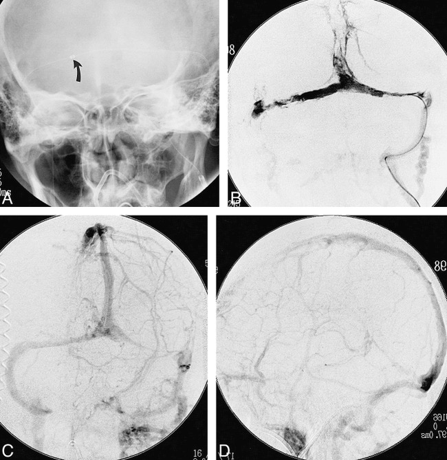
The patient was brought to the neuroangiography suite and was intubated. Arteriography confirmed the presence of occlusion of the superior sagittal, bilateral transverse, and right sigmoid sinuses. After confirming adequate anticoagulation using an activated clotting time, transfemoral venous catheterization of the left jugular bulb was achieved with a 7F guiding catheter. A 0.016-inch–diameter microguidewire was delivered through a standard microcatheter and was navigated through the occluded left transverse sinus, across the torcular and right transverse sinus to the right jugular bulb. The microcatheter was removed and a 5F AngioJet rheolytic thrombectomy catheter (Possis Medical, Minneapolis, MN) was directed over the microguidewire to the mid right transverse sinus (Fig 1A). The AngioJet catheter was activated, and partial sinus thrombolysis was achieved using the hydrodynamic thrombolytic action of the catheter as it was slowly withdrawn to the left jugular bulb. Venography confirmed that partial thrombolysis had been achieved (Fig 1B).
fig 1.

54-year-old woman with dural sinus thrombosis involving the superior sagittal sinus, both transverse sinuses, the right sigmoid sinus, and the right jugular bulb.
A, Plain frontal radiograph shows the AngioJet catheter (navigated through the left internal jugular vein) positioned in the mid right transverse sinus. Arrow indicates catheter tip.
B, Frontal venogram, obtained through the AngioJet catheter after rheolytic thrombectomy, shows partial interval sinus thrombolysis.
C and D, Frontal (C) and lateral (D) venous phases from a left internal carotid angiogram, performed after completion of rheolytic thrombectomy and urokinase thrombolysis, show restored patency of the major dural venous sinuses.
The AngioJet catheter was removed. The microcatheter was repositioned, and bolus deposition of urokinase in the superior sagittal, bilateral transverse, and right sigmoid sinuses was carried out with a total of 750,000 U. The microcatheter was then repositioned into the posterior superior sagittal sinus, and infusion urokinase thrombolysis was carried out for the next 48 hours, to a total of 3,000,000 U.
The AngioJet rheolytic thrombectomy catheter system allows mechanical removal of soft thrombus via percutaneous access. It is currently marketed for use in the treatment of thrombosed dialysis grafts. Additionally, a clinical trial regarding its use in coronary thrombectomy has been completed, and a clinical trial to investigate its use in treating carotid artery occlusion is underway. Anecdotal use of the device in the treatment of pulmonary embolism (3), peripheral arterial thrombosis, and occlusion of a transjugular intrahepatic portosystemic shunt has been described.
The AngioJet rheolytic thrombectomy system permitted accelerated removal of the dural sinus thrombus. The system consists of three component devices: a disposable over-the-wire catheter, a drive unit, and a disposable pump set. The catheter is directed to the site of the thrombus. It is connected to the drive unit via a high-pressure pulsatile pump, which generates the necessary flow of sterile saline to the catheter tip to produce a series of pulsatile high-speed saline jets in this location. This creates a low-pressure zone in the immediate vicinity of the catheter tip. Using the Bernoulli effect, this technique not only dislodges clot but removes it though the catheter and deposits it in a collection bag outside the patient.
The patient became responsive and was able to follow commands on the second day after the initiation of therapy. Repeat angiography on day 3 showed recanalization of the previously occluded dural sinuses (Fig 1C and D). Over the next 5 days, the patient recovered significant left-sided motor function. Heparin infusion was tapered and warfarin therapy was instituted. Six months after her initial presentation, the patient had minimal cognitive deficits and an otherwise normal neurologic examination; she remains on warfarin. A pathogenesis for her sinus thrombosis has not been discovered.
Discussion
Dural sinus thrombosis may present with a range of signs and symptoms, including headache, papilledema, seizure, focal neurologic deficit, coma, and death (1). Diagnosis is complicated by this variable presentation. The natural history without treatment is variable as well, and includes resolution of symptoms with or without restoration of sinus outflow, permanent neurologic deficit, and death from cerebral venous infarction (1, 4). Intravenous heparin therapy remains the treatment of choice for symptomatic dural sinus thrombosis (2), even in the setting of hemorrhagic venous infarction. Transcatheter sinus thrombolysis has become an accepted second-line therapy for patients in whom heparin alone fails (4–13). Most reports describe the use of urokinase as the thrombolytic agent. The goal of thrombolytic therapy is to provide an adequate venous outflow route as quickly as possible, by removing the clot burden within the thrombosed sinuses.
One factor contributing to the technical difficulty of performing transcatheter sinus thrombolysis is the sheer volume of thrombus, which requires elimination in order to reestablish adequate venous drainage. As compared with the clot burden requiring thrombolysis in the emerging procedure of cerebral intraarterial thrombolysis, the volume of thrombus seen in the dural venous sinuses is orders of magnitude greater, given the diameter of most major dural sinuses and the extent of sinus thrombosis seen in symptomatic persons refractory to intravenous heparin therapy. Simple deposition of a thrombolytic agent, using either bolus or infusion technique, even combined with catheter manipulation producing clot maceration, may take hours or days to accomplish. During this time, the brain suffers from impaired venous outflow, venous hypertension, and possibly venous infarction with or without hemorrhagic transformation (4). Therefore, any technique that would eliminate thrombus and restore venous outflow more rapidly would be expected to reduce the risk of venous infarction and subsequent neurologic deficit.
While the AngioJet rheolytic thrombectomy catheter technique may have assisted our patient, several potential limitations are noted. In our case, rheolytic thrombectomy was followed by transcatheter urokinase infusion, both immediately and over the next 2 days. Therefore, while angiographic documentation of partial bilateral transverse sinus thrombolysis was made before urokinase infusion, the isolated effect of the AngioJet system in the patient's overall improvement is difficult to determine. The AngioJet catheter is 5F, tapering to 3.5F proximal to the tip component, and is considerably larger than a standard microcatheter. This creates potential difficulty in navigating the catheter to the site of thrombus within the dural sinuses, as we encountered in our case. Preclinical evaluation of the device (14) suggests that it can result in mild and focal injury to vessels and can produce microscopic particulate debris without significant ischemic effect. Finally, as this is the first report of the use of this device for sinus thrombolysis, an unknown hazard could exist, especially relating to the possible production of local trauma to the dural sinus at the catheter tip. We encountered no such phenomenon.
Conclusion
We have demonstrated the use of a rheolytic thrombectomy catheter system in the successful treatment of a single patient with symptomatic dural sinus thrombosis. While the application of this technique is preliminary, it shows promise in its potential to provide more rapid restoration of venous outflow in a thrombosed dural sinus. Further investigation and experience are needed to define its role.
Acknowledgments
We thank the following people for their invaluable assistance in preparing this manuscript: Van V. Halbach, Randall T. Higashida, David C. Bonovich, S. Claiborne Johnston, Wade S. Smith, and Daryl R. Gress.
Footnotes
Address reprint requests to Christopher F. Dowd, MD, Neurovascular Medical Group, Department of Radiology, University of California San Francisco, 505 Parnassus Ave, Room L-352, San Francisco, CA 94143.
References
- 1.Bousser MG, Chiras J, Bories J, et al. Cerebral venous thrombosis: a review of 38 cases. Stroke 1985;16:1919-213 [DOI] [PubMed] [Google Scholar]
- 2.Eihhaupl KM, Villringer A, Meister W, et al. Heparin treatment in venous sinus thrombosis. Lancet 1991;338:597-600 [DOI] [PubMed] [Google Scholar]
- 3.Koning R, Cribier A, Gerber L, et al. A new treatment for severe pulmonary embolism: percutaneous rheolytic thrombectomy. Circulation 1997;96:2498-2500 [DOI] [PubMed] [Google Scholar]
- 4.Higashida RT, Halbach VV, Barnwell SL, Dowd CF, Hieshima GB. Thrombolytic therapy in acute stroke. J Endovasc Surg 1994;1:4-15 [DOI] [PubMed] [Google Scholar]
- 5.Higashida RT, Helmer E, Halbach VV, Hieshima GB. Direct thrombolytic therapy for superior sagittal sinus thrombosis. AJNR Am J Neuroradiol 1989;10:S4-S6 [PMC free article] [PubMed] [Google Scholar]
- 6.Barnwell SL, Higashida RT, Halbach VV, Dowd CF, Hieshima GB. Direct endovascular thrombolytic therapy for dural sinus thrombosis. Neurosurgery 1991;28:135-142 [DOI] [PubMed] [Google Scholar]
- 7.Tsai FY, Higashida RT, Matovich V, et al. Acute thrombolysis of the intracranial dural sinus: direct thrombolytic treatment. AJNR Am J Neuroradiol 1992;12:1137-1142 [PMC free article] [PubMed] [Google Scholar]
- 8.Gobin YP, Houdart E, Rogopoulos A, Casasco A, Bailly AL, Merland JJ. Percutaneous transvenous embolization through the thrombosed sinus in transverse sinus dural fistula. AJNR Am J Neuroradiol 1993;14:1102-1105 [PMC free article] [PubMed] [Google Scholar]
- 9.Smith TP, Higashida RT, Barnwell SL, et al. Treatment of dural sinus thrombosis by urokinase infusion. AJNR Am J Neuroradiol 1994;15:801-807 [PMC free article] [PubMed] [Google Scholar]
- 10.Higashida RT, Halbach VV, Tsai FY, Dowd CF, Hieshima GB. Interventional neurovascular techniques for cerebral revascularization in the treatment of stroke. AJR Am J Roentgenol 1994;163:793-800 [DOI] [PubMed] [Google Scholar]
- 11.Horowitz M, Purdy P, Unwin H, et al. Treatment of dural sinus thrombosis using selective catheterization and urokinase. Ann Neurol 1995;38:58-67 [DOI] [PubMed] [Google Scholar]
- 12.Rael JR, Orrison WW Jr, Baldwin N, Sell J. Direct thrombolysis of superior sagittal sinus thrombosis with coexisting intracranial hemorrhage. AJNR Am J Neuroradiol 1997;18:1238-1242 [PMC free article] [PubMed] [Google Scholar]
- 13.Gerszten PC, Welch WC, Spearman MP, Jungreis CA, Redner RL. Isolated deep cerebral venous thrombosis treated by direct endovascular thrombolysis. Surg Neurol 1997;48:261-266 [DOI] [PubMed] [Google Scholar]
- 14.Sharafuddin MJA, Hicks ME, Jenson ML, Morris JE, Drasler WJ, Wilson GJ. Rheolytic thrombectomy with use of the Angiojet-F105 catheter: preclinical evaluation of safety. J Vasc Interv Radiol 1997;8:939-945 [DOI] [PubMed] [Google Scholar]


