Abstract
Summary: Thrombosis of the dural venous sinuses is a potentially lethal condition that remains a diagnostic dilemma. Clinical outcome is typically dependent on the timeliness of diagnosis and definitive treatment. We report a case of successful rapid thrombectomy of extensive thrombus within the superior sagittal and transverse sinuses using a rheolytic catheter device. This appears to be a promising treatment option, particularly in those patients who do not respond to other, more established, forms of therapy.
Cerebral venous thrombosis is recognized as a potentially life-threatening condition. It is associated with considerable morbidity, particularly if not recognized and treated promptly. Numerous reports have addressed both systemic and direct treatment regimens, which typically entail a combination of urokinase and systemic anticoagulation (1, 2). We describe a successful case of thrombectomy of acute thrombosis of the superior sagittal and transverse dural venous sinuses using a rheolytic catheter system. To our knowledge, no previous reports have documented the use of such a device for this application.
Case Report
A 19-year-old woman had a 2-day history of nausea, vomiting, and headache. Pertinent medical history included use of oral contraceptives for 1 month; in addition, she had recently learned of a “clotting disorder” in her father that was complicated by multiple thrombotic events and subsequently characterized as being related to anticardiolipin A antibodies. A cranial CT examination was performed and she was transferred to our hospital. Interpretation of the CT study revealed high-density material in the superior sagittal sinus, suggestive of thrombosis.
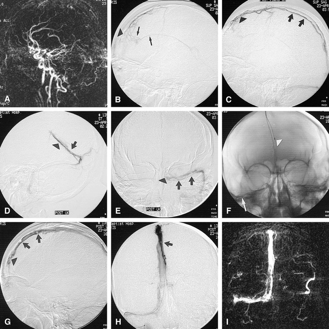
An emergent MR study, performed in conjunction with MR venography upon admission, confirmed the presence of abnormally high T1-weighted signal within the superior sagittal, straight, and bilateral transverse sinuses, indicative of thrombus (Fig 1A). Diffusion-weighted imaging revealed a large area of abnormality throughout the right frontal lobe. Given these findings, she was treated with heparin. Neurologic evaluation on admission documented a Glasgow Coma Scale score of 15, dysarthric but fluent speech, left homonymous hemianopsia, central left cranial nerve VII deficit, with 0/5 motor strength of the left upper extremity, and 4/5 strength of the left lower extremity. Her mental status was fluctuating and she became lethargic before her first treatment.
fig 1.

19-year-old woman with nausea, vomiting, headache, and deteriorating mental status.
A, Initial phase-contrast MR venogram with a velocity-encoding value (VENC) of 20 cm/s shows extensive thrombosis of the superior sagittal sinus, the bilateral transverse sinuses, and the straight sinus.
B, Initial lateral superior sagittal sinus venogram reveals extensive thrombus within the superior sagittal sinus and occlusion of the bilateral transverse sinuses. Flow from the occluded superior sagittal sinus is diverted via transmedullary veins to the inferior sagittal sinus (arrows). Arrowhead indicates microcatheter tip.
C, Lateral venogram of the superior sagittal sinus after direct urokinase treatment shows restored anterograde flow but with lengthy tubular filling defects, consistent with residual thrombus (arrows). Arrowhead indicates microcatheter tip.
D and E, Lateral venogram of the straight sinus (D) and anteroposterior image of the left transverse and sigmoid sinuses (E) after direct endovascular urokinase treatment show restoration of anterograde flow but with residual thrombus in all areas (arrows). Arrowheads indicate microcatheter tip.
F, Anteroposterior image of the AngioJet LF140 rheolytic catheter and 0.014-inch guidewire positioned within the superior sagittal sinus via a right sigmoid and transverse sinus approach. Note the tip of the AngioJet catheter device (arrowhead). The relatively straight course of the right sigmoid sinus probably facilitated catheter manipulation and positioning (arrow).
G and H, Postthrombectomy lateral (G) and anteroposterior (H) venograms obtained via a microcatheter (arrowhead) show marked improvement in the appearance of the superior sagittal sinus with minimum residual thrombus (arrows). Rheolytic thrombectomy was subsequently performed in the bilateral transverse sinuses.
I, Posttreatment MR venogram in the coronal projection with a VENC of 20 cm/s documents flow signal throughout much of the superior sagittal and the dominant right transverse sinus.
Emergency angiographic assessment performed via the right common femoral artery and left common femoral vein showed near complete thrombosis of the superior sagittal sinus and bilateral transverse sinuses and partial thrombosis of the straight sinus (Fig 1B). A 7F introducer catheter was advanced via a left common femoral vein sheath and positioned with its distal tip in the right jugular bulb. A Turbo Tracker 18 microcatheter (Target Therapeutics, Fremont, CA) was advanced over a Transend EX guidewire (Target Therapeutics) through the right transverse sinus and positioned with its tip at the distal straight sinus. In this position and in multiple other sites in the superior sagittal, transverse, and straight sinuses, a total of 750,000 U of urokinase were infused over 90 minutes. Hand injection evaluation of the dural sinuses revealed dissolution of the majority of the thrombus but with partial residual thrombus in many areas (Fig 1C–E). The patient's neurologic status was difficult to assess after treatment owing to the level of sedation used to facilitate the procedure. A CT study performed after the procedure showed no evidence of complicating hemorrhage. The patient was maintained on heparin.
After a brief period of improvement, the patient's neurologic status progressively declined overnight. She was agitated, aphasic, and arousable only to painful stimuli. She traumatically and inadvertently removed her right common femoral artery sheath. Her left upper extremity remained paralytic. She became febrile with a temperature of 101.7°F. Partial thromboplastin time levels remained below the desired range despite periodic boluses of heparin and continuous drip infusion.
The patient was returned for repeat angiographic assessment and treatment. Transvenous catheterization of the dural sinuses with a microcatheter confirmed virtually complete rethrombosis since the first endovascular treatment. Because of her markedly worsening clinical status and these findings, a decision was made to pursue thrombectomy. With the patient under general anesthesia, a rheolytic thrombectomy catheter (AngioJet LF140; Possis Medical, Minneapolis, MN) was advanced over a 300-cm, 0.014-inch ACS Hi-Torque wire (Advanced Cardiovascular Systems, Temecula, CA). Buckling of the introducer catheter within the right side of the heart necessitated the introduction of a long 7F sheath (Cook Inc, Bloomington, IN) through which a 7F Brite Tip catheter (Cordis Corp, Miami, Fl) was advanced. The thrombectomy catheter was then reintroduced coaxially and used to treat the entire length of the superior sagittal sinus as well as the bilateral transverse sinuses (Fig 1F). This resulted in a markedly improved angiographic appearance of the superior sagittal sinus with minimal residual thrombus (Fig 1G and H). The right transverse sinus showed slow anterograde flow. Infusion of 450,000 U of urokinase was then administered via a microcatheter throughout the sinus system. The resulting venous images were quite satisfactory. A posttreatment CT scan showed no evidence of complicating hemorrhage.
The patient's neurologic status improved rapidly during the ensuing 3 days. She was alert and oriented and interacted normally with family members. Weakness of the left lower extremity and facial droop resolved. Full range of motion was present in the left upper extremity, with 4/5 grip strength of the left hand. A follow-up MR venogram obtained 5 days after rheolytic thrombectomy revealed restoration of flow within the superior sagittal sinus and right transverse sinus (Fig 1I). The postprocedural course was complicated by a pseudoaneurysm at the right groin puncture site (subsequently closed with the use of graded-compression sonography) and two chest wall hematomas related to central line placement. One week after thrombectomy, the patient was ambulatory and had completely resolved her motor deficits apart from minimally reduced strength of the left arm.
Results of a thrombophilia screen conducted in our institution documented normal levels of plasminogen activity, protein C activity, free protein S antigen, factor V Leiden, heparin cofactor, and antithrombin III. The patient was converted to daily doses of 2.5 mg warfarin as well as 100 mg phenytoin, and she was discharged on the ninth hospital day. A 6-month follow-up examination showed no evidence of either neurologic or cognitive deficits.
Discussion
Since the original autopsy description of dural venous thrombosis by Ribe in 1825, this entity has remained a quandary to clinicians with regard to timely diagnosis and effective treatment strategies. Clinical presentation is highly variable, depending on both the location and the acuity of thromboses. In their description of 110 patients with cerebral venous thrombosis, Ameri and Bousser (3) found a wide spectrum of physical signs and symptoms, including headache, papilledema, motor or sensory deficits, drowsiness, mental status changes, seizures, confusion, coma, dysphasia, and cranial nerve dysfunction. The diagnostic dilemma is heightened by the relative rarity of this condition as compared with other disorders that manifest many of the above-described findings. Onset of symptoms may be acute, subacute, or chronic. Simulation of benign intracranial hypertension, neoplasm, cerebrovascular accident, encephalitis, or abscess is well recognized (4). A diverse array of etiologic factors has been implicated in cerebral venous thrombosis, including pregnancy, use of oral contraceptives, direct septic trauma, local or disseminated intracranial infection, malignancies, neurosurgical operations, cerebral infarctions and hemorrhages, severe dehydration, systemic lupus erythematosus, and Behçet disease (3). Coagulation disorders have recently received attention in relation to this disorder, specifically deficiencies of antithrombin III, protein C, protein S, and factor V Leiden, plus increased anticardiolipin A antibodies (5). A precise pathogenesis cannot be ascertained in at least 20% to 35% of cases (5). Our patient had begun the use of oral contraceptives 1 month before symptom onset, and possibly this was the origin of her thrombotic event.
The advent of more routinely available CT and MR imaging, supported by their angiographic applications, has significantly improved the likelihood of earlier detection. Conventional catheter angiography has become a confirmatory technique and a platform from which to direct interventional therapy. Despite the prevalence of highly developed diagnostic assets, dural venous thrombosis remains a potentially devastating entity with mortality of approximately 10% in noninfectious forms (4) and permanent neurologic deficits ranging from 15% to 25% (6). Clinical outcome is quite variable but in part is due to the location of thrombus, rapidity of onset, preexisting medical condition of the patient, and timeliness of treatment.
Treatment of cerebral venous thrombosis has been the topic of many recent articles (1, 2). Systemic anticoagulation with heparin has been a mainstay of treatment, although more recently, the direct endovascular administration of thrombolytic agents has gained wider acceptance. Successful endovascular thrombolysis of cerebral venous sinuses has been described in a number of cases in which patients experienced a rapid and precipitous worsening in their clinical status (7). Given these early results, considerable effort has been directed toward achieving rapid endovascular thrombus dissolution as a means to avert the potentially disastrous outcomes associated with cerebral edema and venous congestion, infarction, and herniation.
Controversy exists with regard to optimal administration of endovascular thrombolytics. Some authors (8) have advocated slow continuous drip infusion over prolonged periods, whereas in the experience of others, a rapid high-dose regimen using up to 1 million units of urokinase over 2 to 3 hours has proved efficacious (P.P.M. unpublished data). The latter approach seems favorable in many cases, because the position of the catheter tip can be monitored, thus lessening the likelihood that the infused thrombolytic agent will preferentially disseminate into transmedullary and cortical veins. In addition, when treating a patient displaying signs and symptoms of precipitous clinical decline, such as in the case described here, it seems logical to pursue a strategy that would safely deliver the greatest quantity of a thrombolytic agent into the site of thrombosis as quickly as possible. We pursued this course initially with our patient; however, thrombus quickly reestablished itself, necessitating a more aggressive approach for her second treatment.
A number of recent articles have detailed the technical aspects of rheolytic catheter systems and their modes of operation (9, 10). Succinctly, the AngioJet LF140 device consists of a 140-cm-long double-lumen 5F catheter passed over a 0.018- or 0.014-inch guidewire. The smaller lumen is the source of high-velocity saline jets delivered to the catheter tip via an external pumping unit that generates pressures up to 10,000 psi. The catheter system has no moving parts that could impinge upon the vessel wall. A 1000-mL bag of saline containing 5000 U of heparin serves as the reservoir for the fluid jets. Saline exits the catheter tip at a rate of 50 mL/min via six small jets directed in a retrograde direction. A net negative pressure gradient is developed, which creates a Venturi effect that serves to entrain and break up thrombus and direct particulate debris into an effluent lumen for collection into a disposable bag. The system's pumping mechanism is modulated to ensure isovolumic operation. The system is activated via a foot pedal. In extracranial applications, the catheter is advanced through areas of thrombus at a rate of 0.5 to 1.0 cm/s. We adhered to this guideline during our procedure. An endpoint to the operation of this rheolytic catheter system is reached either with a satisfactory angiographic result or, alternatively, when either the 1000-mL saline supply bag has been exhausted or at the conclusion of 15 minutes of cumulative activation time. The latter restrictions presumably lessen the likelihood of an unacceptable level of hemolysis. We elected to discontinue the use of the rheolytic catheter when there was angiographic evidence of both restoration of anterograde flow within the affected sinuses and near total dissolution of thrombus. This was well before 15 minutes of operating time had been reached.
Interest in rapid endovascular thrombectomy systems is fueled by anticipated benefits. One such benefit is being able to remove the thrombus while reestablishing vessel patency in a significantly more rapid manner than might be achieved by direct endovascular administration of thrombolytic agents. We believe that this was the case in our patient. An additional benefit is the avoidance of thrombolytic drugs, which predictably increase the morbidity associated with endovascular procedures. Currently, rheolytic catheter systems have gained early clinical acceptance in the treatment of acute thromboembolic occlusion of native lower extremity arteries and bypass grafts and hemodialysis access shunts, as well as in the treatment of severe pulmonary embolisms (9, 11). Clinical trials at several centers are examining the utility of the device in the treatment of thrombus within coronary arteries and lysis of thrombus within the carotid arteries in the setting of acute stroke. Currently, efforts are underway to examine the feasibility of using a 3F AngioJet device for rapid thrombectomy within intracranial arteries (12).
Conclusion
We anticipate that rapid thrombolysis of the dural venous sinuses using a rheolytic catheter system will become a more accepted form of therapy, particularly in those cases in which clinical status is worsening. Some of the safety issues that need to be considered for future intracranial arterial and venous applications include any tendency to elicit untoward endothelial damage to treated vessels, distal embolization of fragmented thrombogenic debris, blood loss, and hemolysis. The results of early studies involving extracranial sites indicate a favorable operating profile with regard to these potential concerns (9, 13).
Footnotes
Address reprint requests to Michael J. Opatowsky, MD, Department of Radiology, Wake Forest University School of Medicine, Medical Center Blvd, Winston-Salem, NC 27106
References
- 1.Rael JR, Orrison WW Jr, Baldwin N, Sell J. Direct thrombolysis of superior sagittal sinus thrombosis with coexisting intracranial hemorrhage. AJNR Am J Neuroradiol 1997;18:1238-1242 [PMC free article] [PubMed] [Google Scholar]
- 2.Spearman MP, Jungreis CA, Wehner JJ, Gerszten PC, Welch WC. Endovascular thrombolysis in deep cerebral venous thrombosis. AJNR Am J Neuroradiol 1997;18:502-506 [PMC free article] [PubMed] [Google Scholar]
- 3.Ameri A, Bousser MG. Cerebral venous thrombosis. In: Barnett HJM, Hachinsk VC, eds. Neurologic Clinics. Philadelphia: Saunders; 1992;87-111 [PubMed]
- 4.Ameri A, Bousser MG. Cerebral venous thrombosis: clinical diagnosis. Ann Radiol 1994;37:101-107 [PubMed] [Google Scholar]
- 5.Deschiens MA, Conard J, Horellou MH. et al. Coagulation studies, factor V Leiden, and anticardiolipin antibodies in 40 cases of cerebral venous thrombosis. Stroke 1996;27:1719-1720 [DOI] [PubMed] [Google Scholar]
- 6.Preter M, Tzourio C, Ameri A, Bousser MG. Long-term prognosis in cerebral venous thrombosis: follow-up in 77 patients. Stroke 1996;27:243-246 [DOI] [PubMed] [Google Scholar]
- 7.Holder CA, Bell DA, Lundell AL, Ulmer JL, Glazier SS. Isolated straight sinus and deep venous thrombosis: successful treatment with local infusion of urokinase. J Neurosurg 1997;86:704-707 [DOI] [PubMed] [Google Scholar]
- 8.Smith TP, Higashida RT, Barnwell SL, et al. Treatment of dural sinus thrombosis by urokinase infusion. AJNR Am J Neuroradiol 1994;15:801-807 [PMC free article] [PubMed] [Google Scholar]
- 9.Wagner HJ, Stefan MH, Pitton MB, Weiss W, Wess M. Rapid thrombectomy with a hydrodynamic catheter: results from a prospective, multicenter trial. Radiology 1997;205:675-681 [DOI] [PubMed] [Google Scholar]
- 10.Sharafuddin MJA, Hicks ME. Current status of percutaneous mechanical thrombectomy, I: general principles. J Vasc Interv Radiol 1997;8:911-921 [DOI] [PubMed] [Google Scholar]
- 11.Koning R, Cribier A, Gerber L, et al. A new treatment for severe pulmonary embolism. Circulation 1997;96:2498-2500 [DOI] [PubMed] [Google Scholar]
- 12.Perl J, Morris JM, Setum CM, Le HV. Preclinical evaluation of a prototype 3Fr rheolytic thrombectomy catheter for treatment of cerebrovascular thrombosis. Presented at the annual meeting of the American Society of Neuroradiology, Philadelphia, May 1998
- 13.Sharafuddin MJA, Hicks ME, Jenson ML, Morris JE, Drasler WJ, Wilson GJ. Rheolytic thrombectomy with use of the AngioJet-F105 catheter: preclinical evaluation of safety. J Vasc Interv Radiol 1997;8:939-945 [DOI] [PubMed] [Google Scholar]


