Abstract
Purpose
To analyze the expression of 440 human cytokines in aqueous humor of high myopic patients with cataracts.
Methods
Eighty-five patients with cataracts were recruited in this study. In the screening stage, the RayBio G-Series Human Cytokine Antibody Array 440 was used to assay the aqueous humor samples collected from nine high myopic patients with cataracts and eight non-myopic patients with cataracts right before the surgery. The array was further used for verification of the screened cytokines, with aqueous humor samples obtained from 34 eyes of high myopic patients with cataracts and 34 eyes of non-myopic patients with cataracts.
Results
Compared with the non-myopic patients with cataracts, the expression levels of decorin, receptor activator of NF-kB (RANK), angiopoietin-1 (ANG-1), C-X-C motif ligand 16 (CXCL16), β-inducible gene-h3 (bIG-H3), insulin-like growth factor-binding protein 2 (IGFBP-2), and interleukin-17B (IL-17B) were statistically significantly higher in high myopic patients with cataracts (all p<0.000114). The matrix metalloproteinase-2 (MMP-2) level also increased in the aqueous humor of high myopic patients with cataracts (p = 0.0034). The concentrations of ANG-1 and MMP-2 were also increased in the aqueous humor of the confirmatory stage (all p<0.05).
Conclusions
In this study, numerous cytokines in aqueous humor were detected in high myopic patients with cataracts and non-myopic patients with cataracts, and it was confirmed that the MMP-2 level in the aqueous humor of patients with high myopia was statistically significantly increased. Further verification also revealed the elevation of ANG-1 in the aqueous humor of high myopic patients with cataracts, which suggests that ANG-1 may be related to the pathogenesis of high myopia.
Introduction
The prevalence of myopia has rapidly increased in recent years, becoming a significant global public health issue. Worldwide, 153 million people age 5 and older are affected by visual defects, and about 8 million of these individuals have blindness caused by uncorrected myopia and other refractive errors [1].
High myopia, with a refractive error greater than −6.00 diopters or an axial length greater than 26 mm, is more common in Asian populations [2,3]. There is substantial evidence that high myopia leads to a much higher risk of developing ocular diseases, such as retinal detachment, macular degeneration, and glaucoma, which would seriously impair the vision of patients [4,5]. However, the pathogenesis of high myopia remains unclear. Recently, studies have assessed the aqueous humor in high myopic patients [6,7]. Protein levels in aqueous humor change in several ocular disorders, such as glaucoma and age-related macular degeneration (AMD) [8,9].
High myopia is regarded as an inflammation-related disease [10]. In addition, acute onset myopia is considered a characteristic manifestation of systemic lupus erythematosus [11]. Studies have provided experimental and clinical evidence to support the relationship between inflammation and myopia progression [12]. Previous studies have shown the expression of some inflammatory cytokines in the aqueous humor of high myopic patients [6]. Previously, due to the small sample volume of aqueous humor, only dozens of inflammatory cytokines could be detected at the same time [13]. However, with the latest protein cytokine chip technology, researchers can now detect a large number of cytokines with a small sample volume [14]. Thus, 440 cytokines in the aqueous humor of high myopic patients with cataracts were detected in this study.
Methods
Subjects
This study recruited 43 high myopic patients with cataracts and 42 non-myopic patients with cataracts in the Beijing Tongren Hospital, China, from June 2017 to November 2019. All patients included 58 females and 27 males, with an average age of 63.60 ± 10.16 years old (mean ± SD). All patients were relatively healthy without systemic diseases, such as immune diseases, kidney diseases, diabetes. The Institutional Review Board of Beijing Tongren Hospital approved this case-control study. The protocol (TRECKY2017–018) was approved by the Ethics Committee of Beijing Tongren Hospital. This study was registered (clinical trial accession number: ChiCTR1800014657). Written informed consent was obtained from all patients before participation. All procedures adhered to the Declaration of Helsinki and the ARVO statement on human subjects, and were conducted in accordance with the approved research protocol.
The following inclusion criteria were used: patients with age-related cataracts, older than 40 years old, nuclear and cortical cataract, severity of cataract above grade 3 who need cataract surgery, posterior subcapsular patients with cataracts with best-corrected visual acuity (BCVA) lower than 0.3, severe high myopia cataract with axial length greater than or equal to 26 mm and greater than −6 D, and non-high myopia cataract axial length at 21–25 mm and diopter is less than −6 D. Eyes with glaucoma, uveitis, previous trauma, fundus diseases, or diabetes-related eye diseases were excluded from the study by clinical examination and clinical history. The axial length and diopter of high myopic patients with cataracts were 30.41±1.956mm and -18.14±5.716D. The axial length and diopter of non-high myopic patients with cataracts were 23.05±0.891mm and -0.13±1.654D.
Examination
All patients underwent slit-lamp microscopy (Haag Streit, Berne, Switzerland) to determine the type and severity of cataract. At the same time, a non-mydriatic fundus camera (CR-DGI camera; Canon, Tokyo, Japan) was used to check the fundus and optical coherence tomography (OCT; Optovue, Fremont, CA) to check the patients’ macular condition. The refractive test was performed by using a computer optometer (Topcon, Tokyo, Japan) for preliminary detection, and then by a professional optometrist for shadow optometry to determine the final refractive power. Axial length was measured with IOLmaster (Carl Zeiss, Jena, Germany).
Aqueous humor collection
All cataract surgeries were performed by the same surgeon (WXH). Eyelids and surrounding skin were wiped with disinfectant. Samples of aqueous humor (100–200 μl) were aspirated through corneal paracentesis, by inserting a 26-gauge needle into the anterior chamber just before the surgery. Samples were immediately stored at −80 °C until used for proteomic analysis.
G-series human cytokine antibody array 440
In this study, the G-Series Human Cytokine Antibody Array 440 (product GSH-CAA-440; RayBio; RayBiotech, Norcross, GA) was used for the detection of the cytokines in aqueous humor of high myopic patients with cataracts and non-myopic patients with cataracts in the screening stage.
Four hundred forty cytokines were simultaneously analyzed with an antibody-based protein array. The test was performed following the manufacturer’s instructions. Briefly, the cytokine antibody array follows the sandwich immunoassay principle [14]. A unique set of antibodies was immobilized in specific location spots on the surface of a membrane. Array membranes were incubated with samples of aqueous humor from nine high myopic patients with cataracts (Group 1) and eight non-myopic patients with cataracts (Group 2). As the volume of aqueous humor in one eye cannot meet the requirement for cytokine chip measurement, the aqueous humors in two eyes of the same person were mixed for measurement in this study. Antibodies captured the corresponding cytokines, and then a cocktail of biotinylated antibodies was used to detect bound cytokines. In this study, fluorescent dye-conjugated streptavidin (Cy3 equivalent) was used for the visualization of the signals, and the InnoScan 300 Microarray Scanner (Innopsys, Carbonne, France) was used for the detection of signals. Densitometric analysis of each spot was then performed using a Q-Analyzer program.
Quantibody custom array
To reduce errors and improve accuracy, a quantitative protein detection chip RayBio Quantibody Custom Array (product QAH-CUST; RayBio; RayBiotech, Norcross, GA) was used for the verification of the cytokines of interest in the confirmatory stage. Concentrations of the 11 screened cytokines in the aqueous humor sample obtained from 34 eyes of high myopic patients with cataracts (Group 3) and 34 eyes of non-myopic patients with cataracts (Group 4) were determined using a human 11-plex assay. Antibodies captured corresponding cytokines, and then a cocktail of biotinylated antibodies was used to detect bound cytokines. The test method for the signals was same as that for the G-Series Human Cytokine Antibody Array 440.
Statistical analysis
All data are shown as mean ± standard deviation. A chi-square test was used to examine differences in categorical variables. One-way analysis of variance (ANOVA) followed by Tukey’s test was used to detect statistically significant differences in quantitative variables. Student t test was used to detect differences between two groups. A p value of less than 0.05 was considered statistically significant. As there were 440 different comparisons, p = 0.000114 (i.e., p = 0.05 / 440) was considered statistically significant after Bonferroni correction. All analyses were performed using SPSS software version 20.0 (SPSS, Inc., IBM, New York, NY).
Results
Demographic data
Demographic characteristics of patients are presented in Table 1. There were no statistical differences among the four groups in age (F = 1.932, p = 0.131), gender (p = 0.397), and eye(s) (p = 0.804; all p>0.05, ANOVA and Tukey’s test for age; chi-square tests for gender and eye(s)).
Table 1. Demographic data for all participants.
| Demographic data | G-Series Human Cytokine Antibody Array 440 |
Quantibody Custom Array |
||
|---|---|---|---|---|
| High myopic patients with cataracts | Non-myopic patients with cataracts | High myopic patients with cataracts | Non-myopic patients with cataracts | |
| No. of patients |
9 |
8 |
34 |
34 |
| Age (y) |
59.56±9.13 |
59.75±7.09 |
62.65+12.59 |
66.50±7.57 |
| Gender (No. of males/females) |
4/5 |
4/4 |
11/23 |
8/26 |
| Eye (No. of operated eyes, right eye/left eye) | - | - | 21/13 | 20/14 |
G-Series Human Cytokine Antibody Array 440
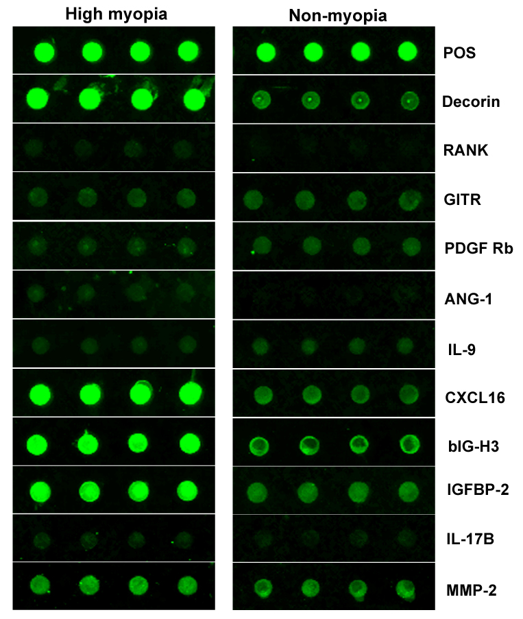
In the test group, the G-Series Human Cytokine Antibody Array 440 was used to determine the expression levels of 440 cytokines in the aqueous humor of high myopic patients with cataracts and non-myopic patients with cataracts. Marked changes in the aqueous humor cytokines were found in the high myopic patients with cataracts compared with the non-myopic patients with cataracts (Appendix 1). After Bonferroni correction (p<0.000114, Student t test with Bonferroni correction), the expression levels of decorin, RANK, ANG-1, CXCL16, bIG-H3, IGFBP-2, and IL-17B in the aqueous humor of the high myopic patients with cataracts were much higher than those in the aqueous humor of the non-myopic patients with cataracts, and the expression levels of GITR, PDGF Rb, and IL-9 were much lower in the aqueous humor of the high myopic patients with cataracts compared with the aqueous humor of the non-myopic patients with cataracts (Table 2). MMP-2 has been shown in previous studies to be associated with high myopia. MMP-2 is elevated in high myopic eyes and can be used as a positive control. Although the p value for MMP-2 was not lower than 0.000114 after Bonferroni correction in this study, the expression of MMP-2 was still increased in the aqueous humor of the high myopic eyes, and there were differences between the two groups. The differences in the fluorescence of these cytokines in the aqueous humor are shown in Figure 1.
Table 2. The expression levels of cytokines marked changes in high myopic patients with cataracts and non-myopic patients with cataracts by G-Series Human Cytokine Antibody Array 440.
| Cytokine | High myopic patients with cataracts (pg/ml) | Non-myopic patients with cataracts (pg/ml) | P value |
|---|---|---|---|
| Decorin |
191,681.41±53365.47 |
41,345.51±39826.20 |
0.00001 |
| RANK |
479.47±92.90 |
201.05±25.96 |
0.00001 |
| GITR |
1989.76±171.27 |
2750.21±272.33 |
0.00002 |
| PDGF Rb |
1632.61±108.24 |
2464.95±279.85 |
0.00003 |
| ANG-1 |
629.33±164.74 |
235.35±95.89 |
0.00004 |
| IL-9 |
1074.26±108.43 |
1342.10±89.40 |
0.00005 |
| CXCL16 |
37,543.94±11339.47 |
10,018.85±9306.51 |
0.00006 |
| bIG-H3 |
31,810.77±8902.45 |
11,394.26±3258.92 |
0.00007 |
| IGFBP-2 |
20,372.86±6343.50 |
6585.80±3547.93 |
0.00009 |
| IL-17B |
625.74±89.12 |
432.29±54.96 |
0.0001 |
| MMP-2 | 9138.34±1160.27 | 7312.61±1010.70 | 0.0034 |
Figure 1.
The differences in the fluorescence of the cytokines with statistical significance in the G-Series Human Cytokine Antibody Array 440. The higher the fluorescent brightness, the higher the concentration of the cytokine in the aqueous humor.
Quantibody custom array
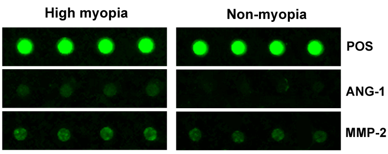
To further validate the results of the G-Series Human Cytokine Antibody Array 440, the Quantibody Custom Array was performed using a human 11-plex assay (including decorin, RANK, ANG-1, CXCL16, bIG-H3, IGFBP-2, IL-17B, GITR, PDGF Rb, IL-9, and MMP-2). Table 3 shows that the expression levels of ANG-1 and MMP-2 had a statistically significant difference in two groups (all p<0.05). The differences in the fluorescence of ANG-1 and MMP-2 are shown in Figure 2.
Table 3. The expression levels of cytokines marked changes in high myopic patients with cataracts and non-myopic patients with cataracts by Quantibody Custom Array.
| Cytokine | High myopic patients with cataracts (pg/ml) | Non-myopic patients with cataracts (pg/ml) | P Value |
|---|---|---|---|
| ANG-1 |
53.83±46.50 |
33.19±20.67 |
0.0209 |
| MMP-2 | 18,297.62±5090.63 | 15,286.09±4990.38 | 0.0164 |
Figure 2.
The differences in the fluorescence of the cytokines with statistical significance in the Quantibody Custom Array. The higher the fluorescent brightness, the higher the concentration of the cytokine in the aqueous humor.
Discussion
In this study, 440 aqueous humor cytokines were detected in patients with high myopia with cataracts and non-myopic patients with cataracts. It was confirmed that the MMP-2 factor in the aqueous humor of patients with high myopia increased significantly, and many distinct aqueous humor cytokines were found in the screening stage. Through further verification, it was found that ANG-1 was elevated in the aqueous humor of high myopic patients with cataracts.
MMPs play an important role in the formation of prolonged axial length in high myopia. Ding et al. found that the mRNA and protein expression levels of MMP-2 are significantly increased in lens-induced myopic eyes compared with normal eyes in guinea pigs [15]. Li et al. also found that the protein expression of MMP-2 is significantly higher in the scleral fibroblasts of lens-induced myopic eyes in vivo [16]. Liu et al. suggested that overexpression of MMP-2 mediated by the IGF-1/STAT3 pathway in sclera accelerates the elongation of axial length in myopic eyes [17]. The level of MMP-2 in the vitreous body was also higher in high myopic patients than in non-myopic patients [18]. In addition, the MMP-2 level in the aqueous humor of high myopic patients was significantly increased [19-21], which was consistent with the present study results at the screening and confirmatory stages.
Several studies confirmed that the level of cytokines in aqueous humor changes markedly in ocular diseases, including high myopia [22-24]. Duan et al. found that, compared with non-myopic eyes, the total protein concentration in aqueous humor is significantly higher in eyes with high myopia, and the protein composition was also different between myopic and non-myopic eyes [7]. Kim et al. found that the oxidative stress in aqueous humor is lower in high myopic eyes, which is negatively correlated with elongated axial length [25]. Multiple studies also confirmed that the TGF-β2 concentration in aqueous humor is higher in high myopic eyes, and TGF-βRII expression is elevated in lens epithelial cells (LECs) simultaneously [2,26]. In addition, high myopia is considered an inflammation-related disease, and highly myopic eyes are believed to have an inflammatory internal microenvironment, which may lead to several inflammation-related complications. Zhu et al. revealed a significant difference in IL-1ra and MCP-1 expression between high myopia and non-myopic eyes, which indicates a proinflammatory status in the anterior chamber of cataract eyes with high myopia [13]. Other studies also disclosed different levels of vascular endothelial growth factor (VEGF) and pigment epithelium-derived factor (PEDF) in high myopic and non-myopic eyes [27,28]. However, due to the low expression level of cytokines in aqueous humor, a large number of cytokines cannot be detected simultaneously given the limited technical equipment and detection conditions [13]. In this study, 440 cytokines in aqueous humor could be detected at the same time, which can better show the expression of cytokines in aqueous humor of high myopic eyes.
Over the past few years, with the development of detection technology, simultaneous detection of multiple cytokines in limited samples became feasible, and the accuracy of detection is consistently improving. In the present study, the expression of MMP-2 in the aqueous humor of high myopic patients with cataracts was increased. At the same time, this study screened out new cytokines related to eyes in high myopic patients with cataracts, including decorin, bIG-H3, and ANG-1. The decorin gene has been found to be an antioncogene with the ability to inhibit the growth or metastasis of solid tumors [29]. Moreover, the decorin gene is considered to be the “most likely” candidate gene for high myopia [30]. Decorin is detected in human sclera which may contribute to scleral disorders, such as high myopia [31]. However, Chen et al. found that a decorin gene polymorphism has no significant correlation with high myopia [32]. At the same time, Yip et al. also indicated that proteoglycan genes are not associated with high myopia in these subjects with single nucleotide polymorphisms, and therefore, they may be controversial in the genetic predisposition to high myopia [33]. The RGD sequence in bIG-H3 protein acts as a recognition site for the interaction between avβ3 and its ligand, and becomes a powerful competitive antagonist for the binding between extracellular matrix and cell integrin. Thus, it can inhibit the formation of new blood vessels and vessel walls [34]. Mutations in the bIG-H3 gene result in amyloid deposition and visual impairment. Joseph et al. revealed that the expression of the bIG-H3 protein in the stroma is reduced compared with that in the normal cornea, but the specific mechanism and pathway are not clear [35]. Therefore, bIG-H3 may not be significantly associated with high myopia. These results were the same as in the present study.
However, in the confirmatory stage in the present study, only MMP-2 and ANG-1 exhibited statistically a significant difference and were consistent with the results in the screening stage. ANG-1 is a critical factor for blood vessel stabilization after the formation of a nascent parent endothelial cells (ECs) tube. ANG-1 induces vessel stabilization by inhibiting endothelial permeability and stimulating EC-dependent release of cytokines, including TGFβ and PDGF-BB, which result in recruitment of pericytes to the nascent vessel. ANG-1-mediated recruitment of pericytes is enhanced by VEGF, which stimulates MMP-1 activity [36]. Wu et al. found increased plasma ANG-2 and MMP-2 levels but not ANG-1 levels in patients with coronary heart disease (CHD), revealing a weak correlation between ANG-1 with MMP-2 and a strong correlation between ANG-2 and MMP-2 in patients with CHD, which indicates the involvement of ANG-2 in atherosclerosis, and its interaction with MMP-2 [37]. Loukovaara et al. found a significant correlation between intravitreal concentrations of ANG-2 with MMP-9, VEGF, erythropoietin (EPO), and TGFβ1 levels in diabetic eyes undergoing vitrectomy. Thus, these cytokines could promote retinal angiogenesis synergistically [38]. In the present study, the expression of ANG-1 was higher in the aqueous humor of highly myopic patients with cataracts, which has not been reported by previous studies. However, whether ANG-1 is involved in the formation of high myopia remains to be studied further.
In this study, as the Quantibody Human Cytokine Antibody Array 440 is expensive, the number of screening samples was relatively small. Future studies should collect more samples and expand the sample volume to further verify the relationship between these cytokines and the pathogenesis of high myopia.
In summary, numerous cytokines in aqueous humor were detected in high myopic patients with cataracts and non-myopic patients with cataracts, and the study confirmed that the MMP-2 level in the aqueous humor of patients with high myopia was significantly increased. Further verification also revealed elevation of ANG-1 in the aqueous humor of high myopic patients with cataracts, which suggests ANG-1 may be related to the pathogenesis of high myopia.
Acknowledgments
Supported by Beijing New Star of Science And Technology (H020821380190), Fund of National Working Committee on Children and Women under State Council (2014108), National Natural Science Foundation of China (30471861), and Beijing Institute of Ophthalmology Leading Programme (201515). Shou Zhi He (heshouzhi@sina.com) and Xiu Hua Wan (xiuhuawan@126.com) are co-correspondence authors.
Appendix 1. The expression levels of 440 cytokines in high myopic cataract patients and non-myopic patients by Human Cytokine Antibody Array 440.
To access the data, click or select the words “Appendix 1.”
References
- 1.Resnikoff S, Pascolini D, Mariotti SP, Pokharel GP. Global magnitude of visual impairment caused by uncorrected refractive errors in 2004. Bull World Health Organ. 2008;86:63–70. doi: 10.2471/BLT.07.041210. [DOI] [PMC free article] [PubMed] [Google Scholar]
- 2.Zhu XJ, Chen MJ, Zhang KK, Yang J, Lu Y. Elevated TGF-β2 level in aqueous humor of cataract patients with high myopia: Potential risk factor for capsule contraction syndrome. J Cataract Refract Surg. 2016;42:232–8. doi: 10.1016/j.jcrs.2015.09.027. [DOI] [PubMed] [Google Scholar]
- 3.Zhu XJ, Zhou P, Zhang KK, Yang J, Luo Y, Lu Y. Epigenetic regulation of αA-crystallin in high myopia-induced dark nuclear cataract. PLoS One. 2013;8:e81900. doi: 10.1371/journal.pone.0081900. [DOI] [PMC free article] [PubMed] [Google Scholar]
- 4.Boutin TS, Charteris DG, Chandra A, Campbell S, Hayward C, Campbell A. UK Biobank Eye & Vision Consortium, Nandakumar P, Hinds D; 23andMe Research Team, Mitry D, Vitart V. Insights into the genetic basis of retinal detachment. Hum Mol Genet. 2019;•••:ddz294. doi: 10.1093/hmg/ddz294. [DOI] [PMC free article] [PubMed] [Google Scholar]
- 5.Lee SH, Lee EJ, Kim TW.Comparison of vascular-function and structure-function correlations in glaucomatous eyes with high myopia. Br J Ophthalmol 2019. 314430 [DOI] [PubMed] [Google Scholar]
- 6.Yuan J, Wu S, Wang Y, Pan S, Wang P, Cheng L. Inflammatory cytokines in highly myopic eyes. Sci Rep. 2019;9:3517. doi: 10.1038/s41598-019-39652-x. [DOI] [PMC free article] [PubMed] [Google Scholar]
- 7.Duan X, Lu Q, Xue P, Zhang H, Dong Z, Yang F, Wang N. Proteomic analysis of aqueous humor from patients with myopia. Mol Vis. 2008;14:370–7. [PMC free article] [PubMed] [Google Scholar]
- 8.Garweg JG, Zandi S, Pfister IB, Skowronska M, Gerhardt C. Comparison of cytokine profiles in the aqueous humor of eyes with pseudoexfoliation syndrome and glaucoma. PLoS One. 2017;12:e0182571. doi: 10.1371/journal.pone.0182571. [DOI] [PMC free article] [PubMed] [Google Scholar]
- 9.Liu F, Ding X, Yang Y, Li J, Tang M, Yuan M, Hu A, Zhan Z, Li Z, Lu L. Aqueous humor cytokine profiling in patients with wet AMD. Mol Vis. 2016;22:352–61. [PMC free article] [PubMed] [Google Scholar]
- 10.Herbort CP, Papadia M, Neri P. Myopia and inflammation. J Ophthalmic Vis Res. 2011;6:270–83. [PMC free article] [PubMed] [Google Scholar]
- 11.Yosar J, Whist E. Acute myopic shift in a patient with systemic lupus erythematosus. Am J Ophthalmol Case Rep. 2019;16:100562. doi: 10.1016/j.ajoc.2019.100562. [DOI] [PMC free article] [PubMed] [Google Scholar]
- 12.Lin HJ, Wei CC, Chang CY, Chen TH, Hsu YA, Hsieh YC, Chen HJ, Wan L. Role of Chronic Inflammation in Myopia Progression: Clinical Evidence and Experimental Validation. EBioMedicine. 2016;10:269–81. doi: 10.1016/j.ebiom.2016.07.021. [DOI] [PMC free article] [PubMed] [Google Scholar]
- 13.Zhu X, Zhang K, He W, Yang J, Sun X, Jiang C, Dai J, Lu Y. Proinflammatory status in the aqueous humor of high myopic cataract eyes. Exp Eye Res. 2016;142:13–8. doi: 10.1016/j.exer.2015.03.017. [DOI] [PubMed] [Google Scholar]
- 14.Chen Y, Li C, Lu Y, Zhuang H, Gu W, Liu B, Liu F, Sun J, Yan B, Weng D, Chen J. IL-10-Producing CD1dhiCD5+ Regulatory B Cells May Play a Critical Role in Modulating Immune Homeostasis in Silicosis Patients. Front Immunol. 2017;8:110. doi: 10.3389/fimmu.2017.00110. [DOI] [PMC free article] [PubMed] [Google Scholar]
- 15.Ding M, Guo D, Wu J, Ye X, Zhang Y, Sha F, Jiang W, Bi H. Effects of glucocorticoid on the eye development in guinea pigs. Steroids. 2018;139:1–9. doi: 10.1016/j.steroids.2018.09.008. [DOI] [PubMed] [Google Scholar]
- 16.Li XJ, Yang XP, Wan GM, Wang YY, Zhang JS. Effects of hepatocyte growth factor on MMP-2 expression in scleral fibroblasts from a guinea pig myopia model. Int J Ophthalmol. 2014;7:239–44. doi: 10.3980/j.issn.2222-3959.2014.02.09. [DOI] [PMC free article] [PubMed] [Google Scholar]
- 17.Liu YX, Sun Y. MMP-2 participates in the sclera of guinea pig with form-deprivation myopia via IGF-1/STAT3 pathway. Eur Rev Med Pharmacol Sci. 2018;22:2541–8. doi: 10.26355/eurrev_201805_14945. [DOI] [PubMed] [Google Scholar]
- 18.Zhuang H, Zhang R, Shu Q, Jiang R, Chang Q, Huang X, Jiang C, Xu G. Changes of TGF-β2, MMP-2, and TIMP-2 levels in the vitreous of patients with high myopia. Graefes Arch Clin Exp Ophthalmol. 2014;252:1763–7. doi: 10.1007/s00417-014-2768-2. [DOI] [PubMed] [Google Scholar]
- 19.Jia Y, Hu DN, Sun J, Zhou J. Correlations Between MMPs and TIMPs Levels in Aqueous Humor from High Myopia and Cataract Patients. Curr Eye Res. 2017;42:600–3. doi: 10.1080/02713683.2016.1223317. [DOI] [PubMed] [Google Scholar]
- 20.Jia Y, Hu DN, Zhu D, Zhang L, Gu P, Fan X, Zhou J. MMP-2, MMP-3, TIMP-1, TIMP-2, and TIMP-3 protein levels in human aqueous humor: relationship with axial length. Invest Ophthalmol Vis Sci. 2014;55:3922–8. doi: 10.1167/iovs.14-13983. [DOI] [PubMed] [Google Scholar]
- 21.Jia Y, Yue Y, Hu DN, Chen JL, Zhou JB. Human aqueous humor levels of transforming growth factor-β2: Association with matrix metalloproteinases/tissue inhibitors of matrix metalloproteinases. Biomed Rep. 2017;7:573–8. doi: 10.3892/br.2017.1004. [DOI] [PMC free article] [PubMed] [Google Scholar]
- 22.Duvesh R, Puthuran G, Srinivasan K, Rengaraj V, Krishnadas SR, Rajendrababu S, Balakrishnan V, Ramulu P, Sundaresan P. Multiplex Cytokine Analysis of Aqueous Humor from the Patients with Chronic Primary Angle Closure Glaucoma. Curr Eye Res. 2017;42:1608–13. doi: 10.1080/02713683.2017.1362003. [DOI] [PubMed] [Google Scholar]
- 23.Houssen ME, El-Hussiny M, El-Kannishy A, Sabry D, El Mahdy R, Shaker ME. Serum and aqueous humor concentrations of interleukin-27 in diabetic retinopathy patients. Int Ophthalmol. 2018;38:1817–23. doi: 10.1007/s10792-017-0655-7. [DOI] [PubMed] [Google Scholar]
- 24.Noma H, Mimura T, Yasuda K, Motohashi R, Kotake O, Shimura M. Aqueous Humor Levels of Soluble Vascular Endothelial Growth Factor Receptor and Inflammatory Factors in Diabetic Macular Edema. Ophthalmologica. 2017;238:81–8. doi: 10.1159/000475603. [DOI] [PubMed] [Google Scholar]
- 25.Kim EB, Kim HK, Hyon JY, Wee WR, Shin YJ. Oxidative Stress Levels in Aqueous Humor from High Myopic Patients. Korean J Ophthalmol. 2016;30:172–9. doi: 10.3341/kjo.2016.30.3.172. [DOI] [PMC free article] [PubMed] [Google Scholar]
- 26.Jia Y, Hu DN, Zhou J. Human aqueous humor levels of TGF- β2: relationship with axial length. BioMed Res Int. 2014;2014:258591. doi: 10.1155/2014/258591. [DOI] [PMC free article] [PubMed] [Google Scholar]
- 27.Shin YJ, Nam WH, Park SE, Kim JH, Kim HK. Aqueous humor concentrations of vascular endothelial growth factor and pigment epithelium-derived factor in high myopic patients. Mol Vis. 2012;18:2265–70. [PMC free article] [PubMed] [Google Scholar]
- 28.Chen W, Song H, Xie S, Han Q, Tang X, Chu Y. Correlation of macular choroidal thickness with concentrations of aqueous vascular endothelial growth factor in high myopia. Curr Eye Res. 2015;40:307–13. doi: 10.3109/02713683.2014.973043. [DOI] [PubMed] [Google Scholar]
- 29.Neill T, Schaefer L, Iozzo RV. Decorin as a multivalent therapeutic agent against cancer. Adv Drug Deliv Rev. 2016;97:174–85. doi: 10.1016/j.addr.2015.10.016. [DOI] [PMC free article] [PubMed] [Google Scholar]
- 30.Young TL Ronan SM, Alvear AB, Wildenberg SC, Oetting WS, Atwood LD, Wilkin DJ, King RA. A second locus for familial high myopia maps to chromosome 12q. Am J Hum Genet. 1998;63:1419–24. doi: 10.1086/302111. [DOI] [PMC free article] [PubMed] [Google Scholar]
- 31.Young TL, Guo XD, King RA, Johnson JM, Rada JA. Identification of genes expressed in a human scleral cDNA library. Mol Vis. 2003;9:508–14. [PubMed] [Google Scholar]
- 32.Chen ZT, Wang IJ, Shih YF, Lin LL. The association of haplotype at the lumican gene with high myopia susceptibility in Taiwanese patients. Ophthalmology. 2009;116:1920–7. doi: 10.1016/j.ophtha.2009.03.023. [DOI] [PubMed] [Google Scholar]
- 33.Yip SP, Leung KH, Ng PW, Fung WY, Sham PC, Yap MK. Evaluation of proteoglycan gene polymorphisms as risk factors in the genetic susceptibility to high myopia. Invest Ophthalmol Vis Sci. 2011;52:6396–403. doi: 10.1167/iovs.11-7639. [DOI] [PubMed] [Google Scholar]
- 34.Ge H, Tian P, Guan L, Yin X, Liu H, Xiao N, Xiong Y, Luo X, Sun Y, Qi D, Ni S, Liu P. A C-terminal fragment BIGH3 protein with an RGDRGD motif inhibits corneal neovascularization in vitro and in vivo. Exp Eye Res. 2013;112:10–20. doi: 10.1016/j.exer.2013.03.014. [DOI] [PubMed] [Google Scholar]
- 35.Joseph R, Srivastava OP, Pfister RR. Differential epithelial and stromal protein profiles in keratoconus and normal human corneas. Exp Eye Res. 2011;92:282–98. doi: 10.1016/j.exer.2011.01.008. [DOI] [PubMed] [Google Scholar]
- 36.Aguilera KY, Brekken RA. Recruitment and retention: factors that affect pericyte migration. Cell Mol Life Sci. 2014;71:299–309. doi: 10.1007/s00018-013-1432-z. [DOI] [PMC free article] [PubMed] [Google Scholar]
- 37.Wu H, Shou X, Liang L, Wang C, Yao X, Cheng G. Correlation between plasma angiopoietin-1, angiopoietin-2 and matrix metalloproteinase-2 in coronary heart disease. Arch Med Sci. 2016;12:1214–9. doi: 10.5114/aoms.2016.62909. [DOI] [PMC free article] [PubMed] [Google Scholar]
- 38.Loukovaara S, Robciuc A, Holopainen JM, Lehti K, Pessi T, Liinamaa J, Kukkonen KT, Jauhiainen M, Koli K, Keski-Oja J, Immonen I. Ang-2 upregulation correlates with increased levels of MMP-9, VEGF, EPO and TGFβ1 in diabetic eyes undergoing vitrectomy. Acta Ophthalmol. 2013;91:531–9. doi: 10.1111/j.1755-3768.2012.02473.x. [DOI] [PubMed] [Google Scholar]


