Abstract
Background
This study is aimed at investigating the feasibility and safety of the laparoscopic radical resection for treating type III and IV hilar cholangiocarcinoma (III/IV Hilar C).
Methods
Six patients with III/IV Hilar C were enrolled in our hospital. All patients underwent total laparoscopic surgery, including basic surgery (laparoscopic gallbladder, hilar bile duct, and common bile duct resection and hepatoduodenal ligament lymph node dissection) combined with left hepatic and caudate lobe resection/portal resection. The tumor size, operation time, intraoperative blood loss, and postoperative complications were observed. The follow-up of the patients after discharge was recorded.
Results
Surgery was successfully completed in 6 patients. We found that the tumor size of 6 patients ranged from 1.5 to 3.6 cm, with 4 lymph nodes. The operation time was 540-660 minutes, and the blood loss was 300-500 ml. One patient developed bile leakage after surgery, healed within 2 weeks after drainage. The postoperative hospital stay was 16 (13-24) days. There were 4 cases of negative bile duct margin tumor, 1 case was positive, and 1 case was not reported. All 6 patients were discharged smoothly without perioperative death. Regular examinations were conducted every 3 months after discharge, and the median duration was 7 months. Only 1 patient had a marginal dysplasia, and 5 patients had no obvious signs of recurrence.
Conclusions
Application of laparoscopic radical resection for III/IV Hilar C is safe and feasible and has good short-term efficacy with adequate preoperative evaluation, appropriate case selection, and precise operative strategy.
1. Background
Cholangiocarcinoma (CC) accounts for 10–20% of primary liver tumors and is the second most common primary hepatic cancer. In particular, 50–67% of CC cases are hilar cholangiocarcinoma (Hilar C); Hilar C is a malignant tumor with poor 5-year survival rate [1]. Moreover, Hilar C can be distinguished in four different types according to the Bismuth-Corlette classification, based on the perihilar longitudinal extension [1]. It was mainly found in the common hepatic duct, left and right hepatic ducts, and confluent bile duct mucosa. And most of the patients are sporadic without any identified susceptibility genes [2, 3]. It is believed that the risk factors of Hilar C include primary sclerosing cholangitis, liver parasitic infections, and cholelithiasis [2, 3].
Nowadays, surgical operation is the only way to improve the long-term survival rate and life quality of patients [4, 5]. Laparoscopic technology has been widely used in almost all of abdominal surgeries and achieved good clinical treatment results [6–8]. However, because of the deep location, small space, complex anatomical structures, and abundant peripheral blood vessels, surgical management of Hilar C is one of the most challenging operations for hepatobiliary surgeons [9]. Accumulation of experience and advanced techniques are needed for it to be recommended. Here, we reported 6 patients who underwent laparoscopic radical resection of type III and IV Hilar C from April 2015 to October 2018. Under adequate preoperative assessment, reasonable case selection, and rigorous surgical planning, we attempted to perform basic procedures (laparoscopic gallbladder, hilar bile duct, and common bile duct resection and hepatoduodenal ligament lymph node dissection). On the basis of considering the involvement of adjacent tissues and organs in the operation, we hope to improve the cure rate of type III and IV hilar cholangiocarcinoma.
2. Methods
2.1. Patients
All studies were approved by the Medical Ethics Committee of our hospital (RS2016909900). Prior to the inclusion in the study, 6 patients who underwent laparoscopic III and IV hilar cholangiocarcinoma were informed and provided written consent. Six patients including 4 males and 2 females, age 35-75 years, with a median age of 53 years, received laparoscopic radical resection from April 2015 to October 2018. There were 1 case of Bismuth IIIA type, 2 cases of Bismuth type IIIB, and 3 cases of Bismuth type IV. There were 1 case of TNM stage and 5 cases of T2N0M0. There were 1 case of stage IIIB in R stage and 5 cases of stage II. The information of patients is shown in Table 1 and .
Table 1.
General data of 6 patients undergoing laparoscopic radical resection of hilar cholangiocarcinoma.
| Number | Sex | Age (Y) | Diagnosis | Bismuth classification | Pre-op | TB (μmol/l) |
|---|---|---|---|---|---|---|
| 1 | Female | 35 | PHCC | IIIB | — | 17.6 |
| 2 | Male | 45 | PHCC | IV | PTCD | 345.1 |
| 3 | Female | 62 | PHCC | IV | PTCD | 325.9 |
| 4 | Male | 37 | PHCC | IIIB | — | 72.3 |
| 5 | Male | 75 | PHCC | IIIA | — | 94.6 |
| 6 | Male | 73 | PHCC | IV | — | 36.7 |
PHCC: perihilar cholangiocarcinoma; PTCD: percutaneous transhepatic cholangio-drainage; —: lacking.
All 6 patients in this study were subjected to rigorous selection and adequate preoperative assessment. Our clinical experience begins with the evaluation of tumor resectability.
Patients underwent liver-enhanced CT or enhanced MRI before surgery, based on imaging findings, to understand the extent of tumor invasion and its relationship with adjacent tissues and to determine whether there is an invasion of the portal vein or hepatic artery and its branches
There are conditional three-dimensional reconstruction and virtual hepatectomy and even 3D model printing
There is a stereoscopic representation of the extent of the tumor and its influence on the liver parenchyma and intrahepatic bile duct
There is an anatomical positional relationship between the tumor and the surrounding blood vessels, in order to establish the residual liver volume which is greater than 40% of total liver volume and R0 resection for the purpose, followed by surgical safety assessment
In addition to preoperative routine examinations and tests, attention should be paid to the assessment of nutrition, physical fitness, respiration, and control of hypertension and diabetes
In patients with chronic hepatitis B, regardless of their HBV DNA copy number, antiviral therapy was performed from the time the surgery was decided. For patients with severe obstructive jaundice, PTCD may be reduced. If jaundice does not decrease or increase after PTCD, it is temporary. There is no need to consider radical surgery
2.2. Surgical Preparation
Total laparoscopic surgery was successfully completed in 6 patients. After general anesthesia, the patient was lying flat with his head up and slightly turned to left side position. The surgeon was on the left of the patient and dissected lymph node around the liver and porta hepatis and performed portojejunal anastomosis. The doctor on the right side performed the removal of the hepatic and caudal lobe. The assistant stood on the opposite side of the surgeon, and the helper stood on the left side of the patient. The port positions were performed as a five-hole method. A 4-5 cm incision was made in the middle of the patient's abdomen to bring the specimen into the abdomen. Pneumoperitoneum was established by 10 mm longitudinal incision for a puncture on the umbilicus and maintained at 10~16 mmHg. After that, 10 mm cone sheath was inserted into the enterocoelia to examine the injury of internal organs. A 12 mm main working cannula was placed into the costal margin under the middle of the left clavicle. Meanwhile, a 5 mm auxiliary operation cannula was placed in the mid of the navel and xiphoid. Then, a 12 mm main liver operation cannula was inserted 2 cm below the costal margin under the middle of the right clavicle. And a 5 mm auxiliary cannula was placed 2 cm below the costal margin under the right anterior axillary line.
2.3. Perihepatic and Lymph Node Dissection
After exploration, the ligamentum teres hepatis was dissected, followed by the secondary porta of the liver and the perihepatic stripping. Then the common hepatic artery was elevated after the removal of 8th and 9th groups of lymph nodes. Subsequently, we went through the skeletonized proper hepatic artery, gastroduodenal artery, and common bile duct in the upper edge of the pancreas crosses. After hepatoduodenal ligament and gallbladder stripping, the hepatic portal was dissected from the bottom to the top. Thereafter, we ligated the left and right hepatic vessel bifurcation, followed by cutting off the lateral hepatic artery, portal vein, and short vein. Finally, the third porta of the liver was dissected.
2.4. Liver Resection
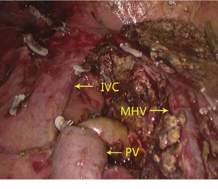
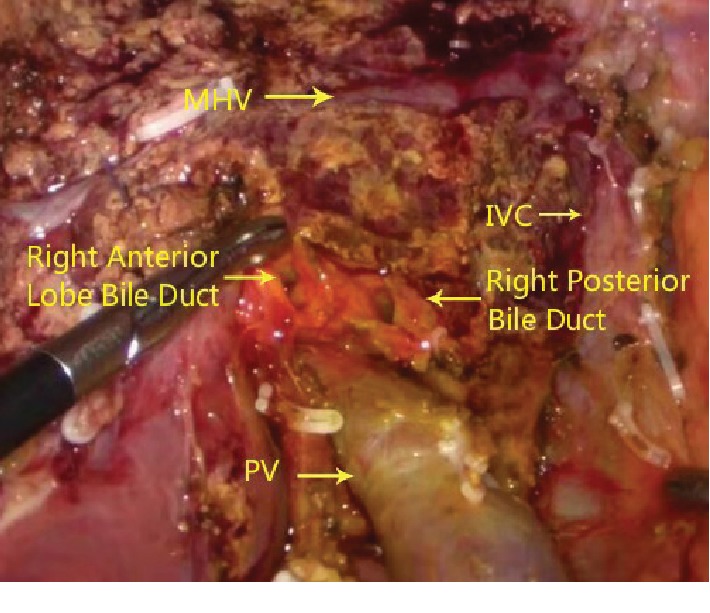
The central venous pressure was maintained at 3 cm water column, and the ischemic boundary line was recorded by the assistant with an electrocoagulation hook. The surgeon dissected the liver to the secondary porta with an ultrasonic small incision, disconnected the hepatic vein with a linear cutting stapler (white nail), and removed the specimen (Figures 1 and 2). During the operation, the blood vessels of 2 mm or less were coagulated, and 2 mm or more were clipped. The proper hepatic artery and portal vein were blocked by endoscopic noninvasive vascular occlusion forceps (15 + 5 mode). Along with the branch, we found and exposed the middle hepatic vein (MHV) until the secondary porta of the liver to protect the healthy lateral hepatic artery and portal vein. Subsequently, the bile duct was cut off as far as possible from the hepatic portal tumor.
Figure 1.

Left vision of postoperative liver section.
Figure 2.

Right vision of postoperative liver section.
2.5. Bile Duct Reconstruction
To achieve R0 status, multiple bile duct cuts were necessary for the specimen to be removed. Hepatic ducts were sutured and closed to form a bile duct basin. The upper jejunum was cut at a distance of 25 cm under the inferior border of the Treitz ligament with a cutter stapler. Then the hepatic duct was anastomosed with the proximal jejunum 50 cm below the distal end using a linear stapler. And the distal jejunum was lifted through the mesentery to anastomose with a bile duct. If there were a large number of tiny cuts, we could directly close the posterior wall bile duct basin. And the anterior wall could be stitched to the nearby liver tissue with a 4-0 prolene suture. To prevent the liver from being cut, we should gather the sutures after the anterior wall is stitched. Then the tube should be placed in the loop for extravasation. Or we could place the abdominal drainage tube behind the hepatic and intestinal anastomosis and near the liver section after water injection test in the 8th urinary catheters.
2.6. Vessel Operation
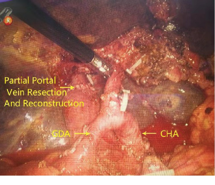
As we know, Hilar C often associates with vascular invasion. It was necessary to perform the vascular resection and reconstruction operation, when the bifurcation of the hepatic portal vein or artery was affected. After disconnecting the hepatoduodenal ligament and cutting off the hepatic artery, the portal branch was separated until invading the site of the bifurcation. Then the end of the common bile duct was pulled up to suspend the right branch of the portal vein at the right posterior side. Subsequently, noninvasive vascular occlusion forceps were used for the portal vein and right branch of the portal vein blocking. Then affected confluence vessels were cut off and washed by heparin sodium solution after the portal vein wall was cut 2-3 mm away from the tumor invasion site. After suturing 4-5 stitches with 5-0 prolene, the suture was closed to make the posterior wall aligned (Figure 3). After operation, the residual vascular wall was about 1/4 of the circumference of the branch of the portal vein.
Figure 3.

Resection and reconstruction of the portal vein.
3. Results
After surgery, we observed that the tumor size of 6 patients was 1.5-3.6 cm, and there were 4 lymph nodes. The operation time was 540-660 minutes, and the blood loss was 300-500 ml. None of the patients needed blood transfusion. Only basic surgery (laparoscopic gallbladder, hilar bile duct, and common bile duct resection and hepatoduodenal ligament lymph node dissection) was combined with left hepatic, caudate lobe resection and portal resection reconstruction. In patients with postoperative bile leakage, drainage healed within 2 weeks. Only patients with basic surgery (laparoscopic gallbladder, hilar bile duct, and common bile duct resection and hepatoduodenal ligament lymph node dissection) were combined with left hepatic, caudate lobe resection and biliary anastomosis-resected marginal atypical hyperplasia. There were 4 cases of negative bile duct margin tumor, 1 case was positive, and 1 case was not reported. The patients were discharged from the hospital (16~24) days after operation, and there was no perioperative death. Six patients were collected. We collected data from multiple outpatient visits after surgery and regularly reviewed for 3 months' time with a median duration of 7 months. Six patients were still alive, 5 patients had no obvious signs of recurrence, and 1 patient lacked clinical data. All patients were able to take care of themselves (Table 2).
Table 2.
Six patients' operation and prognosis.
| Case | Joint surgery | Operation time (min) | Amount of bleeding (ml) | Complication | Bile duct margin | Median size of tumor | Vascular invasion | Atypical hyperplasia |
|---|---|---|---|---|---|---|---|---|
| 1 | Left hepatic+caudate lobe | 540 | 300 | — | P | 2.1 cm | N | N |
| Resection+biliary anastomosis | ||||||||
| 2 | Left hepatic+caudate lobe | 660 | 300 | BL | No | 3.6 cm | Y | N |
| Resection+portal vein resection+biliary anastomosis | Report | |||||||
| 3 | Left hepatic+caudate lobe | 600 | 500 | — | p | 1.5 cm | N | N |
| Resection+biliary anastomosis | ||||||||
| 4 | Left hepatic+caudate lobe | 540 | 300 | — | P | 1.7 cm | N | N |
| Resection+biliary anastomosis | ||||||||
| 5 | Right hepatic+caudate lobe | 660 | 500 | — | P | 2.4 cm | N | N |
| Resection+biliary anastomosis | ||||||||
| 6 | Left hepatic+caudate lobe | 540 | 500 | — | N | 2.0 cm | N | Y |
| Resection+biliary anastomosis |
Basic operation was defined as gallbladder, hilar, and common bile duct resection and lymph node dissection. BT: blood transfusion; LOS: length of stay in hospital; BD: bile ducts; BL: bile leakage; N: negative; P: positive; Y: yes; N: no.
4. Discussion
Laparoscopic surgery is a newly developed minimally invasive method and an inevitable trend in the development of future surgical methods [10]. The advantage of laparoscopic radical resection for cholangiocarcinoma treatment is listed as follows:
With this technique, the surgery could be visible and more precise, easy, and safe
Because of the variable visibility angles and magnification effect, it could be easy for structural discrimination
The establishment of pneumoperitoneum, lower central venous pressure, and hepatic vascular occlusion combined with ultrasonic scalpel could significantly reduce the amount of blood loss during surgery
The application of laparoscopic radical resection could shorten the recovery time and reduce the rate of complication
Previous studies have used nonanatomical repeated laparoscopic hepatectomy for recurrent liver cancer, and this procedure is safe and feasible [11]. In laparoscopic hepatectomy, it may be an effective and feasible method to expose the vein from the trunk to the peripheral branch through the abdominal approach [12]. Single-incision laparoscopic biliary bypass for the treatment of malignant obstructive jaundice is safe and feasible, and the short-term results are satisfactory [13].
As a rare malignant tumor, surgical treatment is currently the only effective treatment for Hilar C. However, due to the complexity of the operation, the application of radical resection for Hilar C is still relatively rare [14].
Six patients with type III and IV hilar cholangiocarcinoma included in this study underwent total laparoscopic surgery. After the operation, only 1 case had postoperative bile leakage and vascular invasion, healed within 2 weeks after drainage. The other 5 patients had no complications after the operation and healed well, with no local or regional recurrence and associated complications. One case with biliary leakage healed within 2 weeks after drainage.
The summary of our clinical experience is that the location and number of the trocar are a critical step for laparoscopic radical resection. Also, the correct position of the patient, surgeon, and assistant and good surgical cooperation could make the surgery more successful and reduce bleeding, injuries, and comorbidities. To find the gap between the vessel and liver parenchyma, we should pay more attention to several important markers, such as ischemic line, hepatic vein, venous ligament fissure, posterior inferior vena cava, and border of liver fibrosis. Control of bleeding is the most important thing during a liver resection period. The easy bleeding sites include 8 ventral and interlobular veins which are close to the secondary porta, 5 main vessels, and 8 side branches which are located in the upper and lower thirds of the middle hepatic vein. During the operation, we disconnected the arteries followed by veins to reduce the risk of bleeding. In addition, we should dissect the liver carefully with the high tension, thin walls, and large number of vessels around the tumor. Bleeding should be treated carefully in the period of operation. However, these tumors are often grey-white scirrhous masses with a poor vascularization, unlike hepatocarcinoma [15]. If the portal vein is affected, portal vein resection and reconstruction are required. Taking advantage of the amplification effect and close observation of laparoscopy, we could control the needle and edge spacing in a better way. Before resection, we must determine the length of the blood vessel dissection to achieve R0 status and facilitate vessel reconstruction. Then we should separate the distal and proximal ends of the blood vessel dissection for vascular occlusion. These techniques can help us complete the surgery successfully. Though disputed, most of the researchers believe that caudate lobe dissection is necessary for Hilar C treatment [16, 17]. The caudate lobe is located between the inferior vena cava and the portal vein, surrounded with dense and important blood vessels [18, 19]. When dealing with the short hepatic vein, slight mistake might lead to tearing of the inferior vena cava and induce uncontrolled bleeding. In the utilization of laparoscopy, we could expose the caudate lobe and left portal vein completely before surgery, which could reduce the difficultly of caudate resection.
In this study, 6 patients with type III and IV hilar cholangiocarcinoma who underwent total laparoscopic hilar cholangiocarcinoma were successfully operated without serious complications and death.
5. Conclusions
Our preliminary clinical practice shows that type III and IV hilar cholangiocarcinoma and combined invasive portal vein resection and reconstruction of type III and IV livers are ensured under the premise of adequate preoperative assessment, reasonable case selection, and rigorous surgical planning. Portal cholangiocarcinoma is safe and feasible in laparoscopic radical surgery, and the short-term effect is good. However, its long-term efficacy, whether it can improve the survival rate of patients, still needs continued follow-up. It should not be overlooked that there are few cases in this study, and the conclusion still needs more laparoscopic surgery to treat cases of type III and IV hilar cholangiocarcinoma.
Acknowledgments
This work was financially supported by the following funders: National Science Funding of China (81902017), Hunan Provincial Natural Science Foundation of China (Grant Nos. 2019JJ50320 and 2019JJ20011), Central Guidance of Local Science and Technology Development Fund (Grant No. 2018CT5008), and Project of Scientific Research of Traditional Chinese Medicine in Hunan (Grant No. 201809).
Abbreviations
- Hilar C:
Hilar cholangiocarcinoma
- MHV:
Middle hepatic vein.
Contributor Information
Chuang Peng, Email: pengchuangcn@163.com.
Bo Jiang, Email: hepatojiangbo@163.com.
Data Availability
Data and materials are included in the manuscript.
Ethical Approval
All applicable international guidelines for the care and use of humans were followed. All procedures performed in studies involving animals were in accordance with the ethical standards of the Hunan Provincial People's Hospital at which the studies were conducted.
Consent
Written informed consent was obtained from the patients for the publication of this case report and any accompanying images. A copy of the written consents is available for a review by the editor of this journal.
Conflicts of Interest
The authors declare that they have no financial or commercial conflict of interest.
Authors' Contributions
All authors agree to the publication of this article.
Supplementary Materials
Table S1: supplementary information of 6 patients undergoing laparoscopic radical resection of hilar cholangiocarcinoma.
References
- 1.Brandi G., Venturi M., Pantaleo M. A., Ercolani G., GICO Cholangiocarcinoma: current opinion on clinical practice diagnostic and therapeutic algorithms: a review of the literature and a long-standing experience of a referral center. Digestive and Liver Disease. 2016;48(3):231–241. doi: 10.1016/j.dld.2015.11.017. [DOI] [PubMed] [Google Scholar]
- 2.Liu S., Jiang B., Li H., et al. Wip1 is associated with tumorigenity and metastasis through MMP-2 in human intrahepatic cholangiocarcinoma. Oncotarget. 2017;8(34):56672–56683. doi: 10.18632/oncotarget.18074. [DOI] [PMC free article] [PubMed] [Google Scholar]
- 3.Liu S., Jiang J., Huang L., et al. iNOS is associated with tumorigenicity as an independent prognosticator in human intrahepatic cholangiocarcinoma. Cancer Management and Research. 2019;11:8005–8022. doi: 10.2147/CMAR.S208773. [DOI] [PMC free article] [PubMed] [Google Scholar] [Retracted]
- 4.Kawasaki S., Imamura H., Kobayashi A., Noike T., Miwa S., Miyagawa S. Results of surgical resection for patients with hilar bile duct cancer: application of extended hepatectomy after biliary drainage and hemihepatic portal vein embolization. Annals of Surgery. 2003;238(1):84–92. doi: 10.1097/01.SLA.0000074984.83031.02. [DOI] [PMC free article] [PubMed] [Google Scholar]
- 5.Yu H., Wu S. D., Chen D. X., Zhu G. Laparoscopic resection of Bismuth type I and II hilar cholangiocarcinoma: an audit of 14 cases from two institutions. Digestive Surgery. 2011;28(1):44–49. doi: 10.1159/000322398. [DOI] [PubMed] [Google Scholar]
- 6.Quadri P., Gonzalez-Heredia R., Masrur M., Sanchez-Johnsen L., Elli E. F. Management of laparoscopic adjustable gastric band erosion. Surgical Endoscopy. 2017;31(4):1505–1512. doi: 10.1007/s00464-016-5183-4. [DOI] [PubMed] [Google Scholar]
- 7.Guan X., Bardawil E., Liu J., Kho R. Transvaginal natural orifice transluminal endoscopic surgery as a rescue for total vaginal hysterectomy. Journal of Minimally Invasive Gynecology. 2018;25(7):1135–1136. doi: 10.1016/j.jmig.2018.01.028. [DOI] [PubMed] [Google Scholar]
- 8.Sarwal A., Khullar R., Sharma A., Soni V., Baijal M., Chowbey P. Case report of ventral hernia complicating bariatric surgery. Journal of Minimal Access Surgery. 2018;14(4):345–348. doi: 10.4103/jmas.JMAS_253_17. [DOI] [PMC free article] [PubMed] [Google Scholar]
- 9.Ratti F., Cipriani F., Ferla F., Catena M., Paganelli M., Aldrighetti L. A. Hilar cholangiocarcinoma: preoperative liver optimization with multidisciplinary approach. Toward a better outcome. World Journal of Surgery. 2013;37(6):1388–1396. doi: 10.1007/s00268-013-1980-2. [DOI] [PubMed] [Google Scholar]
- 10.Lim S., Ghosh S., Niklewski P., Roy S. Laparoscopic suturing as a barrier to broader adoption of laparoscopic surgery. JSLS. 2017;21(3, article e2017.00021) doi: 10.4293/JSLS.2017.00021. [DOI] [PMC free article] [PubMed] [Google Scholar]
- 11.Ogawa H., Nakahira S., Inoue M., et al. Our experience of repeat laparoscopic liver resection in patients with recurrent hepatocellular carcinoma. Surgical Endoscopy. 2019:1–7. doi: 10.1007/s00464-019-06992-8. [DOI] [PubMed] [Google Scholar]
- 12.Kim J. H. Ventral approach to the middle hepatic vein during laparoscopic hemihepatectomy. Annals of Surgical Oncology. 2019;26(1):p. 290. doi: 10.1245/s10434-018-6927-2. [DOI] [PubMed] [Google Scholar]
- 13.Yu H., Wu S., Yu X., Han J., Yao D. Single-incision laparoscopic biliary bypass for malignant obstructive jaundice. Journal of Gastrointestinal Surgery. 2015;19(6):1132–1138. doi: 10.1007/s11605-015-2777-4. [DOI] [PubMed] [Google Scholar]
- 14.Lee W., Han H. S., Yoon Y. S., et al. Laparoscopic resection of hilar cholangiocarcinoma. Annals of Surgical Treatment and Research. 2015;89(4):228–232. doi: 10.4174/astr.2015.89.4.228. [DOI] [PMC free article] [PubMed] [Google Scholar]
- 15.Simone V., Brunetti O., Lupo L., et al. Targeting angiogenesis in biliary tract cancers: an open option. International Journal of Molecular Sciences. 2017;18(2):p. 418. doi: 10.3390/ijms18020418. [DOI] [PMC free article] [PubMed] [Google Scholar]
- 16.Ikeyama T., Nagino M., Oda K., Ebata T., Nishio H., Nimura Y. Surgical approach to Bismuth type I and II hilar cholangiocarcinomas: audit of 54 consecutive cases. Annals of Surgery. 2007;246(6):1052–1057. doi: 10.1097/SLA.0b013e318142d97e. [DOI] [PubMed] [Google Scholar]
- 17.Bhutiani N., Scoggins C. R., McMasters K. M., et al. The impact of caudate lobe resection on margin status and outcomes in patients with hilar cholangiocarcinoma: a multi-institutional analysis from the US Extrahepatic Biliary Malignancy Consortium. Surgery. 2018;163(4):726–731. doi: 10.1016/j.surg.2017.10.028. [DOI] [PMC free article] [PubMed] [Google Scholar]
- 18.Ercolani G., Zanello M., Grazi G. L., et al. Changes in the surgical approach to hilar cholangiocarcinoma during an 18-year period in a Western single center. Journal of Hepato-Biliary-Pancreatic Sciences. 2010;17(3):329–337. doi: 10.1007/s00534-009-0249-5. [DOI] [PubMed] [Google Scholar]
- 19.Perini M. V., Coelho F. F., Kruger J. A., Rocha F. G., Herman P. Extended right hepatectomy with caudate lobe resection using the hilar “en bloc” resection technique with a modified hanging maneuver. Journal of Surgical Oncology. 2016;113(4):427–431. doi: 10.1002/jso.24154. [DOI] [PubMed] [Google Scholar]
Associated Data
This section collects any data citations, data availability statements, or supplementary materials included in this article.
Supplementary Materials
Table S1: supplementary information of 6 patients undergoing laparoscopic radical resection of hilar cholangiocarcinoma.
Data Availability Statement
Data and materials are included in the manuscript.


