Abstract
It is generally accepted that the amyloid β (Aβ) peptide toxicity contributes to neuronal loss and is involved in the initiation and progression of Alzheimer's disease (AD). Cold-inducible RNA-binding protein (CIRBP) is reported to be a general stress-response protein, which is induced by different stress conditions. Previous reports have shown the neuroprotective effects of CIRBP through the suppression of apoptosis via the Akt and ERK pathways. The objective of this study is to examine the effect of CIRBP against Aβ-induced toxicity in cultured rat primary cortical neurons and attempt to uncover its underlying mechanism. Here, MTT, LDH release, and TUNEL assays showed that CIRBP overexpression protected against both intracellular amyloid β- (iAβ-) induced and Aβ25-35-induced cytotoxicity in rat primary cortical neurons. Electrophysiological changes responsible for iAβ-induced neuronal toxicity, including an increase in neuronal resting membrane potentials and a decrease in K+ currents, were reversed by CIRBP overexpression. Western blot results further showed that Aβ25-35 treatment significantly increased the level of proapoptotic protein Bax, cleaved caspase-3, and cleaved caspase-9 and decreased the level of antiapoptotic factor Bcl-2, but were rescued by CIRBP overexpression. Furthermore, CIRBP overexpression prevented the elevation of ROS induced by Aβ25-35 treatment by decreasing the activities of oxidative biomarker and increasing the activities of key enzymes in antioxidant system. Taken together, our findings suggested that CIRBP exerted protective effects against neuronal amyloid toxicity via antioxidative and antiapoptotic pathways, which may provide a promising candidate for amyloid-based AD prevention or therapy.
1. Introduction
Alzheimer's disease (AD) is an irreversible age-related neurodegenerative disorder, mainly characterized by progressive memory loss and cognitive decline [1]. One of the typical histological hallmarks associated with AD brains is the extracellular plaque deposits of the amyloid β (Aβ) peptides, which is produced by the cleavage of the transmembrane amyloid precursor protein (APP) [2–4]. Studies showed that Aβ peptides rapidly self-aggregates into Aβ dimmers, fibrils, and amyloid plaques and induced neuronal apoptosis in the cultured neuron [5, 6]. On the other hand, recent reports also demonstrated the accumulation of intracellular Aβ (iAβ), especially Aβ1–42, at the early stage of AD development, which occurs earlier than the appearance of Aβ plaques [7]. Direct evidence for iAβ cytotoxicity is that microinjection of Aβ1-42 induces neuronal cell death in cultured human primary neurons [8]. This cytotoxicity effect of causing neuronal cell death is found to be more potent than extracellular Aβ [9]. Currently, the toxicity of Aβ peptides is thought to contribute to the neuronal loss in the cerebral cortex and hippocampus and is involved in the initiation and progression of AD [10]. Although the underlying mechanisms by which Aβ production leads to the cytotoxicity and neuronal loss remains elusive, searching for strategies that can ameliorate Aβ toxicity may be potentially beneficial to AD treatment.
Cold-inducible RNA-binding protein (CIRBP) is a stress-responsive gene, which belongs to a family of cold-shock proteins [11]. In addition to upregulation of CIRBP induced by hypothermia, the expression of CIRBP can also be regulated by other stress conditions, such as hypoxia, UV radiation, glucose deprivation, and osmotic pressure [11]. In response to stress, CIRBP generally modulates mRNA stability at the posttranscriptional level through its binding site on the 3′-untranslational region (UTR) of its targeted mRNAs [12, 13]. Recent studies have demonstrated that CIRBP exerts neuroprotective effects against H2O2-induced cell death through the Akt and ERK pathways in primary rat cortical neurons and neuro2a (N2a) cells [14–16]. The aim of the present study is to investigate how CIRBP reacts to the intracellular and extracellular Aβ treatment, and whether CIRBP can protect against Aβ-induced toxicity in cultured rat primary cortical neurons.
In the present study, we found that CIRBP protected against both intracellular amyloid β- (iAβ-) induced and Aβ25-35-induced cytotoxicity in rat primary cortical neurons. These neuroprotective effects of CIRBP were mediated through antioxidative and antiapoptotic pathways.
2. Material and Methods
2.1. Culture of Primary Cortical Neurons
Newborn Sprague-Dawley (SD) rats were used in these experiments. After cervical dislocation and sterilization by immersion in 75% ethanol, the whole brains were taken out from the head. Cortical tissues then were dissected from the brains in Dulbecco's modified Eagle's medium (DMEM) (Invitrogen, Carlsbad, CA). The tissues were mechanically dissociated by gently chopping for about 15 times, and then were digested with 0.25% trypsin (Invitrogen) for 25 minutes at 37°C. After terminating the digestion with DMEM containing 10% fetal bovine serum, the mixture was gently triturated through the pipette to obtain single-cell suspension. The suspension then was filtered through nylon meshes and centrifuged at 500 g for 5 minutes. Single cells were resuspended in DMEM with 10% fetal bovine serum (FBS), 2 g/l HEPES, penicillin G (100 U/ml), and 100 μg/ml streptomycin (Invitrogen, Carlsbad, CA) and plated at a density of 1 × 10⁶/ml on poly-L-lysine-coated plates or coverslips. To inhibit glia cell growth and increase the purity of neurons, 10 μM cytosine arabinoside (Sigma) was added to the medium 24 h after plating. Cells were used for experiments 6 days after culture. All animal experiments were approved by the Animal Care and Use Committee of Harbin Medical University.
2.2. Adenovirus Infection
The rat CIRBP gene was PCR amplified according to the following primers and then was subcloned into the pAdTrack-CMV plasmid (a gift from Bert Vogelstein) [17] through KpnI and XhoI restriction sites. The forward primer is ATGGCATCAGATGAAGGCAA and the reverse primer is TTACTCGTTGTGTGTAGCATA. The human intracellular Aβ1–42 cDNA was synthesized directly, and then was also subcloned into pAdTrack through BglII and XhoI restriction sites. Adenovirus packaging and quality testing were performed in HEK293 cells. The neurons after 6 days in culture were infected by directly adding the adenovirus into the culture medium with an optimized multiplicity of infection (MOI) for 12~24 h. Cellular and biochemical experiments were performed 48 h after infection.
2.3. Aβ25-35 Treatments
Aβ25-35 and a control peptide Aβ35-25 are from Sigma. Before use, 2 mM Aβ25-35 stock solution was prepared by water and aged in a humidified chamber at 37°C for 5 days to obtain aggregates of Aβ peptides. The control peptide Aβ35-25 followed the same procedure. After 12 h infection of adenovirus, the cells were treated with 20 μM Aβ25-35 or Aβ35-25 by directly adding to the medium.
2.4. ELISA Assay
ELISA assay was used to determine the concentrations of Aβ1-42 in the cultured rat cortical neurons after the infection of recombinant adenoviruses using the ELISA kit (R & D Systems) according to the manufacturer's instruction. The microplate reader (Bio-Rad) was used to evaluate the intensity of each well at 480 nm.
2.5. Cell Cytotoxicity Analysis
In this study, the cytotoxicity of the cells after Aβ treatment was assessed by MTT assay and lactate dehydrogenase (LDH) release assay. For MTT assay, cells were seeded in 96-well plates. After treatment, media of the culture neurons were carefully removed by aspiration. After gently washing with PBS, 100 μl cell culture medium containing 0.5 mg/ml MTT was added to each well and incubated at 37°C for 4 h. Then, the medium in the wells was discarded and 150 μl dimethyl sulfoxide was added to each well. The absorbance was measured at 570 nm using a microplate reader (Bio-Rad). LDH release assay was measure using a CytoTox 96® Non-Radioactive Cytotoxicity Assay kit (Promega). This experiment was performed according to the manufacturer's instructions.
2.6. Electrophysiology
The cortical neurons were bathed in an extracellular solution containing (in mM) 140 NaCl, 2.5 KCl, 1.2 MgCl2, 2 CaCl2, 1.2 NaH2PO4, 10 HEPES, and 10 glucose, pH 7.35 adjusted with NaOH. 1 μM TTX and 100 μM CdCl2 were also included in the bath solution to block voltage-gated Na+ and Ca2+ channels. The patch pipette solution contained (in mM) 115 K-gluconate, 5 KCl, 5 Na2-ATP, 2 MgCl2, 1 CaCl2, 10 EGTA, and 10 HEPES, pH 7.2 adjusted with KOH. Pipettes with a resistance of 3–5 M were used in this experiment. The neurons were held at -70 mV, and then depolarized in a whole-cell patch clamp configuration by 1000 ms from -60 to 80 mV with 10 mV steps using an Axon 200B amplifier at room temperature.
2.7. Western Blot Analysis
After washing with PBS for 2 times, the cultured cells were treated with cell lysis buffer to extract the total proteins. A total of 20 μg of protein samples was separated on a 10% SDS-PAGE, and then transferred to a polyvinylidene fluoride (PVDF, Millipore) membrane (Millipore, Bedford, MA, USA). After blocking with 5% nonfat milk in Tris-buffered saline Tween-20 (TBST) for 1 h at room temperature, the PVDF membranes were incubated with the primary antibodies at 4°C overnight. After washing, the membranes were further incubated with horseradish peroxidase- (HRP-) conjugated secondary antibodies for 1 h at room temperature. β-Actin was used as a loading control. The intensities of the lanes were quantified using ImageJ software.
2.8. Cell Apoptosis Analysis
Terminal deoxynucleotidyl transferase-mediated dUTP nick end-labeling (TUNEL) assay (Roche) was used to evaluate the rate of cell apoptosis according to the manufacturer's instructions. Briefly, cells were fixed in 4% parafomaldehyde solution for 30 minutes at room temperature and permeabilized in 0.1% Triton X-100 for 5 min. TUNEL reagents then were added and incubated for 1 h at 37°C. After washing with PBS, the cells were mounted with DAPI in a mounting solution.
2.9. Caspase Activity Measurement
The activities of caspase-9 and caspase-3 were measured by using the fluorometric assay kit according to the manufacturer's instructions (Cell Signaling Technology). The protein samples were incubated with the reaction buffer and initiated by the DEVD-AMC substrate in a 96-well plate. The fluorescence intensity was measured using a fluorescence reader with excitation at 380 nm and emission at 460 nm.
2.10. Intracellular ROS Measurement
The measurement of intracellular ROS level was performed using a 2,7-dichlorofluorescein diacetate (DCF-DA) detection kit (Abcam) according to the manufacturer's instruction. Briefly, after washing twice with PBS buffer, adherent neurons were digested with 0.25% trypsin. Then, the cells were resuspended and incubated with 10 μM DCF-DA at 37°C for 30 min. After staining, the DCF fluorescence was detected using a fluorescence spectroscopy with excitation/emission at 495 nm/529 nm (BD Biosciences).
2.11. ELISA
After different treatments, cortical neurons were homogenized in lysis buffer to obtain the protein extracts. The activities of superoxide dismutase (SOD), catalase (CAT), glutathione peroxidase (GPx), 4-hydroxy-2-nonenal (4-HNE), and malondialdehyde (MDA) were measured using ELISA kits (R&D Systems) according to the manufacturer's instructions.
2.12. Statistical Analysis
Results were expressed as mean ± SE. Statistical analysis was performed using Student's t-test or the Mann–Whitney rank sum test. A value of p < 0.05 was considered significant.
3. Results
3.1. CIRBP Overexpression Reduced Aβ-Induced Neurotoxicity in Rat Primary Cortical Neurons
To examine whether CIRBP could reduce Aβ-induced neurotoxicity, rat primary cortical neurons cultured for 6 days were infected with an adenovirus-carrying CIRBP gene (Ad-CIRBP) at a MOI of 2 for 24 h, and then exposed to 20 μM Aβ25-35 or a control peptide Aβ35-25 for another 24 h. Cell viability was analyzed by MTT assay, and cell cytotoxicity was analyzed by LDH release assay. First, CIRBP expression was analyzed by western blot. In the Ad-CIRBP group, CIRBP expression was 3-fold higher than in the control group (Ad-Con) (Figure 1(a)). Although the biochemical organization of Aβ25-35 (monomeric or oligomeric) was not evaluated, MTT assay results showed that compared to the control peptide Aβ35-25, exposure to 20 μM Aβ25-35 dramatically reduced cell viability to about 60%, which was prevented by CIRBP expression in the Ad-CIRBP group, but not in the Ad-Con group (Figure 1(b)). Notably, CIRBP overexpression itself has no effect on cell viability (Figure 1(b)). LDH release assay showed similar results (Figure 1(c)), suggesting that there is a neuroprotective effect of CIRBP against Aβ-induced toxicity.
Figure 1.
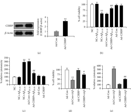
CIRBP protected against Aβ-induced neurotoxicity in rat primary cortical neurons. (a) Western blot result shows CIRBP overexpression in cultured cortical neurons. ∗∗p < 0.01vs. Ad-Con. (b) Cell viability was measured by MTT assay after treatment with 20 μM Aβ25-35. (c) Cell cytotoxicity was tested by LDH release assay after treatment with 20 μM Aβ25-35. ∗∗p < 0.01vs. NC, ##p < 0.01vs. Ad-Con+Aβ25-35. (d) Cell viability was measured by MTT assay after iAβ1-42 adenovirus infection. (e) Cell cytotoxicity was tested by LDH release assay after iAβ1-42 adenovirus infection. ∗∗p < 0.01vs. Ad-Con, #p < 0.05, ##p < 0.01vs. Ad-Con+iAβ.
Here, we also checked the effects of CIRBP on iAβ-induced neurotoxicity. After infection with the adenovirus-carrying CIRBP gene for 24 h, rat primary cortical neurons were infected with another iAβ1-42 adenovirus containing the human Aβ1-42 sequence without any signal peptide for another 24 h. As shown in a previous study [18], this construct mainly expresses Aβ1-42 in the cytosol and has higher toxicity than the construct coding human Aβ1-42 sequence with an addition signal peptide in human neurons. Our result in ELISA assay showed that the production of Aβ1-42 in the cultured rat cortical neurons after the infection of recombinant adenoviruses at the MOI of 0.1 was about 11.2 ng/ml (Supplemental Figure 1). Compared with the results above, iAβ1-42 dramatically reduced cell viability to about 40% tested by MTT and increased cytotoxicity tested by LDH release assay in the Ad-Con+iAβ group, showing more toxicity of iAβ1-42 than Aβ25-35 (Supplemental Figure 2). However, this effects of iAβ1-42 were significantly rescued in the Ad-CIRBP+iAβ group (Figures 1(d) and 1(e)), although the form of intraneuronal Aβ (monomeric or oligomeric) was still unclear. These results indicated that CIRBP overexpression prevented against the iAβ1-42-induced cytotoxicity.
3.2. CIRBP Overexpression Reversed iAβ-Induced Electrophysiological Changes That Are Responsible for Neuronal Toxicity
The change of electrophysiological properties induced by Aβ treatment, such as an increase in neuronal resting membrane potentials and a decrease in K+ currents, is thought to be responsible for neuronal toxicity [18–22]. Here, to test whether CIRBP could reverse the iAβ-induced electrophysiological changes, we recorded the resting membrane potentials and K currents in rat primary cortical neurons that were infected with iAβ1-42 adenovirus with or without CIRBP overexpression. Our results showed that the resting membrane potentials dramatically increased after iAβ1-42 treatment (−38.2 ± 4.5 mV) compared with the Ad-Con group (−64.8 ± 6.2 mV) (Figure 2(a)). Moreover, iAβ1-42 also reduced the whole-cell K+ current density in the Ad-Con+iAβ group (Figures 2(c) and 2(d)) but did not alter the voltage-dependent activation of K+ currents (Figure 2(e)). These results were consistent with previous studies [18, 20–22]. In this study, we found that CIRBP overexpression rescued the loss of resting membrane potentials (−55.4 ± 4.2 mV) and the decrease of K+ currents induced by iAβ1-42 (Figures 2(a) and 2(d)). However, CIRBP overexpression itself had no effect on the resting membrane potentials (−63.1 ± 5.2 mV) and K+ current density (Figures 2(a) and 2(d)). The membrane capacitance did not change either in iAβ treatment or CIRBP overexpression (Figure 2(b)). These results indicated that CIRBP ameliorated Aβ-induced neurotoxicity by rescuing the electrophysiological properties of the neurons.
Figure 2.
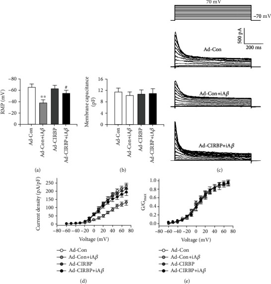
CIRBP reversed iAβ-induced electrophysiological changes that are responsible for neuronal toxicity. (a, b) Resting membrane potentials (RMP) (a) and membrane capacitance (b) were recorded among different groups. ∗∗p < 0.01vs. Ad-Con, #p < 0.05vs. Ad-Con+iAβ. (c) Typical recordings of whole-cell K+ current in the Ad-Con, Ad-Con+iAβ, and Ad-CRIPB+iAβ groups by depolarizing the membrane from -70 mV to +70 mV from a holding potential at -70 mV with 10 mV increasing steps. (d) Current-voltage relationship of the peak K+ current density. ∗∗p < 0.01vs. Ad-Con. (e) Voltage-dependent activation curve of K+ current showing there was no difference among different groups.
3.3. CIRBP Overexpression Prevented Aβ-Induced Activation of Cell Apoptosis Pathway in Cortical Neurons
To elucidate how CIRBP protected against Aβ-induced neurotoxicity in rat primary cortical neurons, we used TUNEL assay to measure the cell apoptosis in each group. Compared with the Ad-Con group, 20 μM Aβ25-35 treatment significantly increased the percentage of cell apoptosis in the Ad-Con+Aβ25-35 group, while overexpression of CIRBP markedly attenuated cell apoptosis induced by Aβ25-35 (Figure 3). However, no significant difference was observed between the Ad-Con and Ad-CIRBP groups (Figure 3).
Figure 3.
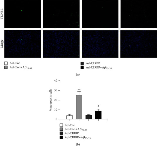
CIRBP prevented Aβ25-35-induced neuronal apoptosis. (a) Apoptotic cells were evaluated by TUNEL staining. Representative TUNEL staining images showing blue nuclear staining by DAPI and green TUNEL staining. (b) The percentage of apoptotic cells was calculated following the formula: Apoptosis index = apoptotic cells/(apoptotic cells + normal cells). ∗∗p < 0.01vs. Ad-Con, ##p < 0.01vs. Ad-Con+Aβ25-35.
Next, western blot was performed to evaluate the expression of some key apoptosis-related genes, such as antiapoptotic factor, B cell leukemia/lymphoma-2 (Bcl-2) and proapoptosis factors, Bcl-2 associated X protein (Bax), cleaved caspase-9, and cleaved caspase-3. As shown in Figures 4(a) and 4(b), the protein level of antiapoptotic factor, Bcl-2, was significantly decreased after Aβ25-35 treatment but was reversed by CIRBP overexpression. In contrast, the proapoptosis factors, Bax, cleaved caspase-9, and cleaved caspase-3, were all upregulated in the Ad-Con+Aβ25-35 group, which were inhibited by CIRBP overexpression (Figures 4(a) and 4(c)). We further measured the activities of caspase-9 and caspase-3 by using commercial fluorescent quantitative detection kit. Consistent with the western bolt result, 20 μM Aβ25-35 treatment significantly increased the activities of caspase-9 and caspase-3, while CIRBP overexpression ameliorate this effect (Figure 4(d)). Thus, these results indicated that the underlying mechanism of CIRBP protective effect against neuronal amyloid toxicity may be mediated by the antiapoptotic pathway.
Figure 4.
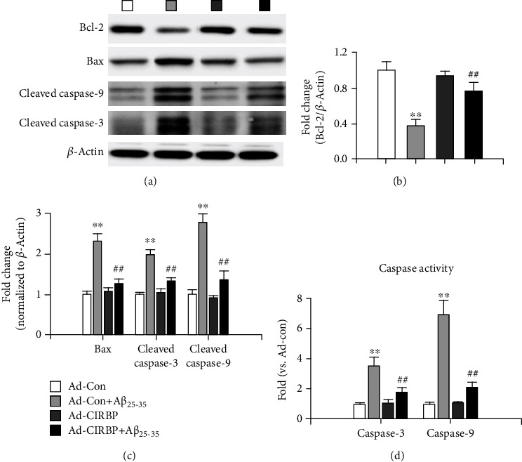
CIRBP inhibited Aβ25-35-induced neurotoxicity via the antiapoptotic pathway. (a) Representative imaging of western blot assay. (b) The expression level of Bcl-2 normalized to the expression level of β-actin. (c) The expression level of Bax, cleaved caspase-3, and cleaved caspase-9. (d) The caspase-3 and caspase-9 activities measured by a commercial fluorescent quantitative detection kit. ∗∗p < 0.01vs. Ad-Con, ##p < 0.01vs. Ad-Con+Aβ25-35.
3.4. CIRBP Overexpression Inhibited Aβ-Induced Oxidative Stress in Cortical Neurons
Oxidative stress, such as reactive oxygen species (ROS) generation, induced by Aβ is one of the major causes that lead to neuronal apoptosis [23]. Therefore, we further examined whether CIRBP could suppress Aβ-induced oxidative stress in cortical neurons. Intracellular ROS level in cortical neurons was assessed by using a ROS-detecting fluorescence dye DCF-DA kit. As expected, the level of ROS in cortical neurons treated with 20 μM Aβ25-35 was significantly elevated to 4.3-fold compared with the Ad-Con group. However, CIRBP overexpression remarkably inhibited the ROS generation induced by Aβ25-35 (Figure 5(a)). Next, the activities of oxidative marker, such as 4-hydroxy-2-nonenal (4-HNE) and malondialdehyde (MDA) and key enzymes in the antioxidant system, such as superoxide dismutase (SOD), catalase (CAT), and glutathione peroxidase (GPx), were measured by ELISA. As shown in Figures 5(b) and 5(c), CIRBP overexpression significantly rescued Aβ25-35-induced upregulation of oxidative marker and Aβ25-35-induced downregulation of antioxidase activities of these three antioxidant enzymes, indicating that the antioxidative pathway mediated the protective effect of CIRBP against Aβ-induced neuronal apoptosis.
Figure 5.
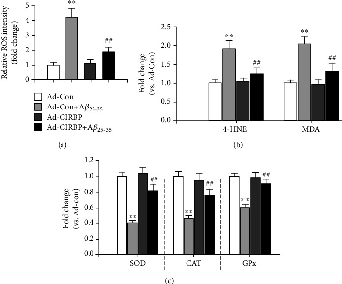
CIRBP inhibited Aβ25-35-induced neurotoxicity via the antioxidative pathway. (a) Intracellular ROS level was measured using a ROS-detecting fluorescence dye DCF-DA kit. (b) The activities of oxidative stress biomarkers, such as 4-HNE and MDA, measured by ELISA assay. (c) The activities of key enzymes in an antioxidant system, such as SOD, CAT, and GPx, were measured by ELISA. ∗∗p < 0.01vs. Ad-Con, ##p < 0.01vs. Ad-Con+Aβ25-35.
4. Discussion
AD is one of the neurodegenerative disorders in the elderly with extremely deficient clinical therapies and is associated with high morbidity and mortality [1]. AD is clinically characterized by progressive memory loss and cognitive decline [1]. One of the major pathological hallmarks in AD is the appearance of amyloid plaques that is enriched in Aβ [2–4]. Extensive studies suggest that the neurotoxicity of Aβ contributes to the neuronal loss and involves in the pathogenesis of neuronal dysfunction in AD [5, 24]. Although the underlying mechanisms are still largely unknown, accumulated evidences have showed that natural products or genetical manipulation that protect against Aβ-induced neuronal loss are beneficial to the treatment of AD [18, 25]. In the present study, we found that CIRBP, a stress-response protein, attenuated Aβ-induced cytotoxicity via antioxidative and antiapoptotic pathways in cultured rat primary cortical neurons, which may provide a promising candidate for amyloid-based AD prevention or therapy.
Aβ-mediated electrophysiological changes have been extensively studied in human primary neurons, rodent neurons, and different cell lines [18–22]. Dysfunction of neuronal excitability is responsible for the neuronal toxicity induced by Aβ [26]. Here, we found that iAβ induced significant increase in resting membrane potential and decrease in K+ current density in the Ad-Con+iAβ group, which is consistent with previous reports [18, 20–22]. Suppression of K+ current by Aβ was reported to trigger a large increase of Ca2+ influx by activating voltage-gated Ca2+ channels in the distal dendrites of rat hippocampal neurons [20]. The Ca2+ imbalance is suggested to initiate neuronal dysfunction and lead to cell death in rat hippocampus cells. In the present study, CIRBP overexpression rescued the resting membrane potential, probably by increasing the K+ current density, to maintain normal neuronal excitability. A previous report in the heart showed that CIRBP regulated cardiac repolarization by reducing the expression and function of transient outward K+ current [27]. Our present study identified a role of CIRBP in regulating another K+ channel, although the underlying mechanism still needs further investigation.
CIRBP, a cold-shock protein found in mammals, expressed in various cell types and is involved in multiple biological and cellular processes, such as cell survival, apoptosis, cell proliferation, circadian rhythm, immune response, reproduction, and cancer [11]. Upregulation of CIRBP at mild hypothermia is reported to suppress cell death and contribute to cell survival [16, 28, 29]. Li et al. first demonstrated that CIRBP upregulation in rat cortical neurons at low temperature inhibited H2O2-induced neuronal apoptosis [16]. Liu et al. also found that CIRBP protected against oxidative stress via the Akt and ERK signal transduction pathways in N2a cells [15]. Sakurai et al. observed that mild hypothermia-induced elevated CIRP levels inhibited tumor necrosis factor-alpha-induced apoptosis by caspase-8 activation and ERK phosphorylation [30]. Zhang et al. revealed a neuroprotective effect of CIRBP during mild hypothermia by inhibiting neuron apoptosis via the suppression of the mitochondria apoptosis pathway [14]. Consistent with these previous reports, we found that CIRBP overexpression significantly attenuated Aβ-induced neuronal apoptosis and exerted a neuroprotective effect in cultured rat cortical neurons. Mitochondrial signaling pathway plays an important role in promoting apoptotic cell death [31]. In our study, CIRBP overexpression rescued the decreased protein expression of antiapoptotic factor-Bcl-2 and also inhibited the increased protein expression of proapoptosis factor Bax, cleaved caspase-9, and cleaved caspase-3. All these results indicated that CIRBP suppressed Aβ-induced neuronal apoptosis, probably via mitochondrial signaling pathway.
Aβ-induced neuronal apoptosis is associated with the generation of ROS and plays an important role in the pathogenesis of AD [23]. Various compounds with antioxidant ability have been proposed to attenuate Aβ-induced oxidative stress in studies done in vitro and in vivo [32]. In this study, we showed that Aβ25-35 treatment dramatically elevated intracellular ROS levels in culture primary cortical neurons, which is consistent with previous studies [23, 32]. However, CIRBP overexpression largely prevented Aβ25-35-induced excessive ROS release, confirming the antioxidative activity of CIRBP. In addition, we evaluated the activities of primary antioxidant enzyme, such as SOD, CAT, and GPx. SOD can scavenge superoxide anions by catalyzing them to hydrogen peroxide and molecular oxygen. CAT can detoxify hydrogen peroxide by converting them to oxygen and water. GPx can remove hydrogen peroxide by catalyzing them to water. Our results suggested that CIRBP overexpression significantly suppressed the downregulation of SOD, CAT, and GPx activities induced by Aβ. Thus, CIRBP inhibits oxidative damage-induced mitochondrial dysfunction and cell apoptosis.
5. Conclusions
In conclusion, we demonstrate a neuroprotective effect of CIRBP against Aβ induced-cytotoxicity through antioxidative and antiapoptotic pathways in rat primary cortical neurons, which may provide a novel therapeutic strategy for amyloid-based AD prevention or therapy. To the best of our knowledge, this is the first study to evaluate the function of CIRBP against Aβ-induced neurotoxicity.
Acknowledgments
This work was supported by Natural Science Foundation of Heilongjiang Province of China (Grant No. H2018031).
Contributor Information
Zhengyi Qu, Email: quzhengyi@hrbmu.edu.cn.
Hong Zhao, Email: zhaohong123@hrbmu.edu.cn.
Data Availability
The appropriate data used to support the findings of this study are included within the article.
Conflicts of Interest
All authors declare no conflict of interest.
Authors' Contributions
Fang Su and Shanshan Yang contributed equally to the work.
Supplementary Materials
Supplemental Figure 1: The production of Aβ1-42 in the cultured rat cortical neurons after the infection of recombinant adenoviruses at the MOI of 0.1. ND: no detection. ∗∗p < 0.01 vs. Control. Supplemental Figure 2: MTT results showed no toxicity in the control peptide Aβ35-25, and more toxicity is in the intraneuronal Aβ1-42 than Aβ25-35. ∗∗p < 0.01 vs. NC, ##p < 0.01 vs. NC+Aβ25-35.
References
- 1.Citron M. Alzheimer’s disease: strategies for disease modification. Nature Reviews Drug Discovery. 2010;9(5):387–398. doi: 10.1038/nrd2896. [DOI] [PubMed] [Google Scholar]
- 2.Wenk G. L. Neuropathologic changes in Alzheimer’s disease. Journal of Clinical Psychiatry. 2003;64:7–10. [PubMed] [Google Scholar]
- 3.Wenk G. L. Neuropathologic changes in Alzheimer’s disease: potential targets for treatment. Journal of Clinical Psychiatry. 2006;67:3–7. [PubMed] [Google Scholar]
- 4.Price D. L., Sisodia S. S. Mutant genes in familial Alzheimer’s disease and transgenic models. Annual Review of Neuroscience. 1998;21:479–505. doi: 10.1146/annurev.neuro.21.1.479. [DOI] [PubMed] [Google Scholar]
- 5.Benilova I., Karran E., De Strooper B. The toxic Aβ oligomer and Alzheimer’s disease: an emperor in need of clothes. Nature Neuroscience. 2012;15(3):349–357. doi: 10.1038/nn.3028. [DOI] [PubMed] [Google Scholar]
- 6.Pike C. J., Burdick D., Walencewicz A. J., Glabe C. G., Cotman C. W. Neurodegeneration induced by beta-amyloid peptides in vitro: the role of peptide assembly state. Journal of Neuroscience. 1993;13(4):1676–1687. doi: 10.1523/JNEUROSCI.13-04-01676.1993. [DOI] [PMC free article] [PubMed] [Google Scholar]
- 7.Li M., Chen L., Lee D. H. S., Yu L. C., Zhang Y. The role of intracellular amyloid β in Alzheimer's disease. Progress in Neurobiology. 2007;83(3):131–139. doi: 10.1016/j.pneurobio.2007.08.002. [DOI] [PubMed] [Google Scholar]
- 8.Zhang Y., McLaughlin R., Goodyer C., LeBlanc A. Selective cytotoxicity of intracellular amyloid beta peptide1-42 through p53 and Bax in cultured primary human neurons. Journal of Cell Biology. 2002;156(3):519–529. doi: 10.1083/jcb.200110119. [DOI] [PMC free article] [PubMed] [Google Scholar]
- 9.Kienlen-Campard P., Miolet S., Tasiaux B., Octave J. N. Intracellular amyloid-beta 1-42, but not extracellular soluble amyloid-beta peptides, induces neuronal apoptosis. Journal of Biological Chemistry. 2002;277(18):15666–15670. doi: 10.1074/jbc.M200887200. [DOI] [PubMed] [Google Scholar]
- 10.Serrano-Pozo A., Frosch M. P., Masliah E., Hyman B. T. Neuropathological alterations in Alzheimer disease. Cold Spring Harbor Perspectives in Medicine. 2011;1(1, article a006189) doi: 10.1101/cshperspect.a006189. [DOI] [PMC free article] [PubMed] [Google Scholar]
- 11.Zhu X. Z., Buhrer C., Wellmann S. Cold-inducible proteins CIRP and RBM3, a unique couple with activities far beyond the cold. Cellular and Molecular Life Sciences. 2016;73(20):3839–3859. doi: 10.1007/s00018-016-2253-7. [DOI] [PMC free article] [PubMed] [Google Scholar]
- 12.Xia Z., Zheng X., Zheng H., Liu X., Yang Z., Wang X. Cold-inducible RNA-binding protein (CIRP) regulates target mRNA stabilization in the mouse testis. FEBS Letters. 2012;586(19):3299–3308. doi: 10.1016/j.febslet.2012.07.004. [DOI] [PubMed] [Google Scholar]
- 13.Morf J., Rey G., Schneider K., et al. Cold-inducible RNA-binding protein modulates circadian gene expression posttranscriptionally. Science. 2012;338(6105):379–383. doi: 10.1126/science.1217726. [DOI] [PubMed] [Google Scholar]
- 14.Zhang H. T., Xue J. H., Zhang Z. W., et al. Cold-inducible RNA-binding protein inhibits neuron apoptosis through the suppression of mitochondrial apoptosis. Brain Research. 2015;1622:474–483. doi: 10.1016/j.brainres.2015.07.004. [DOI] [PubMed] [Google Scholar]
- 15.Liu J., Xue J., Zhang H., et al. Cloning, expression, and purification of cold inducible RNA-binding protein and its neuroprotective mechanism of action. Brain Research. 2015;1597:189–195. doi: 10.1016/j.brainres.2014.11.061. [DOI] [PubMed] [Google Scholar]
- 16.Li S., Zhang Z., Xue J., Liu A., Zhang H. Cold-inducible RNA binding protein inhibits H2O2-induced apoptosis in rat cortical neurons. Brain Research. 2012;1441:47–52. doi: 10.1016/j.brainres.2011.12.053. [DOI] [PubMed] [Google Scholar]
- 17.He T.-C., Zhou S., da Costa L. T., Yu J., Kinzler K. W., Vogelstein B. A simplified system for generating recombinant adenoviruses. Proceedings of the National Academy of Sciences. 1998;95(5):2509–2514. doi: 10.1073/pnas.95.5.2509. [DOI] [PMC free article] [PubMed] [Google Scholar]
- 18.Cui J., Wang Y., Dong Q., et al. Morphine protects against intracellular amyloid toxicity by inducing estradiol release and upregulation of Hsp70. Journal of Neuroscience. 2011;31(45):16227–16240. doi: 10.1523/JNEUROSCI.3915-11.2011. [DOI] [PMC free article] [PubMed] [Google Scholar]
- 19.Qi J. S., Ye L., Qiao J. T. Amyloid beta-protein fragment 31-35 suppresses delayed rectifying potassium channels in membrane patches excised from hippocampal neurons in rats. Synapse. 2004;51(3):165–172. doi: 10.1002/syn.10299. [DOI] [PubMed] [Google Scholar]
- 20.Chen C. Beta-amyloid increases dendritic Ca2+ influx by inhibiting the A-type K+ current in hippocampal CA1 pyramidal neurons. Biochemical and Biophysical Research Communications. 2005;338(4):1913–1919. doi: 10.1016/j.bbrc.2005.10.169. [DOI] [PubMed] [Google Scholar]
- 21.Good T. A., Smith D. O., Murphy R. M. Beta-amyloid peptide blocks the fast-inactivating K+ current in rat hippocampal neurons. Biophysical Journal. 1996;70(1):296–304. doi: 10.1016/S0006-3495(96)79570-X. [DOI] [PMC free article] [PubMed] [Google Scholar]
- 22.Hou J. F., Cui J., Yu L. C., Zhang Y. Intracellular amyloid induces impairments on electrophysiological properties of cultured human neurons. Neuroscience Letters. 2009;462(3):294–299. doi: 10.1016/j.neulet.2009.07.031. [DOI] [PubMed] [Google Scholar]
- 23.Uttara B., Singh A. V., Zamboni P., Mahajan R. T. Oxidative stress and neurodegenerative diseases: a review of upstream and downstream antioxidant therapeutic options. Current Neuropharmacology. 2009;7(1):65–74. doi: 10.2174/157015909787602823. [DOI] [PMC free article] [PubMed] [Google Scholar]
- 24.Murphy M. P., LeVine H. Alzheimer’s disease and the amyloid-beta peptide. Journal of Alzheimers Disease. 2010;19(1):311–323. doi: 10.3233/JAD-2010-1221. [DOI] [PMC free article] [PubMed] [Google Scholar]
- 25.Singh S. K., Srivastav S., Yadav A. K., Srikrishna S., Perry G. Overview of Alzheimer's Disease and Some Therapeutic Approaches Targeting A β by Using Several Synthetic and Herbal Compounds. Oxidative Medicine and Cellular Longevity. 2016;2016:22. doi: 10.1155/2016/7361613.7361613 [DOI] [PMC free article] [PubMed] [Google Scholar]
- 26.Shankar G. M., Walsh D. M. Alzheimer’s disease: synaptic dysfunction and Aβ. Molecular Neurodegeneration. 2009;4(1):p. 48. doi: 10.1186/1750-1326-4-48. [DOI] [PMC free article] [PubMed] [Google Scholar]
- 27.Li J., Xie D., Huang J., et al. Cold-inducible RNA-binding protein regulates cardiac repolarization by targeting transient outward potassium channels. Circulation Research. 2015;116(10):1655–1659. doi: 10.1161/CIRCRESAHA.116.306287. [DOI] [PubMed] [Google Scholar]
- 28.Liu A., Zhang Z., Li A., Xue J. Effects of hypothermia and cerebral ischemia on cold-inducible RNA-binding protein mRNA expression in rat brain. Brain Research. 2010;1347:104–110. doi: 10.1016/j.brainres.2010.05.029. [DOI] [PubMed] [Google Scholar]
- 29.Chip S., Zelmer A., Ogunshola O. O., et al. The RNA-binding protein RBM3 is involved in hypothermia induced neuroprotection. Neurobiology of Disease. 2011;43(2):388–396. doi: 10.1016/j.nbd.2011.04.010. [DOI] [PubMed] [Google Scholar]
- 30.Sakurai T., Itoh K., Higashitsuji H., et al. Cirp protects against tumor necrosis factor-α-induced apoptosis via activation of extracellular signal-regulated kinase. Biochimica et Biophysica Acta-Molecular Cell Research. 2006;1763(3):290–295. doi: 10.1016/j.bbamcr.2006.02.007. [DOI] [PubMed] [Google Scholar]
- 31.Wang C., Youle R. J. The role of mitochondria in apoptosis. Annual Review of Genetics. 2009;43(1):95–118. doi: 10.1146/annurev-genet-102108-134850. [DOI] [PMC free article] [PubMed] [Google Scholar]
- 32.Ono K., Hamaguchi T., Naiki H., Yamada M. Anti-amyloidogenic effects of antioxidants: Implications for the prevention and therapeutics of Alzheimer's disease. Biochimica et Biophysica Acta-Molecular Basis of Disease. 2006;1762(6):575–586. doi: 10.1016/j.bbadis.2006.03.002. [DOI] [PubMed] [Google Scholar]
Associated Data
This section collects any data citations, data availability statements, or supplementary materials included in this article.
Supplementary Materials
Supplemental Figure 1: The production of Aβ1-42 in the cultured rat cortical neurons after the infection of recombinant adenoviruses at the MOI of 0.1. ND: no detection. ∗∗p < 0.01 vs. Control. Supplemental Figure 2: MTT results showed no toxicity in the control peptide Aβ35-25, and more toxicity is in the intraneuronal Aβ1-42 than Aβ25-35. ∗∗p < 0.01 vs. NC, ##p < 0.01 vs. NC+Aβ25-35.
Data Availability Statement
The appropriate data used to support the findings of this study are included within the article.


