

Abstract
Human and animal research has shown that the ventral striatum, including the nucleus accumbens, may play a critical role in mediating positive emotions. Recently we described a subject with obsessive-compulsive disorder who intra-operatively exhibited the acute onset of an asymmetric smile and acute positive emotional change with contralateral deep brain stimulation (DBS) in either the right or left nucleus accumbens and anterior limb of the internal capsule region. The purpose of the present study was to examine the stability of the stimulation-induced smile(s) over a 12-month period. Custom computer software objectively quantified left and right facial movement during DBS. Although stimulation-induced smiles were elicited at one and two months post-surgery, they were no longer present from 3–12 months following chronic high frequency DBS. The smiles could not be elicited even with long washout periods. These findings imply potential long-term habituation and changes in the neural chemistry (possibly neuroplasticity) induced by chronic DBS.
Introduction
Deep brain stimulation (DBS) has been used to treat a variety of movement disorders by modulating abnormal patterns of neural activity in motor and non-motor circuitry of basal ganglia. (Benabid et al., 1994; Koller, et al., 2000; Krack and Vercueil, 2001). DBS has also shown promise in alleviating the symptoms of certain neuropsychiatric afflictions such as obsessive-compulsive disorder (OCD), Tourette’s syndrome, and refractory depression (Greenberg and Rezai, 2003; Mayberg et al., 2005; Nuttin et al., 2003; Temel and Visser-Vandewalle, 2004).
Recently, a subject with severe “contamination” OCD underwent DBS surgery at the University of Florida as part of an NIH-sponsored study. The complete details of the surgical procedure and intra-operative results from this case are described by Okun and colleagues (Okun et al., 2004). The patient’s leads were bilaterally implanted in the anterior limbs of the internal capsule and the nucleus accumbens region. During intra-operative testing, she developed asymmetric smiles contralateral to the side of stimulation (i.e., left DBS – right asymmetric smile, right DBS – left asymmetric smile). These responses were reproducible under blinded on/off conditions and were accompanied by spontaneous reports of euphoria and giddiness.
During the intra-operative procedure, the smile and the affective response were reproducible with acute stimulation. The smile response was non-linear, appearing between 2 and 6 V and disappearing at higher levels of stimulation. We hypothesized (Okun et al., 2004) that the smile resulted from stimulation of a limbic-motor loop. In this current study, we sought to determine whether the smile and affective response would habituate with chronic DBS.
Details of the Study Subject
A 34-year-old right-handed woman with a 10-year history of OCD was evaluated at the University of Florida. Her OCD symptoms manifested with a fear of contamination of bodily fluids, especially blood (with the exception of her own and that of immediate family members), complex cleaning rituals, and avoidance of contact with people and objects that she perceived to be potentially contaminated. Early in her history, she had responded to paroxetine (20 mg), but this medication was discontinued 3 years prior to being admitted (before she became pregnant). During pregnancy, her symptoms reappeared and worsened until she essentially became housebound. She became refractory to multiple pharmacological agents and failed treatment with cognitive-behavioral therapy including exposure to symptom-triggering stimuli while encouraging resistance to compulsive urges (exposure and response prevention).
The patient met the diagnostic criteria for major depression upon presentation at the University of Florida. Her Hamilton Depression Scale (HAM-D), however, was only mildly elevated at 12 points. Her severe OCD was evident both on clinical examination and with the administration of the Yale-Brown Obsessive-Compulsive Scale, on which she scored 38 out of a possible 40 points, indicating an extremely severe illness. Her neurological examination was normal. She signed informed consents for DBS surgery and for subsequent testing sessions, as part of a NIH-supported study to examine the effectiveness of DBS in treating severe intractable OCD. She was implanted with DBS devices bilaterally in the anterior limb of the internal capsule and shell of the nucleus accumbens (see Figure 1). The procedure was previously described (Okun, et al. 2004).
Fig. 1.

Anatomical localization of bilateral DBS electrode in the anterior limb of the internal capsule (AIC). The distal end of the deepest contact is depicted by the crosshairs in each view. The deepest contact lies within the nucleus accumbens and the more superficial contacts span the inferior half of the AIC. A post-operative CT was performed and fused to the MRI. The figure shows the reader a reconstruction of one DBS lead in its anatomical position. The lateral (X), antero-posterior (Y), and axial (Z) measurements for both distal lead tips relative to the mid-commissural point were: 6.1 mm (X), 13.6 mm (Y), −3.6 mm (Z) (left lead), and 7.5 mm (X), 13.4 mm (Y), −4 mm (Z) (right lead).
Methods
Procedure
The following sections overview the methods used to objectively quantify facial movement in this subject, as well describe the protocol utilized to experimentally characterize and track habituation of the smile response across testing sessions spanning approximately 1 year.
Facial Movement Analysis
During each experimental testing session, the patient sat comfortably in a chair and was continuously videotaped using a black-and-white Pulnix camera and Sony videorecorder. A Polaris light meter was used to uniformly balance the incident light upon the patient’s left and right face to within 1 lux of brightness. The patient’s head was secured within a rigid immobilization cushion in order to minimize out-of-plane head movement. Before commencement of videorecording, the experimenter ensured that the patient’s face encompassed the camera’s full field of view and that no part of the patient’s face was obstructed by the cushion. The testing neurologist (MSO) directed the parameters and timing of the DBS stimulation by written cues to a DBS programmer, who sat out of view (behind a barrier) of the patient. In this way, the patient was blinded both visually and aurally to changes in DBS settings during each testing session.
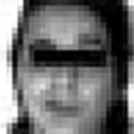
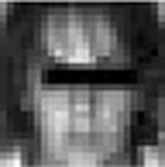
The testing sessions involved alternating conditions of “on” and “off” stimulation for 1- or 5-minute periods. The right lead alone was activated first (Contacts 0 then 1, 135 Hz, 2 V, 210/330 μs) and then the left lead alone (Contact 0 then 1, 135 Hz, 4 V, 210/330 μs). Monopolar stimulation was used for all trials. To analyze the changes in facial movement in response to DBS activation at these settings, methods and procedures described by Bowers and colleagues (Bowers et al., 2003; Leonard et al., 1991; Okun et al., 2004; Richardson et al., 2000) were employed. After the videotapes were reviewed, video segments containing facial movement were extracted, and time locked to the onset of stimulation. One stimulation-induced facial expression per side of stimulation (Left-DBS, Right-DBS), per testing session (at 2, 3, 6, and 12 months) was selected and extracted for comparison to two facial expressions (Left-DBS, Right-DBS) previously extracted and analyzed from the intra-operative procedure (Okun et al., 2004). Ten total stimulation-induced videosegments of the patient’s face, then, were used in the present analysis. The peak intensities of the patient’s facial expression in each of the representative videosegments are provided in Table 1, as are the associated DBS settings and maximum percent change in facial movement (i.e., entropy) from a resting state.
Table 1.
Maximal facial responsesa and corresponding DBS settings
| Optimization Sessions | Surgery | 1 month | 2 months | |||
|---|---|---|---|---|---|---|
| RDBS | LDBS | RDBS | LDBS | RDBS | LDBS | |
| Peak expressivity | 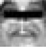 |
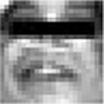 |
no video available | no video available |  |
 |
| LROI, entropy change | 22.1% | 29.0% | n/a | n/a | 12.8% | 2.4% |
| RROI, entropy change | 18.9% | 43.5% | n/a | n/a | 9.3% | 6.4% |
| Contacts | 0− C+ | 0− C+ | 1− C+ | 0− C+ | 0− C+ | 0− C+ |
| Frequency (Hz) | 130 | 130 | 135 | 135 | 135 | 135 |
| Pulse width (μs) | 210 | 210 | 210 | 210 | 330 | 210 |
| Amplitude (V) | 2 | 2 | 6 | 4 | 2 | 4 |
| Habituation Testings | 3 months | 6 monthsb | 12 monthsc | |||
| RDBS | LDBS | RDBS | LDBS | RDBS | LDBS | |
| Peak expressivity |  |
 |
 |
 |
 |
 |
| LROI, entropy change | 0.45% | 0.97% | 1.28% | 1.74% | 0.92% | 0.82% |
| RROI, entropy change | 0.60% | 0.70% | 1.53% | 1.07% | 0.78% | 0.81% |
| Contacts | 1− C+ | 0− C+ | 1− C+ | 0− C+ | 1− C+ | 0− C+ |
| Frequency (Hz) | 135 | 135 | 135 | 135 | 135 | 135 |
| Pulse width (μs) | 210 | 210 | 210 | 210 | 210 | 210 |
| Amplitude (V) | 5 | 4 | 5 | 4 | 5 | 4 |
as agreed upon by visual consensus of all investigators.
after 16 hours in bilateral DBS-OFF condition.
after 2+ weeks in bilateral DBS-OFF condition
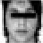
Extracted video segments were digitized and saved onto a PC using an Iscan-PCI video card and EYEVIEW software (Imaging Technology). Each segment consisted of a series of 30 digitized images or frames. Each frame was approximately 30.75 ms in duration (i.e., the industry standard for a video frame) with a resolution of 640 × 480 pixels at 256 levels of grayscale. Custom software, written in PV-Wave (Gokcay, personal communication), was used to extract the lower face region, defined by the area between the chin and region below the lower eyelid. The program used anatomic landmarks to automatically define this area and separate it into equally proportioned right and left regions (see Figure 2). After the facial expressions were extracted from the videotapes, the software computed the summed changes in the intensities of the pixels over the left and right lower face regions for each frame following the onset of deep brain stimulation. Changes in the intensities of the pixels across these regions corresponded to changes in light reflectance patterns that occurred over the face during an expressive movement. The difference of the summed pixel intensity values between each adjacent pair of frames was calculated across the entire expression videosegment in order to create an array of entropy values (one entropy value for each frame in the videosegment). This array of entropy values can be displayed graphically as an “entropy curve,” which represents changes in facial movement over the course of a given expression (i.e., higher entropy values correspond to greater change in facial movement). Separate entropy curves were thus obtained for the left and for the right lower face, as well as for the left and right lower face regions combined, for each of the ten videosegments. Figure 3 graphically depicts an example of the process of entropy curve calculation.
Fig. 2.

Anatomic landmarks and ROI extraction.
Fig. 3.

Frame-by-frame calculation of entropy for lower left and right ROIs. Rows, from top to bottom: original video frames, difference images, and entropy over time. An example of entropy induced by right-DBS is shown.
For each of the selected ten videosegments following the onset of DBS, a corresponding “baseline” condition was also identified, extracted, and analyzed, for comparison purposes. Baseline conditions were defined as the 1 s period (30 frames) just prior to the onset of stimulation. Each baseline condition was associated with 0 V DBS, and the subject’s face was visually verified as containing no movement throughout the 30 frames prior to extraction. Entropy curves were calculated for each baseline condition, as was the case with the representative videosegments, post-onset DBS.
Entropy curves for both baseline conditions and facial expressions post-onset DBS were transformed into entropy percent change values for standardization purposes. The entropy values over the entire entropy curve of a given videosegment were divided by the average of the first five entropy values in the videosegment. This yielded a series of 30 values per videosegment, baseline or post-DBS, corresponding to units of percentage change in entropy over the course of the videosegment.
Emotion Analysis
In addition to her affective facial responses to DBS, we measured her emotional responses. This was achieved through the patient’s qualitative self-report of any changes in her mood or anxiety state following DBS onset at each setting.
Results
Means and standard deviations for percent change in entropy for all captured expressions, corresponding to both post-DBS onset expressions and their corresponding baseline conditions, are provided in Table 2. An initial analysis was conducted to compare the amount of change in facial movement (i.e., percent entropy change) over the course of all post-DBS videosegments with that of each corresponding baseline condition. Paired t-tests were conducted using 30 frames of percent entropy change over the total region of the lower face (i.e., both left and right regions combined) from the baseline and post-DBS onset expressions. The expressions captured at the intra-operative session and two-month testing session showed significant facial movement upon analysis of both Left-DBS and Right-DBS conditions. Comparing mean percent entropy change for post-DBS onset and baseline videosegments, respectively: Intra-operative, Left-DBS [11.54% vs. −0.63%; t(29) = 9.74, p < .001]; Intra-operative, Right-DBS [8.38% vs. −0.26%; t(29) = 11.97, p < .001]; 2-months, Left-DBS [0.69% vs. −0.33%; t(30) = 8.275, p < .001]; 2-months, Right-DBS[4.74% vs. 0.04%; t(30) = 2.43, p < .05].
Table 2.
Stimulation-induced movement (entropy change) over lower-face ROIs
| Left ROI | Right ROI | Total ROI | |||||||||||
|---|---|---|---|---|---|---|---|---|---|---|---|---|---|
| Expression | Baseline | Expression | Baseline | Expression | Baseline | ||||||||
| Mean | SD | Mean | SD | Mean | SD | Mean | SD | Mean | SD | Mean | SD | ||
| Surgery | Right DBS | 8.68% | 4.61% | −0.43% | 1.02% | 7.97% | 4.22% | −0.10% | 1.18% | 8.38% | 4.38% | −0.25% | 1.07% |
| Left DBS | 10.89% | 7.08% | −0.65% | 0.56% | 12.11% | 6.71% | −0.62% | 0.57% | 11.54% | 6.89% | −0.63% | 0.50% | |
| 2 months | Right DBS | 1.24% | 1.80% | −0.39% | 0.38% | 0.09% | 2.37% | −0.31% | 0.54% | 0.69% | 2.04% | −0.33% | 0.36% |
| Left DBS | 3.69% | 3.11% | 0.02% | 0.49% | 5.75% | 2.80% | 0.12% | 0.49% | 4.74% | 2.87% | 0.04% | 0.47% | |
| 3 months | Right DBS | −0.16% | 0.36% | 0.03% | 0.35% | −0.12% | 0.45% | −0.21% | 0.36% | −0.14% | 0.37% | −0.09% | 0.32% |
| Left DBS | −0.65% | 0.47% | 0.41% | 0.72% | −1.09% | 0.73% | 0.56% | 1.04% | −0.89% | 0.53% | 0.49% | 0.82% | |
| 6 months | Right DBS | 0.00% | 0.58% | 0.25% | 0.79% | −0.21% | 0.67% | 0.70% | 0.99% | −0.10% | 0.55% | 0.48% | 0.85% |
| Left DBS | −0.19% | 0.49% | 0.37% | 0.65% | 0.08% | 0.54% | 0.15% | 0.76% | 0.05% | 1.13% | 0.08% | 0.91% | |
| 12 months | Right DBS | 0.48% | 1.44% | 0.23% | 0.67% | 0.48% | 1.27% | 0.12% | 0.96% | 0.48% | 1.33% | 0.17% | 0.80% |
| Left DBS | 0.03% | 0.35% | 0.00% | 0.38% | 0.07% | 0.31% | 0.22% | 0.39% | 0.04% | 0.25% | 0.11% | 0.33% | |
A second analysis was conducted in order to determine whether for every facial expression identified as having significant movement, more facial movement occurred on the side contralateral to stimulation. Paired t-tests contrasting percent entropy change between left and right hemifaces for each expression indicated that, for every facial expression identified as having significant movement, more facial movement occurred on the side contralateral to stimulation: Intra-operative, Left-DBS [Right ROI = 12.11% vs. Left ROI = 10.89%; t(29) = 4.08, p < .001]; Intra-Operative, Right-DBS [Right ROI = 7.97% vs. Left ROI = 8.68%; t(29) = 2.27, p < .05]; 2-month optimization session, Left-DBS [Right ROI = 5.75% vs. Left ROI = 3.69%; t(30) = 2.36, p < .05]; 2-month optimization session, Right-DBS [Right ROI = 0.09% vs. Left ROI = 1.24%; t(29) = 4.65, p < .001]. Means and standard deviations by hemiface, for left and right unilateral stimulation, are also provided in Table 2.
Along with the patient’s facial response to DBS at 2 months, the patient reported her acute changes in mood. Prior to the testing session, she characterized her mood as “anticipatory fear” and rated her anxiety at 30/100. In response to unilateral Right-DBS, she described her mood as “better, more upbeat”, and in response to Left-DBS, she reported that she felt “in a good mood”. Anxiety remained at 30/100 after DBS onset for each lead.
Three Months Post-Surgery
No significant facial movement relative to baseline was observed as a result of any stimulation at 3 months, in contrast to the optimization session at 2 months. We contrasted the percent change in entropy of the post-onset DBS videosegment from the entire facial ROI from this session (3 months) with that observed during the optimization session at 2 months. Paired t-tests were used for unilateral Left-DBS and unilateral Right-DBS conditions. Significantly less movement occurred over the face in this testing session under both left and right DBS conditions (Left-DBS at 3 months, −0.89% vs. at 2 months, 4.74%; t(28) = 10.45, p < .001; Right-DBS at 3 months, −0.14% vs. at 2 months, 0.69%; t(22) = 2.24, p < .05). Qualitatively the patient also reported no acute changes in mood as a result of stimulation during this session, although she did report feeling “slightly less anxious” in response to DBS as well as some improvement in alertness with settings previously shown to elicit the smile response.
Six Months Post-surgery
Six months following her surgery, she was evaluated with routine clinical testing of her DBS leads. We hypothesized that a longer washout period (i.e., greater than 5 minutes) was necessary to restore the affective smile response. Both leads were deactivated overnight (16 hours between inactivation and testing). The right lead was activated alone first, and then was turned off, and then the left lead was tested alone in an identical manner. Neither the left nor right lead was associated with significant facial movement. These analyses were performed using paired t-tests contrasting facial entropy associated with DBS to facial entropy during baseline conditions without stimulation. Means and standard deviations are displayed in Table 2. The patient self-reported that upon onset of Left-DBS, she felt “better, more pepped up”. With Right-DBS, she stated that she felt “more alert”.
Twelve Months Post-surgery
One year following the date of the patient’s surgery, it was necessary to replace her batteries. Interviews, examinations and testing suggested that the right lead had remained in the “off-stimulation” state for approximately 2–4 weeks. We hypothesized that this longer washout period (i.e., weeks vs. hours) may be associated with a restoration of the stimulation-induced smile. The right lead, and then the left lead, were activated and tested using an identical procedure as described above. Despite the longer (2 to 4 weeks) washout period, reactivation was not associated with significant facial movement in any ROI. Reactivation of the leads was also not associated with any induced change in mood, affect, or arousal.
Discussion
As was previously described in detail by Okun et al. (2004), acute DBS in the region of the anterior limb of the internal capsule and nucleus accumbens had resulted in a contralateral smile and affective change during intra-operative testing. In the present study however chronic DBS resulted in habituation of the smile response. This habituation was characterized by the loss in intensity of the facial response during successive testing over the initial 3 months following surgery. At 6 months and a year following the operative procedure, longer washout periods in the “DBS-off” condition of 16 hours and 2–4 weeks respectively (on the right side), failed to restore the smile or affective response. Reasons for this habituation raise important questions for consideration of changes in the neural circuitry induced by DBS.
The habituation of the smile response was accompanied by the loss of acute positive mood shifts, previously induced by lead activation. These emotions (“euphoria”, “giddiness”), observed during intra-operative testing were diminished to feeling “better” and “more upbeat” at 2 months post-surgery, to simply feeling “slightly more awake”, and “slightly less anxious” when the smile response disappeared at three months post-surgery. It is interesting to note that what appeared initially together, also disappeared together providing further evidence for stimulation of a limbic-motor loop (Okun et al., 2004). Although the acute affective changes (i.e., the smile response and accompanying positive mood shifts) had disappeared in response to chronic DBS, the subject’s overall mood had improved since her initial, preoperative evaluation, as reflected in the maintenance of her HAM-D scores near 0 over time.
Similarly to the habituation of the smile response, the unpleasant side effects (i.e. nausea, olfactory and gustatory hallucinations) also habituated over time (Okun et al., 2004). Interestingly, the unpleasant side effects seemed to reappear with longer washout periods of 2–4 weeks, which was characteristically different than what was seen with the smile and mood responses. This raises the question as to whether the habituation of these responses was a form of neural plasticity (i.e., the alteration of neurochemical or neuroreceptor sensitivity) induced by DBS. Future studies will need to examine this question more carefully.
One consideration regarding possible mechanisms of habituation in these stimulation-induced responses is the influence of stimulation on the subject’s general level of arousal. It is possible that her intra-operative euphoria might have been primed if she had anticipated her surgery to be a great success. If this were the case, greater levels of arousal due to stimulation may have magnified feelings, which in turn may have translated into intense expressions of giddiness. In other words, excitement due to hopeful feelings of a possible “OCD cure” could have been amplified upon stimulation. Likewise, the observed expressions of feeling intensely embarrassed might have been inflated from being watched or even the thought that something was “different” and perhaps wrong. This explanation is plausible but unlikely given the on/off blinding utilized in this study. Furthermore, a contralateral smile is not a normal physiologic response; it would be impossible for her to will it no matter how strong her expectational bias.
A simpler, more plausible explanation to consider regarding the possible mechanism of habituation in this subject is the kinetics of acute versus chronic stimulation on euphoria. It is well known from stimulant and opioid abuse literature that the rate of administration is crucial to the experience of euphoria. Giving the same dose and even attaining the same peak dose but over a longer interval does not produce the rush of a rapid bolus as when given intravenously or inhaled. However, this would not explain why re-administration of DBS failed to produce euphoria in the post-operative studies.
One other potentially useful piece of information from this study was the observation that the facial response elicited by right-DBS was always a bit weaker than with left-DBS and that this may be due to hemispheric differences in the limbic structures mediating positive and negative affect. It is well established that left frontal cortical areas and subcortical ventral striatum mediate emotions with positive valence, while right correlates mediate negative emotion (for a review, see Harmon-Jones, 2003). Our observations that the facial responses due to right DBS were never as strong as those due to left DBS are in accord with developing theory regarding hemispheric differences in mediating positive and negative emotion. However, the observed differences could just as well have been due to differences in lead placement between the left and right sides. Finally, the amplitude of DBS with the right lead was increased to 6 V at one month post-surgery. It is unclear whether the higher amplitude on the right during this time impacted right hemisphere emotional systems to play some causal role in the eventual disappearance of the smile response. We are as sure as we can be from a radiographic and clinical perspective that the patient’s leads did not drift over time, as verified by a CT scan and CT-MRI fusion study as well as our observation that the clinical effect of DBS has remained constant with an excellent result (i.e., consistent, overall improvement of her mood).
Was the “smile response” in fact a smile or simply a motor contraction? A “true” emotional smile arguably connotes genuine positive affect. If the response were purely motoric in nature, on the other hand, strong positive mood shifts would not have occurred. As the case stands, the patient clearly reported strong, positive emotions (e.g., euphoria, laughter) coincident with the contralateral smile when it was first observed during her intra-operative testing. Orbicularis oculi contraction characterized by the “genuine” Duchenne smile was also observed at that time. When weaker facial responses were elicited in follow-up sessions, weaker positive mood shifts were also reported. Finally, the disappearance of the motor response was concomitant with the disappearance of changes in mood. The close relationship between the motoric and positive limbic responses strongly suggests that this phenomenon was a truly emotional smile, and not simply a motor contraction. Moreover, the habituation response would most likely have been concomitant with muscle fatigue if the smile response to stimulation were only motoric in nature; however, the patient never reported this. On the other hand, the habituation of motor responses in response to emotional stimuli has been described by the literature (e.g., Kandel, 1976).
The habituation of the smile and mood change with chronic DBS is an interesting observation that will need to be followed up and replicated. The notion that chronic high frequency DBS can lead to persistent changes in neuronal functioning may have potential implications for treatment of many diseases. Neuromodulation by DBS may have neuroplastic effects; however, these changes will need to be proven by larger and more carefully performed studies.
Acknowledgments
This study was supported by grants from NIMH (R01MH62539, PI:Bowers; R21MH064161, PI:Goodman) and NINDS (R01N550633, PI: Bowers). We would also like to acknowledge the DBS for OCD Study Group and the University of Florida Movement Disorders Center.
References
- Benabid AL, Pollak P, Gross C, Hoffmann D, Benazzouz A, Gao DM, et al. Acute and long-term effects of subthalamic nucleus stimulation in Parkinson’s disease. Stereotact Funct Neurosurg 1994; 62(1–4): 76–84. [DOI] [PubMed] [Google Scholar]
- Bowers D, Bosch W, Peden C, Triggs W, Gokcay D. Faces of emotion in Parkinson’s disease: Microexpressivity and bradykinesia during voluntary emotions. Journal of the International Neuropsychological Society (2003), 9(2): 135–330. Abstract. [DOI] [PubMed] [Google Scholar]
- Greenberg BD, Rezai AR. Mechanisms and the current state of deep brain stimulation in neuropsychiatry. CNS Spectr 2003; 8(7): 522–6. [DOI] [PubMed] [Google Scholar]
- Harmon-Jones E Early Career Award. Clarifying the emotive functions of asymmetrical frontal cortical activity. Psychophysiology 2003; 40(6): 838–48. [DOI] [PubMed] [Google Scholar]
- Kandel ER, Brunelli M, Byrne J, Castellucci V. A common presynaptic locus for the synaptic changes underlying short-term habituation and sensitization of the gill-withdrawal reflex in Aplysia. Cold Spring Harbor Symposia on Quantitative Biology 1976; 40: 465–82. [DOI] [PubMed] [Google Scholar]
- Koller WC, Pahwa PR, Lyons KE, Wilkinson SB. Deep brain stimulation of the Vim nucleus of the thalamus for the treatment of tremor. Neurology 2000; 55(12 Suppl 6): S29–33. [PubMed] [Google Scholar]
- Krack P, Vercueil L. Review of the functional surgical treatment of dystonia. Eur J Neurol 2001; 8(5): 389–399. [DOI] [PubMed] [Google Scholar]
- Leonard C, Voeller KKS, Kuldau JM. When’s a smile a smile? Or how to detect a message by digitizing the signal. Psychological Science 1991; 2(3): 166–172. [Google Scholar]
- Mayberg HS, Lozano AM, Voon V, McNeely HE, Seminowicz D, Hamani C, Schwalb, JM, Kennedy SH. Deep brain stimulation for treatment-resistant depression. Neuron 2005; 45(5): 651–660. [DOI] [PubMed] [Google Scholar]
- Nuttin BJ, Gabriels L, van Kuyck K, Cosyns P. Electrical stimulation of the anterior limbs of the internal capsules in patients with severe obsessive-compulsive disorder: anecdotal reports. Neurosurg Clin N Am 2003; 14(2): 267–74. [DOI] [PubMed] [Google Scholar]
- Okun MS, Bowers D, Springer U, Shapira NA, Malone D, Rezai AR, Nuttin B, Heilman KM, Morecraft RJ, Rasmussen SA, Greenberg BD, Foote KD, Goodman WK. What’s in a “smile?” Intra-operative observations of contralateral smiles induced by deep brain stimulation. Neurocase 2004; 10(4): 271–279. [DOI] [PMC free article] [PubMed] [Google Scholar]
- Richardson CK, Bowers D, Bauer RM, Heilman KM, Leonard CM. Digitizing the moving face during dynamic displays of emotion. Neuropsychologia 2000; 38(7): 1028–39. [DOI] [PubMed] [Google Scholar]
- Temel Y, Visser-Vandewalle V. Surgery in Tourette syndrome. Mov Disord 2004; 19(1): 3–14. [DOI] [PubMed] [Google Scholar]