PLOS ONE logoLink to PLOS ONE
. 2020 Mar 10;15(3):e0229558. doi: 10.1371/journal.pone.0229558

Pneumococcal colonisation is an asymptomatic event in healthy adults using an experimental human colonisation model

Ashleigh Trimble 1,, Victoria Connor 1,2,, Ryan E Robinson 1,2, Daniella McLenaghan 1,2, Carole A Hancock 2, Duolao Wang 1, Stephen B Gordon 1,3, Daniela M Ferreira 1, Angela D Wright 1,3,, Andrea M Collins 1,2,‡,*
Editor: Ray Borrow4
PMCID: PMC7064211  PMID: 32155176

Abstract

Introduction

Pneumococcal colonisation is regarded as a pre-requisite for developing pneumococcal disease. In children previous studies have reported pneumococcal colonisation to be a symptomatic event and described a relationship between symptom severity/frequency and colonisation density. The evidence for this in adults is lacking in the literature. This study uses the experimental human pneumococcal challenge (EHPC) model to explore whether pneumococcal colonisation is a symptomatic event in healthy adults.

Methods

Healthy participants aged 18–50 were recruited and inoculated intra-nasally with either Streptococcus pneumoniae (serotypes 6B, 23F) or saline as a control. Respiratory viral swabs were obtained prior to inoculation. Nasal and non-nasal symptoms were then assessed using a modified Likert score between 1 (no symptoms) to 7 (cannot function). The rate of symptoms reported between the two groups was compared and a correlation analysis performed.

Results

Data from 54 participants were analysed. 46 were inoculated with S. pneumoniae (29 with serotype 6B, 17 with serotype 23F) and 8 received saline (control). In total, 14 became experimentally colonised (30.4%), all of which were inoculated with serotype 6B. There was no statistically significant difference in nasal (p = 0.45) or non-nasal symptoms (p = 0.28) between the inoculation group and the control group. In those who were colonised there was no direct correlation between colonisation density and symptom severity. In the 22% (12/52) who were co-colonised, with pneumococcus and respiratory viruses, there was no statistical difference in either nasal or non-nasal symptoms (virus positive p = 0.74 and virus negative p = 1.0).

Conclusion

Pneumococcal colonisation using the EHPC model is asymptomatic in healthy adults, regardless of pneumococcal density or viral co-colonisation.

Introduction

Streptococcus pneumoniae (pneumococcus, SPN) frequently colonises the human nasopharynx, with 40–95% of infants and 10–25% of adults being colonised at any one time[1]. Pneumococcal/SPN colonisation rates also vary with geographical location, genetics and socioeconomic background[2]. SPN colonisation is a dynamic process. Although multiple SPN serotypes can both simultaneously and sequentially colonise, one serotype is usually the predominant current coloniser[3]. In addition interspecies competition occurs between resident flora and potential colonisers including S.pneumoniae, H.influenzae and S.aureus[4].

Colonisation of the nasopharynx is considered a pre-requisite for SPN infections including pneumonia, sepsis, meningitis and otitis media. However, most colonisation episodes will not lead to subsequent disease. Pneumococcal colonisation is also thought to be the predominant source of immunological boosting against SPN infection in both children and adults[5, 6].

SPN colonisation appears to be asymptomatic in murine models[7] and in adults, however the current data are limited[8]. Previous studies in children have demonstrated mild nasal symptoms following SPN colonisation however when adjusted for age this relationship was weak[9]. Other studies have reported a relationship between symptom severity, pneumococcal density and pneumococcal/viral co-colonisation in children[10]. Pneumococcal colonisation may cause nasal symptoms in two ways; the bacteria could induce host secretions and inflammatory responses or in co-colonised individuals (pneumococcus and virus) due to viral proliferation inducing rhinitis[9]. Some studies have concluded that the presence of respiratory viruses and/or other bacteria within the nasopharynx are the main cause of symptoms; this colonisation in turn increases the rate of pneumococcal colonisation[9].

The novel experimental pneumococcal challenge model (EHPC) model mimics natural pneumococcal colonisation in healthy human adults and has been used to effectively study mucosal immunity and as a platform to test the efficacy of pneumococcal vaccines in randomised control trials[11, 12]. We aimed to use the EHPC model to investigate if the process of nasopharyngeal pneumococcal colonisation leads to symptoms.

Methods

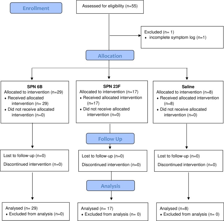
We recruited non-smoking healthy participants aged 18–60 years old (self-selection) as part of a larger EHPC dose ranging study between 24th May 2012 to 23rd October 2012, with follow up until January 2013. A modified CONSORT flow diagram is shown in Fig 1. Specimen collection and sample processing were conducted in Liverpool, UK. All participants gave written, informed consent. Ethical permission was granted by local NHS Research and Ethics Committee (REC) (11/NW/0592 Liverpool-East, date 11/10/2011). This study was retrospectively registered on the ISRCTN database (ISRCTN85403723) as this was not a mandatory requirement at the time of recruitment. All ongoing and related trials for this intervention are now prospectively registered with ISRCTN. Exclusion criteria included natural pneumococcal colonisation at baseline, any chronic medical condition or regular medication (study participation could put the participant at increased risk of pneumococcal disease) and regular contact with an at-risk individual such as young children (study participation could put the at-risk individual at increased risk of pneumococcal disease).

Fig 1. Modified CONSORT diagram.

Fig 1

Participants were nasally inoculated with 8x10⁴, 1.6x10⁵, or 3.2x10⁵ mid-log phase colony forming units (CFU) S. pneumoniae (prepared as previously described)[6]. Bacterial inoculation density was confirmed by serial dilutions of the inoculation stock onto blood agar (Oxoid). Two serotypes were used; 6B and 23F, both were fully sensitive to penicillin. 46 participants were allocated to be inoculated with S. pneumoniae (SPN 6B or 23F) as part of a dose-ranging study and 8 participants inoculated with saline as a control group. They were blinded to their groups.

Pre-inoculation oropharyngeal swabs were assayed for respiratory viruses using multiplex Polymerase Chain reaction (PCR) as previously published [13]. The PCR assay panel detected Influenza A and B, Respiratory syncytial virus, Human metapneumovirus, Human rhinovirus, Parainfluenza viruses 1–4 and Coronaviruses OC43, NL63, 229E and HKU1. Nasopharyngeal colonisation was assessed in nasal washes (Nacleiro technique, as previously described) collected at day 2, 7 and 14 post inoculation[14]. Pneumococcal colonisation status and density in nasal washes was determined by classical culture as previously described[6, 14].

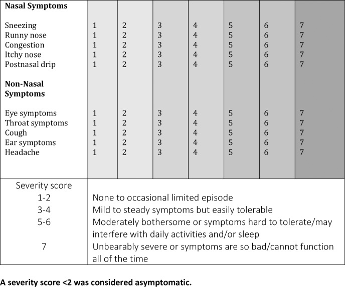
Participants were prompted to complete a daily symptom log on the day of inoculation (baseline) and daily for 7 days post-inoculation. The symptom log consisted of a 7-point visual analogue scale (a type of Likert scale) which assessed five nasal and five non-nasal symptoms[15]. The only modification to the validated questionnaire was the removal of ‘mental function’ as a non-nasal symptom (Fig 2). Scores ≥2 were considered ‘symptomatic’. The score awarded at inoculation (day 0) was considered their baseline score, the participant was considered symptomatic if the score went above baseline and ≥ 2.

Fig 2. Participant symptom log.

Fig 2

A severity score <2 was considered asymptomatic.

Graphical and statistical analyses were performed using GraphPad version 5.0 (GraphPad Software, La Jolla, CA, USA) and Microsoft Excel, with a p-value of <0.05 considered significant. Rates of symptoms reported between groups were compared using Fisher’s exact tests and Chi square where appropriate. Correlation analysis was performed using Spearman’s rank text. The daily symptom logs were collected at the next scheduled visit following completion.

Results

Fifty-five participants were recruited with an age range of 19–49 years old over a 6- month period from24/04/2012-23/10/2012. Participants with incomplete symptom severity score logs were excluded, therefore data from 54 participants were analysed. 46 participants were inoculated with SPN (29 with 6B, 17 with 23F) and 8 with saline (control group). Participants inoculated with 6B, 23F and saline were similar in age and gender distribution. In total, 14 participants became experimentally colonised (30.4%), all of which were inoculated with serotype 6B. None of the participants in the control group developed natural SPN colonisation during the study. Overall 72% (39/54) of participants reported either or both nasal or non-nasal symptoms during the 7 days post-inoculation. Of these symptoms, similar rates of nasal and non-nasal symptoms were reported; 59% (32/54) of participants reported nasal symptoms and 56% (30/54) reported non-nasal symptoms.

No statistical difference was seen between number of participants who reported symptoms in the experimental SPN positive or negative group. Similar rates of experimental SPN positive participants reported nasal symptoms (71%, 10/14) and non-nasal symptoms (57%, 8/14) compared to experimental SPN negative participants (50%, 16/32 in nasal (OR 2.50 [95% CI: 0.65–9.66] P = 0.212) and non-nasal (OR 1.33 [95% CI: 0.38–4.73] P = 0.754). See Fig 3.

Fig 3. Comparison of nasal, non-nasal and all symptoms between experimental SPN positive and negative participants.
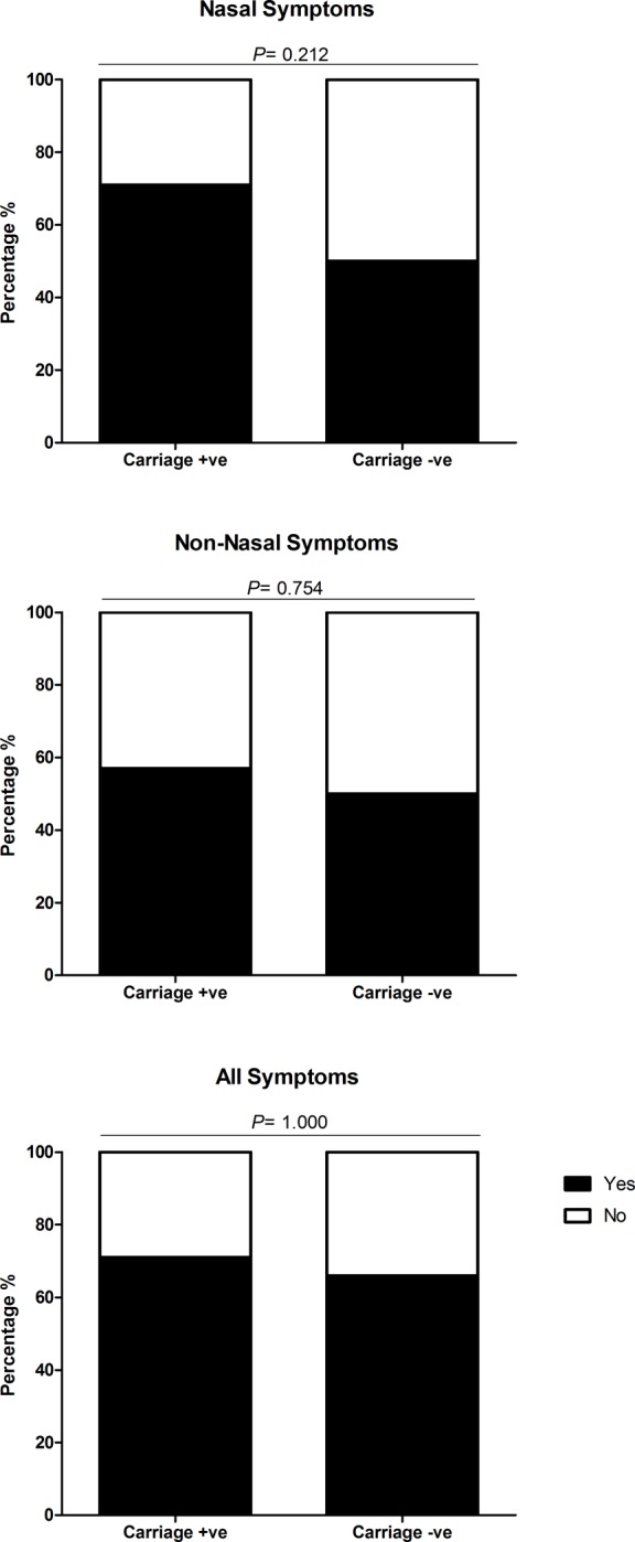
Fig 3

Each bar chart shows the percentage of carriage positive (N = 14) and carriage negative (N = 32) participants, who reported symptoms (nasal, non-nasal and all symptoms) after inoculation with Streptococcus pneumoniae serotypes 6B or 23F. Participants were deemed symptomatic if they scored >2, or >1 point above baseline for any of the five nasal or non-nasal symptoms in the visual analogue scale. The number of participants reporting symptoms between carriage positive and negative status were statistically compared using Fishers Exact and deemed significant if P = ≤0.05. There was no significant difference in the number of participants reporting nasal (OR 2.50 [95% CI: 0.65–9.66] P = 0.212), non-nasal (OR 1.33 [95% CI: 0.38–4.73] P = 0.754) or all symptoms (OR 1.31 [95% CI: 0.33 to 5.16] P = 1.000) between carriage positive and carriage negative participants.

Nasal SPN inoculation did not lead to greater rates of reported symptoms when compared to the control (saline inoculation) group, as show in Fig 4. Nasal symptoms were reported by 75% (6/8) of participants inoculated with saline compared to 57% (26/46) of those who were inoculated with SPN, no statistical difference was seen (p = 0.45). Similarly, no statistical difference was seen with the reporting of non-nasal symptoms 24/46 (52%) post-SPN inoculation compared to post-saline inoculation 6/8 (75%), (p = 0.28). Participants that reported ‘any symptom’ were higher in the control group 100% (8/8) compared to 67% (31/46) in the inoculation group, this was not statistically significant (p = 0.09).

Fig 4. Comparison of nasal, non-nasal and all symptoms between participants inoculated with S. pneumoniae compared to those inoculated with normal saline (control).
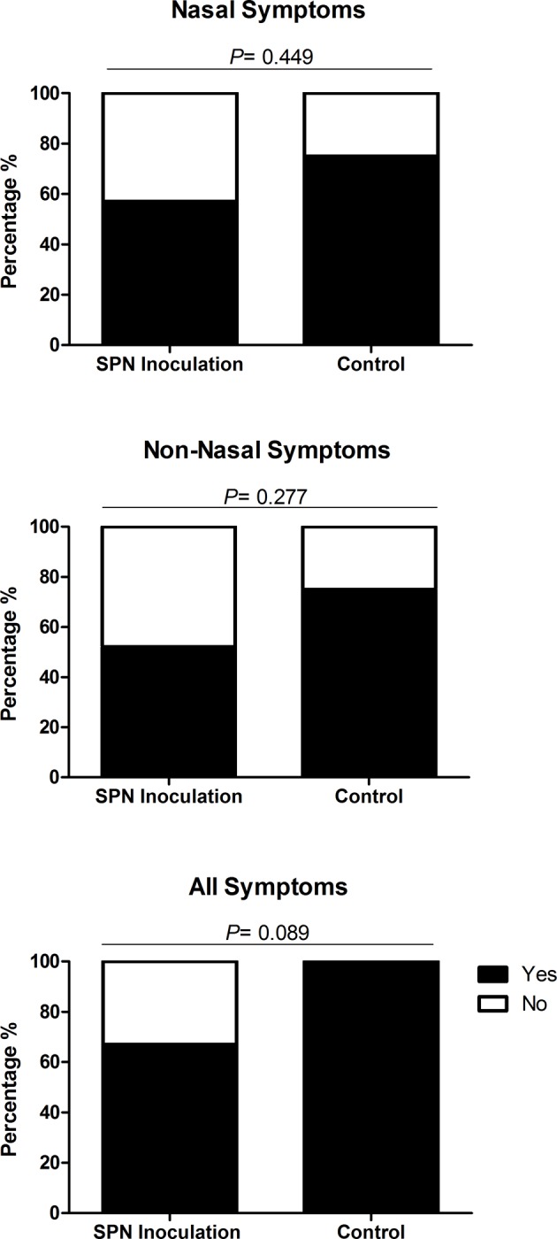
Fig 4

Each bar chart shows the percentage of participants inoculated with S. pneumoniae (SPN) serotypes 6B and 23F (N = 46) and normal saline (control) (N = 8) reporting symptoms (nasal, non- nasal and all symptoms) after inoculation. Participants were deemed symptomatic if they scored >2, or >1 point above baseline for any of the five nasal or non-nasal symptoms on the visual analogue scale. The number of participants reporting symptoms between inoculation with SPN and control were statistically compared using Fishers Exact and deemed significant if P = ≤0.05. There was no significant difference in the number of participants reporting nasal (OR 0.43 [95% CI: 0.08–2.38] P = 0.449), non-nasal (OR 0.36 [95% CI: 0.07–2.00] P = 0.277) and all symptoms (OR 0.12 [95% CI: 0.01–2.21] P = 0.089).

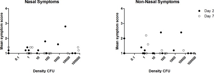
Of the 14 participants colonised with SPN, colonisation density was measured at days 2 and 7. No direct correlation was seen between SPN density and the mean symptom severity score at day 2 and day 7 for nasal (p = 0.86 Spearman’s correlation) and non-nasal symptoms (p = 0.83 Spearman’s correlation), Fig 5.

Fig 5. Correlation between pneumococcal colonisation density (SPN positive participants) and mean nasal symptom severity scores at days 2 and 7.

Fig 5

Spearman’s correlation was used to statistically analyse the correlation between bacterial colonisation density and the participants symptoms score on the visual analogue scale. Participants were deemed symptomatic if they scored >2, or >1 point above baseline for any of the five nasal or non-nasal symptoms in the visual analogue scale. No direct correlation was seen between SPN density and the mean symptom severity score at day 2 and day 7 for nasal (p = 0.86) or non-nasal symptoms (p = 0.83).

Viral colonisation data was available for 96% (52/54) participants at baseline. Viral colonisation was detected in 22% (12/52) of participants, 2 were inoculated with saline and 10 with SPN [serotype 23F (n = 2) and 6B (n = 8)]. There was no increase in nasal or non-nasal symptoms respectively in virus positive 8/12 (67%) and 7/12 (58%) respectively compared to virus negative participants 23/40 (58% for both symptoms), p = 0.74 and p = 1.0. Experimental SPN colonisation rates were higher in the presence of virus 6/10 (60%) compared to 8/35 (23%) in virus negative participants (p = <0.05). Virus and SPN positive (Co-colonised) participants did not report greater rates of nasal or non-nasal symptoms [4/6, (60%) for both symptoms],when compared to SPN positive only [nasal symptoms 6/8 (75%), OR 0.67 [95% CI: 0.06–6.88], P = 1.000), non-nasal symptoms 4/8 (50%), OR 2.00 [95% CI: 0.22–17.90] P = 0.627] and virus positive only [nasal symptoms 3/4 (75%) OR 3.23 [0.30–35.13] P = 0.600, non-nasal symptoms 2/4 (50%] OR 0.93 [95% CI: 0.11–7.59] P = 1.000). This is shown in Fig 6.

Fig 6. Comparison of nasal, non-nasal and all symptoms in carriage positive and negative participants with and without viral infection.
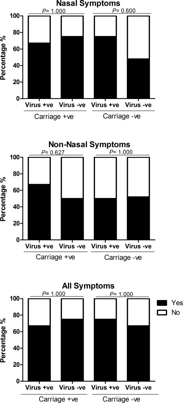
Fig 6

Each bar chart shows a comparison of the percentage of carriage positive and negative participants, after inoculation with S. pneumoniae serotype 6B or 23F, who reported symptoms (nasal, non-nasal and all symptoms), between those infected with a virus and those without viral infection. Participants were deemed symptomatic if they scored >2, or >1 point above baseline for any of the five nasal or non-nasal symptoms on the visual analogue scale. The number of participants reporting symptoms between those infected with a virus and those without viral infection were statistically compared using Fishers Exact and deemed significant if P = ≤0.05. There was no significant difference in the number of participants reporting nasal, non-nasal and all symptoms between those with viral infection and those without viral infection in both the carriage positive (nasal symptoms OR 0.67 [95% CI: 0.06–6.88], P = 1.000, non-nasal symptoms OR 2.00 [95% CI: 0.22–17.90] P = 0.627, all symptoms OR 0.67 [95% CI: 0.06–6.88] P = 1.000) and carriage negative groups (nasal symptoms OR 3.23 [0.30–35.13] P = 0.600, non-nasal symptoms OR 0.93 [95% CI: 0.11–7.59] P = 1.000, all symptoms OR 1.50 [95% CI: 0.14–16.55] P = 1.000).

Discussion

This study, with a clear methodology, provides evidence supporting the hypothesis that experimental pneumococcal (SPN) colonisation in adults is an asymptomatic event. This novel use of a human challenge model allowed the study of pneumococcal colonisation and symptomology in a controlled environment. The carriage rate of pneumococcus in this study was 30.4% and all with SPN6B. This is higher than the ‘natural rate’, due to the artificial introduction of the bacteria directly into the nasopharynx. This SPN 6B (BHN418) is known from epidemiological studies to have a very low prevalence in the community, therefore the participants are unlikely to have been exposed to it previously. The carriage rate for those inoculated with 23F was 0%. The reasons for the variability in carriage rate between serotypes is unclear but thought to be related to the evasion of mucociliary clearance, host nutrient availability and niche competition[8].

The strengths of this study are the robust methodology used to assess symptom severity[15], the lack of recall bias (due to prospective daily data log completion) and the use of a control group. Using this novel human challenge model, the exact day of pneumococcal inoculation and the onset and termination of each SPN colonisation episode was known allowing association between symptoms and pneumococcal presence and density over time. The main limitations of our study was the total sample size (n = 54), the lack of randomisation for group allocation and the use of a single serotype of SPN.

Although a previous study in adults used a small sample size (n = 14) and did not include the methods used to support this conclusion[16], it agrees with our data that pneumococcal colonisation in healthy adults is indeed asymptomatic. Higher symptom severity scores were not a predictor for colonisation.

SPN colonisation is more common in children; therefore, a limitation of this work is the lack of generalisability of results to all age groups, however reasonable evidence exists that SPN colonisation in children does cause nasal symptoms[10, 17]. Another limitation is that only one serotype was assessed in this study, SPN 6B. This particular serotype is not thought to be present in the community and therefore it is very unlikely that the participants would have pre-existing immunity from previous exposure. A previous study suggested that the presence of symptoms could be dependent on the serotype of pneumococcus[17]. The authors reported that colonisation with serotype 19F was strongly associated with symptoms such as coryza, sneezing, cough and expectoration. However, these children were recruited from a paediatric hospital emergency room, the study did not report on the diagnosis given to these patients therefore upper or lower respiratory infection may have been the cause of these symptoms rather than solely SPN colonisation[17].

Rodrigues et al found that rhinitis symptoms, rates of colonisation with SPN and Haemophilus Influenzae (Hi) in pre-school children decreased with age. Symptoms of rhinitis were reported using the Symptoms of Nasal Outflow Tally (SNOT) score. Both SPN and Hi colonisation were strongly associated with increased SNOT scores in children <5 years (p = 0.002 and 0.001) whereas colonisation with S. aureus was negatively associated with SNOT scores (p = 0.04). Interestingly, 40% of asymptomatic children (low SNOT score) were in fact SPN colonised. However, when the data was analysed considering age, the association between SPN colonisation and SNOT scores was weak (p = 0.06) whereas the association between SNOT scores and Hi colonisation remained strong (p = 0.003). This suggest that Hi may stimulate rhinitis in children to increase transmission[9]. The study by Rodrigues et al does not, however, report the effect of co-colonisation (SPN and virus) on symptoms.

Our results suggest that in adults co-colonisation (SPN and virus) is also an asymptomatic process with similar rates of nasal and non-nasal symptoms reported in all groups. Our results did show that asymptomatic viral infection at baseline was associated with the acquisition of SPN colonisation in adults. This is in keeping with results in children which found that asymptomatic viral infections had a large effect on SPN colonisation[18]. They reported that the proportion of children with SPN colonisation was higher during prompted visits for review of upper respiratory tract infections (URTI) symptoms rather than for regular planned study follow up visits. Due to the small sample size of SPN and virus co-colonisers (n = 6), it is difficult to make strong assumptions about the symptomology of co-infection from our study. Viral swabs were also only performed at baseline (up to 7 days prior to inoculation) therefore we cannot assess correlation between symptoms and viral status at each point, nor was density measured.

In conclusion we have provided evidence to support the hypothesis that neither nasopharyngeal inoculation nor experimental pneumococcal colonisation cause nasal or non-nasal symptoms in adults. Our results suggest that asymptomatic viral infection prior to nasopharyngeal inoculation or experimental SPN colonisation does not increase nasal or non-nasal symptoms. A better understanding of the process of viral co-infection in adults and the symptoms caused by viral infection prior to or following acquisition of SPN colonisation is needed and would add to this study’s preliminary data. A key question, given the difference between adults and children, is the association between colonisation symptoms and transmission; our study indicates that pneumococcal colonisation in adults is asymptomatic, but does not address transmission dynamics.

Supporting information

S1 Fig. TREND checklist.

(TIF)

S2 Fig. Experimental human pneumococcal carriage (EHPC) study protocol.

(PDF)

S3 Fig. Symptoms study flowchart.

(DOCX)

Acknowledgments

We would like to thank the members of our DMSC—Professor Robert C Read, University of Southampton (Chair), Professor David Lalloo and Dr Brian Faragher, Liverpool School of Tropical Medicine (LSTM), the respiratory research nurses, Clinical Research Unit staff, the Infectious diseases team, RLBUHT laboratories and clinical trials pharmacy.

This work would not have been possible without members of the LSTM respiratory team; Debbie Jenkins for data input, Jessica Owugha and Shaun Pennington for their involvement with statistical analysis and Jenna Gritzfeld for involvement in bacterial inoculum preparation, sample processing, storage and interpretation of samples and results.

We would like to acknowledge the Comprehensive Local Research Network for their support.

Data Availability

All relevant data are within the paper and its Supporting Information files.

Funding Statement

This work received support from the Bill and Melinda Gates Grand Challenges Exploration Programme II to DF, SG, descriptor number: 10.12. The funders did not play any role in the study design, data collection and analysis, decision to publish, or preparation of the manuscript.

References

  • 1.Goldblatt D, Hussain M, Andrews N, Ashton L, Virta C, Melegaro A, et al. Antibody responses to nasopharyngeal carriage of Streptococcus pneumoniae in adults: a longitudinal household study. JID. 2005;192(3):387–93. 10.1086/431524 [DOI] [PubMed] [Google Scholar]
  • 2.Bogaert D, De Groot R, Hermans PW. Streptococcus pneumoniae colonisation: the key to pneumococcal disease. Lancet Infect Dis. 2004;4(3):144–54. 10.1016/S1473-3099(04)00938-7 [DOI] [PubMed] [Google Scholar]
  • 3.Cobey S, Lipsitch M. Niche and neutral effects of acquired immunity permit coexistence of pneumococcal serotypes. Science. 2012;335(6074):1376–80. 10.1126/science.1215947 [DOI] [PMC free article] [PubMed] [Google Scholar]
  • 4.Margolis E, Yates A, Levin BR. The ecology of nasal colonization of Streptococcus pneumoniae, Haemophilus influenzae and Staphylococcus aureus: the role of competition and interactions with host's immune response. BMC microbiol. 2010;10(1):59. [DOI] [PMC free article] [PubMed] [Google Scholar]
  • 5.Simell B, Auranen K, Kayhty H, Goldblatt D, Dagan R, O'Brien KL. The fundamental link between pneumococcal carriage and disease. Expert Rev Vaccines. 2012;11(7):841–55. 10.1586/erv.12.53 [DOI] [PubMed] [Google Scholar]
  • 6.Ferreira DM, Neill DR, Bangert M, Gritzfeld JF, Green N, Wright AK, et al. Controlled human infection and rechallenge with Streptococcus pneumoniae reveals the protective efficacy of carriage in healthy adults. Am J Respir Crit Care Med. 2013;187(8):855–64. 10.1164/rccm.201212-2277OC [DOI] [PMC free article] [PubMed] [Google Scholar]
  • 7.McCool TL, Weiser JNJI, immunity. Limited role of antibody in clearance of Streptococcus pneumoniae in a murine model of colonization. J Exp Med. 2004;72(10):5807–13. [DOI] [PMC free article] [PubMed] [Google Scholar]
  • 8.Jochems SP, Weiser JN, Malley R, Ferreira. The immunological mechanisms that control pneumococcal carriage. PLoS Pathog. 2017;13(12):e1006665 10.1371/journal.ppat.1006665 [DOI] [PMC free article] [PubMed] [Google Scholar]
  • 9.Rodrigues F, Foster D, Nicoli E, Trotter C, Vipond B, Muir P, et al. Relationships between rhinitis symptoms, respiratory viral infections and nasopharyngeal colonization with Streptococcus pneumoniae, Haemophilus influenzae and Staphylococcus aureus in children attending daycare. Pediatr Infect Dis J. 2013;32(3):227–32. 10.1097/INF.0b013e31827687fc [DOI] [PubMed] [Google Scholar]
  • 10.Fan RR, Howard LM, Griffin MR, Edwards KM, Zhu Y, Williams JV, et al. Nasopharyngeal pneumococcal density and evolution of acute respiratory illnesses in young children, Peru, 2009–2011. EID. 2016;22(11):1996. [DOI] [PMC free article] [PubMed] [Google Scholar]
  • 11.Collins AM, Wright AD, Mitsi E, Gritzfeld JF, Hancock CA, Pennington SH, et al. First Human Challenge Testing of a Pneumococcal Vaccine—Double Blind Randomised Controlled Trial. Am J Respir Crit Care Med. 2015. [DOI] [PubMed] [Google Scholar]
  • 12.Rylance J, de Steenhuijsen Piters WA, Mina MJ, Bogaert D, French N, Ferreira DM. Two Randomized Trials of the Effect of Live Attenuated Influenza Vaccine on Pneumococcal Colonization. Am J Respir. 2019;199(9):1160–3. [DOI] [PMC free article] [PubMed] [Google Scholar]
  • 13.Glennie S, Gritzfeld JF, Pennington SH, Garner-Jones M, Coombes N, Hopkins MJ, et al. Modulation of nasopharyngeal innate defenses by viral coinfection predisposes individuals to experimental pneumococcal carriage. Mucosal Immunol. 2016;9(1):56–67. 10.1038/mi.2015.35 [DOI] [PMC free article] [PubMed] [Google Scholar]
  • 14.Gritzfeld JF, Wright AD, Collins AM, Pennington SH, Wright AK, Kadioglu A, et al. Experimental human pneumococcal carriage. Journal of visualized experiments: JoVE. 2013(72). [DOI] [PMC free article] [PubMed] [Google Scholar]
  • 15.Spector SL, Nicklas RA, Chapman JA, Bernstein IL, Berger WE, Blessing-Moore J, et al. Symptom severity assessment of allergic rhinitis: part 1. Ann Allergy Asthma Immunol. 2003;91(2):105–14. 10.1016/s1081-1206(10)62160-6 [DOI] [PubMed] [Google Scholar]
  • 16.McCool TL, Cate TR, Moy G, Weiser JN. The immune response to pneumococcal proteins during experimental human carriage. J Exp Med. 2002;195(3):359–65. 10.1084/jem.20011576 [DOI] [PMC free article] [PubMed] [Google Scholar]
  • 17.Neves FP, Pinto TC, Correa MA, dos Anjos Barreto R, de Souza Gouveia Moreira L, Rodrigues HG, et al. Nasopharyngeal carriage, serotype distribution and antimicrobial resistance of Streptococcus pneumoniae among children from Brazil before the introduction of the 10-valent conjugate vaccine. BMC Infect Dis. 2013;13:318 10.1186/1471-2334-13-318 [DOI] [PMC free article] [PubMed] [Google Scholar]
  • 18.DeMuri GP, Gern JE, Eickhoff JC, Lynch SV, Wald ER. Dynamics of Bacterial Colonization With Streptococcus pneumoniae, Haemophilus influenzae, and Moraxella catarrhalis During Symptomatic and Asymptomatic Viral Upper Respiratory Tract Infection. Clin Infect Dis. 2018;66(7):1045–53. 10.1093/cid/cix941 [DOI] [PMC free article] [PubMed] [Google Scholar]

Decision Letter 0

Ray Borrow

1 Dec 2019

PONE-D-19-25985

Pneumococcal Colonisation is an Asymptomatic Event in Healthy Adults using an Experimental Human Colonisation Model.

PLOS ONE

Dear Dr Robinson,

Thank you for submitting your manuscript to PLOS ONE. After careful consideration, we feel that it has merit but does not fully meet PLOS ONE’s publication criteria as it currently stands. Therefore, we invite you to submit a revised version of the manuscript that addresses the points raised below during the review process.

We would appreciate receiving your revised manuscript by Dec 30 2019 11:59PM. When you are ready to submit your revision, log on to https://www.editorialmanager.com/pone/ and select the 'Submissions Needing Revision' folder to locate your manuscript file.

If you would like to make changes to your financial disclosure, please include your updated statement in your cover letter.

To enhance the reproducibility of your results, we recommend that if applicable you deposit your laboratory protocols in protocols.io, where a protocol can be assigned its own identifier (DOI) such that it can be cited independently in the future. For instructions see: http://journals.plos.org/plosone/s/submission-guidelines#loc-laboratory-protocols

Please include the following items when submitting your revised manuscript:

  • A rebuttal letter that responds to each point raised by the academic editor and reviewer(s). This letter should be uploaded as separate file and labeled 'Response to Reviewers'.

  • A marked-up copy of your manuscript that highlights changes made to the original version. This file should be uploaded as separate file and labeled 'Revised Manuscript with Track Changes'.

  • An unmarked version of your revised paper without tracked changes. This file should be uploaded as separate file and labeled 'Manuscript'.

Please note while forming your response, if your article is accepted, you may have the opportunity to make the peer review history publicly available. The record will include editor decision letters (with reviews) and your responses to reviewer comments. If eligible, we will contact you to opt in or out.

We look forward to receiving your revised manuscript.

Kind regards,

Ray Borrow, Ph.D., FRCPath

Academic Editor

PLOS ONE

Journal Requirements:

1. When submitting your revision, we need you to address these additional requirements.

Please ensure that your manuscript meets PLOS ONE's style requirements, including those for file naming. The PLOS ONE style templates can be found at

http://www.journals.plos.org/plosone/s/file?id=wjVg/PLOSOne_formatting_sample_main_body.pdf and http://www.journals.plos.org/plosone/s/file?id=ba62/PLOSOne_formatting_sample_title_authors_affiliations.pdf

2.  Please include captions for your Supporting Information files at the end of your manuscript, and update any in-text citations to match accordingly. Please see our Supporting Information guidelines for more information: http://journals.plos.org/plosone/s/supporting-information.

3. Thank you for submitting your clinical trial to PLOS ONE and for providing the name of the registry and the registration number. The information in the registry entry suggests that your trial was registered after patient recruitment began. PLOS ONE strongly encourages authors to register all trials before recruiting the first participant in a study.

As per the journal’s editorial policy, please include in the Methods section of your paper:

1) your reasons for your delay in registering this study (after enrolment of participants started);

2) confirmation that all related trials are registered by stating: “The authors confirm that all ongoing and related trials for this drug/intervention are registered”.

Please also ensure you report the date at which the ethics committee approved the study as well as the complete date range for patient recruitment and follow-up in the Methods section of your manuscript.

4. Please ensure that you refer to Figure 5 in your text as, if accepted, production will need this reference to link the reader to the figure.

[Note: HTML markup is below. Please do not edit.]

Reviewers' comments:

Reviewer's Responses to Questions

Comments to the Author

1. Is the manuscript technically sound, and do the data support the conclusions?

The manuscript must describe a technically sound piece of scientific research with data that supports the conclusions. Experiments must have been conducted rigorously, with appropriate controls, replication, and sample sizes. The conclusions must be drawn appropriately based on the data presented.

Reviewer #1: Yes

Reviewer #2: Yes

**********

2. Has the statistical analysis been performed appropriately and rigorously?

Reviewer #1: Yes

Reviewer #2: Yes

**********

3. Have the authors made all data underlying the findings in their manuscript fully available?

The PLOS Data policy requires authors to make all data underlying the findings described in their manuscript fully available without restriction, with rare exception (please refer to the Data Availability Statement in the manuscript PDF file). The data should be provided as part of the manuscript or its supporting information, or deposited to a public repository. For example, in addition to summary statistics, the data points behind means, medians and variance measures should be available. If there are restrictions on publicly sharing data—e.g. participant privacy or use of data from a third party—those must be specified.

Reviewer #1: Yes

Reviewer #2: Yes

**********

4. Is the manuscript presented in an intelligible fashion and written in standard English?

PLOS ONE does not copyedit accepted manuscripts, so the language in submitted articles must be clear, correct, and unambiguous. Any typographical or grammatical errors should be corrected at revision, so please note any specific errors here.

Reviewer #1: Yes

Reviewer #2: Yes

**********

5. Review Comments to the Author

Please use the space provided to explain your answers to the questions above. You may also include additional comments for the author, including concerns about dual publication, research ethics, or publication ethics. (Please upload your review as an attachment if it exceeds 20,000 characters)

Reviewer #1: The manuscript by Trimble and colleagues present additional results from an experimental human challenge model, describing whether experimental pneumococcal colonisation in healthy adults is a symptomatic event. This manuscript is a resubmission from 2016 (but due to time limits is now a new submission) which has addressed some of the queries from the initial review. This study reports some interesting data but is nevertheless limited mainly due to the small sample size, especially for the secondary analyses.

Some points for consideration:

1. The pneumococcal colonisation rate was 30.4% (14/46), which was solely due to serotype 6B, and actually 48% if only those who were inoculated with 6B are included. This seems quite high for an otherwise healthy adult population. The authors state that this model mimics natural pneumococcal exposure but the high carriage rate may suggest otherwise. Can the authors provide some comment on the colonisation rate they observed with respect to what has been reported among UK healthy adults or more generally? Some explanation of this would greatly benefit the manuscript. The authors also argued against providing some comment on the lack of 23F colonisation but I tend to think that this would also be worth noting in the discussion, given that both these are commonly carried serotypes.

2. Figures 2 and 3 could be combined since they are both essentially reporting the same data but with a different comparison. Also, it should be Fisher’s exact test, not Fischer’s exact test.

3. Can the authors comment on the apparent high rate of nasal and non-nasal symptoms in the control group and whether they believe this could have contributed to the lack of any clinical symptoms being demonstrated for the other groups? An independent assessor may have been useful here.

4. There are a couple of instances where what is reported in the results is inconsistent with the data or discussion. For example, on lines 192-193 it states that “This study does not however report the effect of co-colonisation (SPN and virus) on symptoms” when this is clearly shown in Figure 5. Also on lines 207-208, it states that “…experimental SPN colonisation does not increase nasal or non-nasal symptoms” when in the results (lines 156-157) it states that “Experimental SPN colonisation rates were higher in the presence of virus….(p<0.05)”. Please correct. The p-value, if indeed significant, should be included in the Figure.

5. What were the viruses that were detected, and were there any associations between specific viruses?

6. For most of the reported p-values there are no = sign. Please include.

Reviewer #2: General comments

This study uses a human challenge model to examine if symptoms are experienced in the context of pneumococcal colonisation among adults. The structure of the paper is acceptable, however there could be improvement made in the finer details, e.g. error bars/confidence intervals and raw numbers

I think it is also important to mention the limitations of this study e.g. not an RCT, low sample size, and only one serotype successfully colonised. Consequently I think some of the statements in the discussion should have the language softened, as this study is not the definitive study which demonstrates that colonisation is asymptomatic among adults. Rather it might suggest this, but further work using more serotypes, in larger RCTs is needed.

Specific comments

#Abstract

Methods section line 46

" ... reported between groups was compared ..." to groups were compared

Results section line 49

S. pneumoniae (29 with serotype 6B, 17 with serotype 23F ...

Conclusion section line 58

Pneumococcal colonisation using the EHPC was asymptomatic ...

#Introduction

line 69 - correct typo, H. influenzae

line 70-73 - could be better worded

lines 83-87 - suggest rearranging this paragraph e.g.

The experimental pneumococcal challenge model (EHPC) mimics natural colonisation in healthy ....vaccines in randomised control trials. We aimed to use the EHPC to investigate if the process is symptomatic....

#Methods

Was a sample size calculation conducted?

#Results

line 134-137

please provide actual numbers, proportions, confidence intervals, and p-values.

figures - please add in confidence intervals.

#Discussion

Lines 164 and 212

I would tone down the language here e.g.

This study provides evidence supporting the hypothesis that SPN colonisation among adults is asymptomatic

line 171

Other than sample size, key limitations also include: the study wasn't randomised, only one serotype was assessed in this study, and pre-existing immunity.

line 213

I would suggest adding a sentence to say that larger randomised studies using a wider variety of

**********

6. PLOS authors have the option to publish the peer review history of their article (what does this mean?). If published, this will include your full peer review and any attached files.

If you choose “no”, your identity will remain anonymous but your review may still be made public.

Do you want your identity to be public for this peer review? For information about this choice, including consent withdrawal, please see our Privacy Policy.

Reviewer #1: No

Reviewer #2: Yes: Rama Kandasamy

[NOTE: If reviewer comments were submitted as an attachment file, they will be attached to this email and accessible via the submission site. Please log into your account, locate the manuscript record, and check for the action link "View Attachments". If this link does not appear, there are no attachment files to be viewed.]

While revising your submission, please upload your figure files to the Preflight Analysis and Conversion Engine (PACE) digital diagnostic tool, https://pacev2.apexcovantage.com/. PACE helps ensure that figures meet PLOS requirements. To use PACE, you must first register as a user. Registration is free. Then, login and navigate to the UPLOAD tab, where you will find detailed instructions on how to use the tool. If you encounter any issues or have any questions when using PACE, please email us at figures@plos.org. Please note that Supporting Information files do not need this step.

PLoS One. 2020 Mar 10;15(3):e0229558. doi: 10.1371/journal.pone.0229558.r003

Author response to Decision Letter 0


6 Jan 2020

Review Comments to the Author

Reviewer #1:

The manuscript by Trimble and colleagues present additional results from an experimental human challenge model, describing whether experimental pneumococcal colonisation in healthy adults is a symptomatic event. This manuscript is a resubmission from 2016 (but due to time limits is now a new submission) which has addressed some of the queries from the initial review. This study reports some interesting data but is nevertheless limited mainly due to the small sample size, especially for the secondary analyses.

Some points for consideration:

1. The pneumococcal colonisation rate was 30.4% (14/46), which was solely due to serotype 6B, and actually 48% if only those who were inoculated with 6B are included. This seems quite high for an otherwise healthy adult population. The authors state that this model mimics natural pneumococcal exposure but the high carriage rate may suggest otherwise. Can the authors provide some comment on the colonisation rate they observed with respect to what has been reported among UK healthy adults or more generally? Some explanation of this would greatly benefit the manuscript. The authors also argued against providing some comment on the lack of 23F colonisation but I tend to think that this would also be worth noting in the discussion, given that both these are commonly carried serotypes.

Thank you for your feedback, the EHPC model is able to artificially induce the otherwise naturally occurring phenomenon of pneumococcal carriage by pipetting the live bacteria directly into the participant’s nasopharynx. As the EHPC inoculation is more efficient than the ‘natural process’ the rate of colonisation is higher. This has now been added to the discussion. We have added an explanation for the difference in colonisation rate between serotypes as requested.

2. Figures 2 and 3 could be combined since they are both essentially reporting the same data but with a different comparison. Also, it should be Fisher’s exact test, not Fischer’s exact test.

Thank you, we have now corrected the spelling of Fisher’s exact test. We feel that combining the two figures together may affect the clarity of the underlying message, and therefore have respectfully chosen to keep them separate. We have however added a legend to all figures to make them easier to interpret.

3. Can the authors comment on the apparent high rate of nasal and non-nasal symptoms in the control group and whether they believe this could have contributed to the lack of any clinical symptoms being demonstrated for the other groups? An independent assessor may have been useful here.

We recognise that there is a high number of symptoms in the control group. We believe this may be due in part to the small study sample size but also reflects the high sensitivity of the Likert scale used to record any symptoms. Since the symptoms were self-reported by participants and they were blinded to their intervention group, we feel that an independent assessor would have been unlikely to improve this.

4. There are a couple of instances where what is reported in the results is inconsistent with the data or discussion. For example, on lines 192-193 it states that “This study does not however report the effect of co-colonisation (SPN and virus) on symptoms” when this is clearly shown in Figure 5. Also on lines 207-208, it states that “…experimental SPN colonisation does not increase nasal or non-nasal symptoms” when in the results (lines 156-157) it states that “Experimental SPN colonisation rates were higher in the presence of virus….(p<0.05)”. Please correct. The p-value, if indeed significant, should be included in the Figure.

Thank you, the comment regarding symptoms from co-colonisation refers to the Rodrigues at al study. This has been made clearer in the text to avoid confusion.

In the discussion we note that experimental SPN colonisation does not increase nasal or non-nasal symptoms. In the results section we note that experimental SPN colonisation was higher in the presence of virus. This is referring to two separate points. Participants who are positive for the presence of a virus and SPN (‘co-colonised’) did not appear to have greater rates of symptoms. The figure relating to this data (Figure 5) has been edited to include the relevant p- values and a legend has been added for clarity.

5. What were the viruses that were detected, and were there any associations between specific viruses?

A variety of viruses were detected, however since these were only present in very small numbers, we were unable to ascertain any relationship between symptoms and these specific viruses. In future studies we aim to further investigate this relationship in more detail.

For most of the reported p-values there are no = sign. Please include.

Thank you, this has now been added.

Reviewer #2: General comments

This study uses a human challenge model to examine if symptoms are experienced in the context of pneumococcal colonisation among adults. The structure of the paper is acceptable, however there could be improvement made in the finer details, e.g. error bars/confidence intervals and raw numbers

I think it is also important to mention the limitations of this study e.g. not an RCT, low sample size, and only one serotype successfully colonised. Consequently I think some of the statements in the discussion should have the language softened, as this study is not the definitive study which demonstrates that colonisation is asymptomatic among adults. Rather it might suggest this, but further work using more serotypes, in larger RCTs is needed.

Thank you, we have modified the manuscript to take this into account. Changes have been made to the abstract, discussion and conclusion to recognise this.

Specific comments

Abstract

Methods section line 46

" ... reported between groups was compared ..." to groups were compared

Thank you, this has now been updated.

Results section line 49

S. pneumoniae (29 with serotype 6B, 17 with serotype 23F ...

Thanks, this has been updated

Conclusion section line 58

Pneumococcal colonisation using the EHPC was asymptomatic ...

Thanks, this has also been updated as requested.

Introduction

line 69 - correct typo, H. influenzae

Thanks, this has now been updated

line 70-73 - could be better worded

This has been re-worded for clarity.

lines 83-87 - suggest rearranging this paragraph e.g.

The experimental pneumococcal challenge model (EHPC) mimics natural colonisation in healthy ....vaccines in randomised control trials. We aimed to use the EHPC to investigate if

the process is symptomatic....

Thank you this has now been updated.

Methods

Was a sample size calculation conducted?

Thank you for this feedback. We have not performed a sample size calculation as this was performed as a pilot study. In a larger follow up study we will ensure that a formal sample size calculation is done.

Results

line 134-137

please provide actual numbers, proportions, confidence intervals, and p-values.

These have been added where required.

figures - please add in confidence intervals.

Thank you, these have been included where required. A legend has been added to aid the interpretation.

Discussion

Lines 164 and 212

I would tone down the language here e.g.

This study provides evidence supporting the hypothesis that SPN colonisation among adults is asymptomatic

Thank you, this has been updated. We have taken this into account and altered the discussion and conclusion to reflect this.

line 171

Other than sample size, key limitations also include: the study wasn't randomised, only one serotype was assessed in this study, and pre-existing immunity.

The lack of randomisation to the allocated group and the use of a single successful serotype has been added as key limitations. As we used SPN 6B, a serotype that is not otherwise present in the community, it is unlikely that the participants have pre-existing immunity to this serotype. A line has been added to the discussion in order to address this.

line 213

Attachment

Submitted filename: PLOS one symptoms rebuttal letter 16.12.19.docx

Decision Letter 1

Ray Borrow

11 Feb 2020

Pneumococcal Colonisation is an Asymptomatic Event in Healthy Adults using an Experimental Human Colonisation Model.

PONE-D-19-25985R1

Dear Dr. Robinson,

We are pleased to inform you that your manuscript has been judged scientifically suitable for publication and will be formally accepted for publication once it complies with all outstanding technical requirements.

Within one week, you will receive an e-mail containing information on the amendments required prior to publication. When all required modifications have been addressed, you will receive a formal acceptance letter and your manuscript will proceed to our production department and be scheduled for publication.

Shortly after the formal acceptance letter is sent, an invoice for payment will follow. To ensure an efficient production and billing process, please log into Editorial Manager at https://www.editorialmanager.com/pone/, click the "Update My Information" link at the top of the page, and update your user information. If you have any billing related questions, please contact our Author Billing department directly at authorbilling@plos.org.

If your institution or institutions have a press office, please notify them about your upcoming paper to enable them to help maximize its impact. If they will be preparing press materials for this manuscript, you must inform our press team as soon as possible and no later than 48 hours after receiving the formal acceptance. Your manuscript will remain under strict press embargo until 2 pm Eastern Time on the date of publication. For more information, please contact onepress@plos.org.

With kind regards,

Ray Borrow, Ph.D., FRCPath

Academic Editor

PLOS ONE

Additional Editor Comments (optional):

Reviewers' comments:

Reviewer's Responses to Questions

Comments to the Author

1. If the authors have adequately addressed your comments raised in a previous round of review and you feel that this manuscript is now acceptable for publication, you may indicate that here to bypass the “Comments to the Author” section, enter your conflict of interest statement in the “Confidential to Editor” section, and submit your "Accept" recommendation.

Reviewer #1: All comments have been addressed

**********

2. Is the manuscript technically sound, and do the data support the conclusions?

The manuscript must describe a technically sound piece of scientific research with data that supports the conclusions. Experiments must have been conducted rigorously, with appropriate controls, replication, and sample sizes. The conclusions must be drawn appropriately based on the data presented.

Reviewer #1: Yes

**********

3. Has the statistical analysis been performed appropriately and rigorously?

Reviewer #1: Yes

**********

4. Have the authors made all data underlying the findings in their manuscript fully available?

The PLOS Data policy requires authors to make all data underlying the findings described in their manuscript fully available without restriction, with rare exception (please refer to the Data Availability Statement in the manuscript PDF file). The data should be provided as part of the manuscript or its supporting information, or deposited to a public repository. For example, in addition to summary statistics, the data points behind means, medians and variance measures should be available. If there are restrictions on publicly sharing data—e.g. participant privacy or use of data from a third party—those must be specified.

Reviewer #1: Yes

**********

5. Is the manuscript presented in an intelligible fashion and written in standard English?

PLOS ONE does not copyedit accepted manuscripts, so the language in submitted articles must be clear, correct, and unambiguous. Any typographical or grammatical errors should be corrected at revision, so please note any specific errors here.

Reviewer #1: Yes

**********

6. Review Comments to the Author

Please use the space provided to explain your answers to the questions above. You may also include additional comments for the author, including concerns about dual publication, research ethics, or publication ethics. (Please upload your review as an attachment if it exceeds 20,000 characters)

Reviewer #1: (No Response)

**********

7. PLOS authors have the option to publish the peer review history of their article (what does this mean?). If published, this will include your full peer review and any attached files.

If you choose “no”, your identity will remain anonymous but your review may still be made public.

Do you want your identity to be public for this peer review? For information about this choice, including consent withdrawal, please see our Privacy Policy.

Reviewer #1: No

Acceptance letter

Ray Borrow

18 Feb 2020

PONE-D-19-25985R1

Pneumococcal Colonisation is an Asymptomatic Event in Healthy Adults using an Experimental Human Colonisation Model.

Dear Dr. Robinson:

I am pleased to inform you that your manuscript has been deemed suitable for publication in PLOS ONE. Congratulations! Your manuscript is now with our production department.

If your institution or institutions have a press office, please notify them about your upcoming paper at this point, to enable them to help maximize its impact. If they will be preparing press materials for this manuscript, please inform our press team within the next 48 hours. Your manuscript will remain under strict press embargo until 2 pm Eastern Time on the date of publication. For more information please contact onepress@plos.org.

For any other questions or concerns, please email plosone@plos.org.

Thank you for submitting your work to PLOS ONE.

With kind regards,

PLOS ONE Editorial Office Staff

on behalf of

Prof. Ray Borrow

Academic Editor

PLOS ONE

Associated Data

    This section collects any data citations, data availability statements, or supplementary materials included in this article.

    Supplementary Materials

    S1 Fig. TREND checklist.

    (TIF)

    S2 Fig. Experimental human pneumococcal carriage (EHPC) study protocol.

    (PDF)

    S3 Fig. Symptoms study flowchart.

    (DOCX)

    Attachment

    Submitted filename: Response to reviewers.docx

    Attachment

    Submitted filename: PLOS one symptoms rebuttal letter 16.12.19.docx

    Data Availability Statement

    All relevant data are within the paper and its Supporting Information files.


    Articles from PLoS ONE are provided here courtesy of PLOS

    RESOURCES