Abstract
Background
Long noncoding RNAs (lncRNAs) play critical and complex roles in regulating various biological processes of cancers. Our study aimed to investigate the involvement of lncRNA NCK1-AS1 in urinary bladder cancer (UBC).
Methods
qRT-PCR was used to detect the expression of lncRNA NCK1-AS1 and miR-143 in UBC tissues and cells. The dual-luciferase reporter system assays were used to confirm the interaction between NCK1-AS1 and miR-143, and flow cytometry assays were applied to examine the behavioral changes in HT-1376 and HT-1197 cell lines.
Results
It was observed that NCK1-AS1 was up-regulated, while miR-143 was down-regulated in tumor tissues than in adjacent healthy tissues of urinary bladder cancer (UBC) patients. A 5-year survival analysis showed that the survival rate of patients with high NCK1-AS1 level or low miR-143 level in tumor tissues appears relatively low. Correlation analysis revealed a significant inverse correlation between NCK1-AS1 and miR-143 in tumor tissues. Over-expression NCK1-AS1 reduced the expression level of miR-143, while elevating the level of miR-143 failed to affect NCK1-AS1 expression. NCK1-AS1 over-expression led to promoted proliferation and increased percentage of CD133+ (stemness) cells.
Conclusion
Therefore, NCK1-AS1 promotes cancer cell proliferation and increases cell stemness in UBC patients by down-regulating miR-143.
Keywords: NCK1-AS1, miR-143, urinary bladder cancer, survival, proliferation, stemness
Introduction
Urinary bladder cancer is the 10th most commonly diagnosed malignancy.1 According to the latest GLOBOCAN statics, urinary bladder cancer (UBC) caused 549,393 new causes, accounting for 3.0% of all new cases, and caused 199,922 deaths, accounting for 2.1% all cancer mortalities.2 Incidence rate of UBC is significantly affected by gender. It is estimated that incidence of UBC is 4 times higher in males than in females.3 Besides gender, water contaminants, chemical exposure and cigarette smoking are the main risk factors of UBC.4 Most UBC patients are diagnosed at advanced stages and radical cystectomy is usually performed to treat advanced UBC.5 However, recurrence is common and prognosis is poor.6 Moreover, several lines of evidence suggest the contribution of cancer stem cells (CSCs) to the tumorigenicity of bladder cancer.7 CD24, CD44, CD133, Oct4, ALDH1A1 and Nanog were always known as the UBC stem cell markers, and most studies reported CD133 is adopted for detecting the UBC stem cells.8 To better understand the molecular mechanisms of the development of UBC, it requires more efforts to investigate this field from multi-lever and multi-angle.
A wealth of evidence has shown that non-coding RNAs (ncRNAs) are critical players in cancer biology.9 With protein-coding capacities, ncRNAs, such as long (>200 nt) non-coding RNAs (lncRNAs) and miRNAs, participate in cancer progression mainly by regulating gene expression.10 Besides the roles in the regulation of protein-coding gene expression, different lncRNAs can also interact with each other to play their roles.11 NCK1 divergent transcript (NCK1-DT, also named NCK1-AS1) is a recently identified oncogenic lncRNA in cervical cancer.12,13 Previous studies have revealed that LncRNA NCK1-AS1 promotes proliferation and induces cell cycle progression.12 More studies indicated that NCK1-AS1 was involved in many signaling pathways, such as CDK and TGF-β1 signaling.12,14 By analysis of TCGA dataset, we observed the significantly upregulated NCK1-AS1 in urothelial bladder carcinoma (TCGA-BLCA, see results: http://gepia.cancer-pku.cn/detail.php?gene=NCK1-AS1). MicroRNAs (miRNAs, ~20 nt) are a class of non-coding small RNAs that are highly conserved in evolution.15,16 There is increasing evidence that miRNAs regulate gene expression at the post-transcriptional level and play an important role in cell proliferation and tumor formation.17 MiR-143 is a tumor suppressor in UBC.18 Previous studies revealed miR-143 expression profiles in urinary bladder cancer always were correlated with clinical and epidemiological parameters.19 Meanwhile, the induction of miR-143 resulted the reductions of MMP9, CD44, Sox2 in UBC.20 Further study on the mechanism of the development of UBC will explore new research directions for the clinical directive significance. This study aimed to investigate the interaction between NCK1-AS1 and miR-143 in UBC.
Materials and Methods
UBC Patients and Specimens
This study passed the review board of The Affiliated Cancer Hospital of Harbin Medical University Ethics Committee. UBC and adjacent (2cm around tumors) non-tumor tissues were collected from 60 UBC patients (40 males and 20 females; 30–68 years; 50.2±6.1 years) through biopsy, which was performed before therapies and under the guidance of MRI. The 60 UBC patients were selected from the 122 cases of UBC admitted to aforementioned hospital between March 2011 and May 2014. Inclusion criteria: 1) patients diagnosed by histopathological biopsy; 2) newly diagnosed cases. Exclusion criteria: 1) any therapies for any clinical disorders were performed within 100 days before admission; 2) recurrent UBC; 3) patients transferred from other hospital; 4) multiple disorders were diagnosed. All tissue samples were kept in a liquid nitrogen sink before subsequent experiments.
Treatment and Follow-Up
The 60 UBC patients were staged according to the criteria established by AJCC. Based on clinical findings, the 60 patients included 16, 25 and 19 cases at stage II–IV, respectively. According to patients’ conditions, radical cystectomy in combination with radiotherapy or chemotherapy, or radiotherapy or chemotherapy alone was performed.
From the day of admission, the 60 patients were followed up for 5 years or until their deaths to record the survival. Follow-up visit was performed in a monthly manner through telephone. The patients died of other causes or the one who were lost during follow-up were not included in the survival analysis.
UBC Cells and Transient Transfections
HT-1376 and HT-1197 two UBC cell lines (ATCC, USA) were included. The cell culture medium was a mixture of 10% FBS and 90% Eagle’s Minimum Essential Medium. Cells were cultivated under the following conditions: 37°C, 95% humidity and 5% CO2.
To performed overexpression experiments, negative control (NC) miRNA and miR-143 mimic were purchased from Sigma-Aldrich (USA). Vector expressing NCK1-AS1 was constructed using pcDNA3.1 vector (Sangon, Shanghai, China). Lipofectamine 2000 (Sigma-Aldrich) was used to transfect 10 nM vector (empty vector as NC group) or 50 nM miRNA (NC miRNA as NC group) into 9×10⁵ cells, which were harvested at confluence of 80%. Untransfected cells were control (C) cells. All following experiments were performed using cells harvested at 48h post-transfection.
RNA Extractions and qPCR
TRIzol reagent (Thermo Fisher Scientific, Inc.) was used to extract total RNAs from 0.02g tissue samples or 105 cells. Tissues were ground in liquid nitrogen and cells were harvested at 48h post-transfection. In order to harvest miRNAs, RNA samples were precipitated using 85% ethanol.
In order to measure the expression levels of NCK1-AS1, PrimeScript RT Reagent Kit (Takara) was used to perform reverse transcriptions and all qPCR assays were performed using QuantiTect SYBR Green PCR Kit (QIAGEN). The expression level of NCK1-AS1 was normalized to endogenous control 18S rRNA.
To measure the expression level of mature miR-143, All-in-OneTM miRNA qRT-PCR Detection Kit (GeneCopoeia) was used to perform all steps including addition of poly (A), miRNA reverse transcription and qPCR assays.
All PCR reactions were repeated 3 times. After PCR, 2−ΔΔCt method was used to process all Ct values.
Cell Proliferation Analysis
HT-1376 and HT-1197 cells were harvested at 48h post-transfection. Cells were counted after trypsinization. To prepare single-cell suspensions, 1 mL aforementioned cell culture medium was used to resuspend cell pellets containing 3×10⁴ cells. Cell plates (96-well, 0.1mL per well) were used to cultivate cells under aforementioned hospital. Each well was added with 10 μL CCK-8 solution (Sigma-Aldrich) at 3 hrs before the end of cell culture. After cell culture was terminated, OD values were measured at 450 nm.
Apoptosis
Cells were collected, washed and suspended. Then, it was, incubated with Annexin V at 37°C for 10 mins, and got stained by propidium iodide. Flow cytometry was employed to measure the cell apoptosis rate.
Cell Stemness Analysis
HT-1376 and HT-1197 cells were harvested at 48hrs post-transfection. Cells were counted after trypsinization. IgG1-PE or CD133-PE antibody (Meltenyi Biotec) was used to stain 105 cells for 15mins at 4°C. After that, signals were detected using ACS Aria system (BD Immunocytometry Systems).
Statistical Analysis
All experiments were performed in 3 replicates and mean values of the 3 replicates were calculated and used in all data analyses. Correlations were analyzed by Pearson’s correlation coefficient. Differences between UBC and non-tumor tissues were explored using paired t-test. Differences among multiple cell groups were explored using ANOVA (one-way) combined with Tukey’s test. With the mean NCK1-AS1/miR-143 expression level in UBC as cutoff value, the 60 UBC patients were divided into high- and low-level groups (n=30). Survival curve plotting and comparison was performed by K-M plotter and Log-rank test, respectively. p<0.05 was statistically significant.
Results
NCK1-AS1 and miR-143 Showed Opposite Expression Patterns in UBC
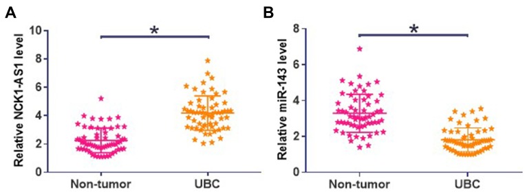
NCK1-AS1 and miR-143 expression level measurements and comparisons (UBC vs non-tumor) were performed by qPCR and paired t-test, respectively. Comparing to non-tumor tissues, expression levels of NCK1-AS1 were significantly higher in UBC tissues (Figure 1A, p<0.05). In contrast, significantly lower expression levels of miR-143 were significantly lower in UBC tissues than in non-tumor tissues (Figure 1B, p<0.05).
Figure 1.
NCK1-AS1 and miR-143 showed opposite expression patterns in UBC NCK1-AS1 (A) and miR-143 (B) expression level measurements and comparisons (UBC vs non-tumor) were performed by qPCR and paired t test, respectively. Three replicates were included and mean values were presented, *p<0.05.
Alter Expression Levels of NCK1-AS1 and miR-143 in UBC Tissues Predicted Poor Survival
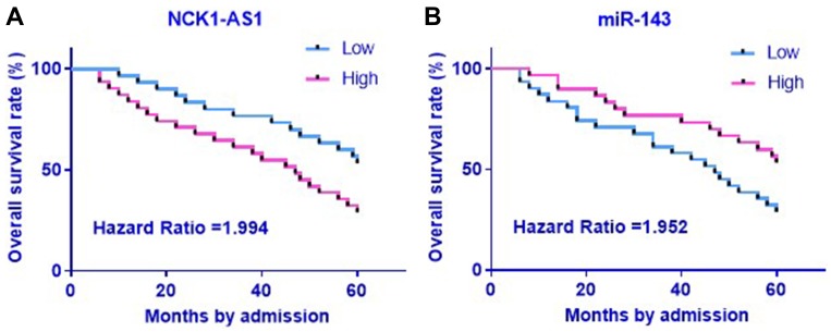
Survival curves were plotted and compared between high- and low-level groups using the aforementioned methods. Comparing to patients in low NCK1-AS1 level group, the overall survival rate of patients in high NCK1-AS1 level group was significantly lower (Figure 2A). Moreover, significantly lower overall survival rate was observed in low miR-143 level group comparing to high miR-143 level group (Figure 2B).
Figure 2.
Alter expression levels of NCK1-AS1 and miR-143 in UBC tissues predicted poor survival with the mean NCK1-AS1 (A)/miR-143 (B) expression level in UBC as cutoff value, the 60 UBC patients were divided into high- and low-level groups (n=30). Survival curve plotting and comparison were performed by K-M plotter and Log-rank test, respectively.
NCK1-AS1 and miR-143 Were Significantly and Inversely Correlated in UBC Tissues
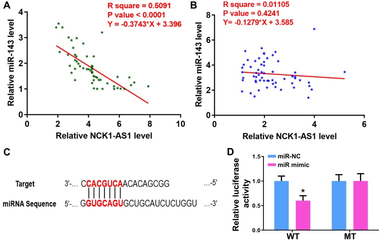
Correlations between the expression levels of NCK1-AS1 and miR-143 were analyzed by Pearson’s correlation coefficient. Expression levels of NCK1-AS1 were significantly and inversely correlated with the expression levels of miR-143 across UBC tissues (Figure 3A). However, no significant correlation between NCK1-AS1 and miR-143 was found across non-tumor tissues (Figure 3B). Analyzing the relation between the miR-143 and NCK1-AS1 by the miRDB (mirdb.org), the binding site is shown in Figure 3C. Furthermore, the luciferase reporter system confirmed this interaction in Figure 3D.
Figure 3.
NCK1-AS1 and miR-143 were significantly and inversely correlated in UBC tissues correlations between the expression levels of NCK1-AS1 and miR-143 across UBC tissues (A) and non-tumor tissues (B) were analyzed by Pearson’s correlation coefficient. (C). The binding site between NCK1-AS1 and miR-143. (D). The luciferase reporter system was used to detect the interaction. *p<0.05.
NCK1-AS1 Overexpression Mediated the Downregulation of miR-143 in UBC Cells
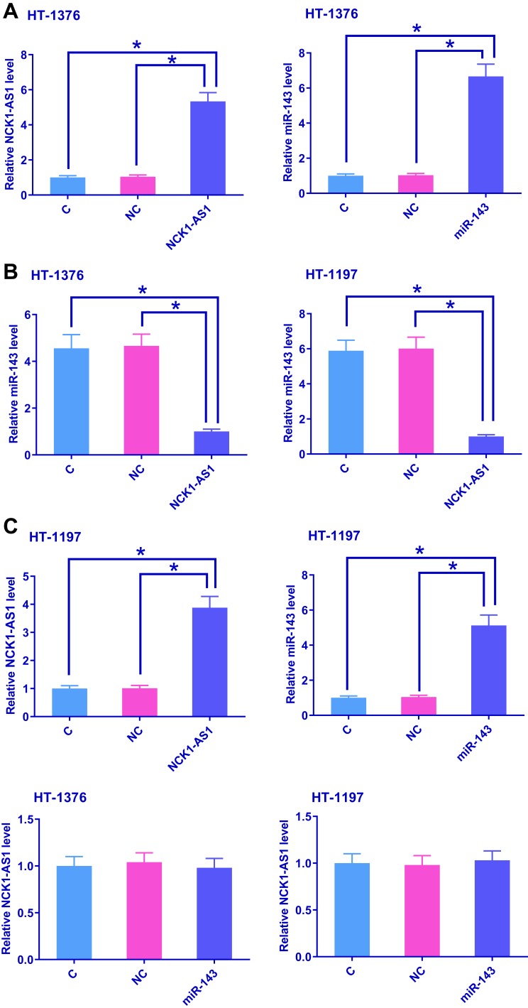
HT-1376 and HT-1197 cells were transfected with NCK1-AS1 expression vector and miR-143 mimic. NCK1-AS1 and miR-143 overexpression were confirmed by qPCR at 48hrs post-transfection. Comparing to C (untransfected cells) and NC (empty vector or NC miRNA transfection) groups, expression levels of NCK1-AS1 and miR-143 were significantly increased after transfections (Figure 4A, p<0.05). In addition, compared to two controls, NCK1-AS1 overexpression mediated the downregulation of miR-143 (Figure 4B, p<0.05), while miR-143 overexpression failed to significantly affect NCK1-AS1 expression (Figure 4C).
Figure 4.
NCK1-AS1 overexpression mediated the downregulation of miR-143 in UBC cells HT-1376 and HT-1197 cells was transfected with NCK1-AS1 expression vector and miR-143 mimic. NCK1-AS1 and miR-143 overexpression were confirmed by qPCR at 48hrs post-transfection (A). The effects of NCK1-AS1 overexpression on miR-143 expression (B) and the effects of miR-143 overexpression on NCK1-AS1 expression were analyzed by qPCR (C). Three replicates were included and mean values were presented, *p<0.05.
NCK1-AS1 Regulated UBC Cell Proliferation and Stemness Through miR-143
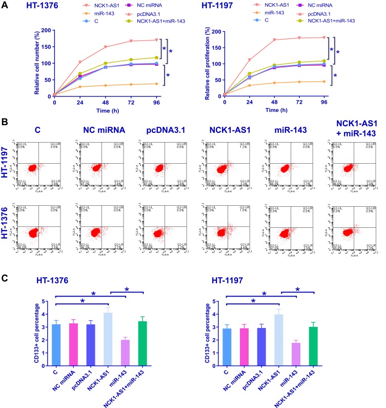
Cell proliferation and stemness assays were used to analyze the effects of NCK1-AS1 and miR-143 overexpression on the proliferation and stemness of HT-1376 and HT-1197 cells. Comparing to C (untransfected cells) and NC (empty vector or NC miRNA transfection) groups, NCK1-AS1 overexpression led to promoted proliferation (Figure 5A, p<0.05) and increased percentage of CD133+ (stemness) cells (Figure 5B and C, p<0.05). Also in Figure 5B and C, overexpression of miR-143 played an opposite role and attenuated the effects of NCK1-AS1 overexpression (p<0.05).
Figure 5.
NCK1-AS1 regulated UBC cell proliferation and stemness through miR-143. Cell proliferation and stemness assays were used to analyze the effects of NCK1-AS1 and miR-143 overexpression on the proliferation (A) and CD133+ (B, C) of HT-1376 and HT-1197 cells. Three replicates were included and mean values were presented, *p<0.05.
Discussion
The functions of NCK1-AS1 in UBC were analyzed in this study. We observed that NCK1-AS1 was upregulated in UBC and downregulate miR-143 to promote cancer cell proliferation and increase cell stemness. Moreover, altered expression levels of NCK1-AS1 and miR-143 predicted survival of UBC patients.
NCK1-AS1 is a recently identified oncogenic lncRNA and its functionality has only been investigated in cervical cancer. It has been reported that NCK1-AS1is upregulated in cervical cancer and can promote the proliferation of cancer cells by inducing cell phase transition.12 Besides that, the overexpression of NCK1-AS1 is also closely correlated with the development of chemoresistance in cervical cancer cells.13 In this study, we observed the upregulation of NCK1-AS1 in UBC. Consistent with the previous study we have proved that NCK1-AS1 can also increase the proliferation rate of UBC cells. Cancer stemness determines cancer progression potentials.21,22 In this study, we found that NCK1-AS1 overexpression increased the stemness of UBC cells. Therefore, our study reported the new functions of NCK1-AS1 in cancer biology.
MiR-143 is a well-characterized tumor-suppressive miRNA in different types of cancers such as colon cancer and glioblastoma.23,24 MiR-143 inhibits cancer progression by altering cancer cell properties, such as cell proliferation and stemness.23,24 In a previous study, Noguchi et al proved miR-143 as a tumor-suppressive miRNA in UBC.18 Consistently, our study also observed the downregulated miR-143 in UBC and the decreased cell proliferation rate and stemness after miR-143 overexpression. Our study confirmed the tumor-suppressive role of miR-143 in UBC.
We showed that NCK1-AS1 can downregulate miR-143 to regulate UBC cell proliferation and stemness. However, the mechanism is still unclear. It is known that lncRNAs can regulate the expression of miRNAs through epigenetic pathways. However, our preliminary methylation-specific PCR results revealed no significant effects of NCK1-AS1 overexpression on the methylation of miR-143 gene. In view the fact that NCK1-AS1 and miR-143 were only significantly correlated across UBC tissues not non-tumor tissues. The interaction between NCK1-AS1 and miR-143 is likely mediated by certain oncological factors.
In conclusion, NCK1-AS1 is upregulated in UBC and can downregulate miR-143 to promote UBC cell proliferation and increase stemness.
Acknowledgments
We thank the financial support from The Affiliated Cancer Hospital of Harbin Medical University, JJ2006-02.
Ethical Statement
All patients provided written informed consent, and that this study was conducted in accordance with the Declaration of Helsinki.
Disclosure
The authors report no conflicts of interest in this work.
References
- 1.Antoni S, Ferlay J, Soerjomataram I, et al. Bladder cancer incidence and mortality: a global overview and recent trends. Eur Urol. 2017;71(1):96–108. doi: 10.1016/j.eururo.2016.06.010 [DOI] [PubMed] [Google Scholar]
- 2.Bray F, Ferlay J, Soerjomataram I, et al. Global cancer statistics 2018: GLOBOCAN estimates of incidence and mortality worldwide for 36 cancers in 185 countries. CA Cancer J Clin. 2018;68(6):394–424. doi: 10.3322/caac.21492 [DOI] [PubMed] [Google Scholar]
- 3.Dobruch J, Daneshmand S, Fisch M, et al. Gender and bladder cancer: a collaborative review of etiology, biology, and outcomes. Eur Urol. 2016;69(2):300–310. doi: 10.1016/j.eururo.2015.08.037 [DOI] [PubMed] [Google Scholar]
- 4.Burger M, Catto JWF, Dalbagni G, et al. Epidemiology and risk factors of urothelial bladder cancer. Eur Urol. 2013;63(2):234–241. doi: 10.1016/j.eururo.2012.07.033 [DOI] [PubMed] [Google Scholar]
- 5.Stein JP, Lieskovsky G, Cote R, et al. Radical cystectomy in the treatment of invasive bladder cancer: long-term results in 1054 patients. J Clin Oncol. 2001;19(3):666–675. doi: 10.1200/JCO.2001.19.3.666 [DOI] [PubMed] [Google Scholar]
- 6.Westhoff E, Witjes JA, Fleshner NE, et al. Body mass index, diet-related factors, and bladder cancer prognosis: a systematic review and meta-analysis. Bladder Cancer. 2018;4(1):91–112. doi: 10.3233/BLC-170147 [DOI] [PMC free article] [PubMed] [Google Scholar]
- 7.Farid RM, Sammour SA-E, Shehab EZA, et al. Expression of CD133 and CD24 and their different phenotypes in urinary bladder carcinoma. Cancer Manag Res. 2019;11:4677–4690. doi: 10.2147/CMAR.S198348 [DOI] [PMC free article] [PubMed] [Google Scholar]
- 8.Qin J, Wang Y, Bai Yet al,. Epigallocatechin-3-gallate inhibits bladder cancer cell invasion via suppression of nf-κb?mediated matrix metalloproteinase-9 expression. Mol Med Rep.2012;6:1040–1044. [DOI] [PubMed] [Google Scholar]
- 9.Anastasiadou E, L S J, Slack FJ. Non-coding RNA networks in cancer. Nat Rev Cancer. 2018;18(1):5–18. doi: 10.1038/nrc.2017.99 [DOI] [PMC free article] [PubMed] [Google Scholar]
- 10.Esteller M. Non-coding RNAs in human disease. Nat Rev Cancer. 2011;12(12):861–874. doi: 10.1038/nrg3074 [DOI] [PubMed] [Google Scholar]
- 11.Paraskevopoulou MD, Hatzigeorgiou AG. Analyzing Mirna–Lncrna Interactions In Long Non-Coding RNAs. New York (NY): Humana Press; 2016:271–286. [DOI] [PubMed] [Google Scholar]
- 12.Li H, Jia Y, Cheng J, et al. LncRNA NCK1-AS1 promotes proliferation and induces cell cycle progression by crosstalk NCK1-AS1/miR-6857/CDK1 pathway. Cell Death Dis. 2018;9(2):198. doi: 10.1038/s41419-017-0249-3 [DOI] [PMC free article] [PubMed] [Google Scholar]
- 13.Zhang WY, Liu YJ, He Y, et al. Suppression of long noncoding RNA NCK1-AS1 increases chemosensitivity to cisplatin in cervical cancer. J Cell Physiol. 2019;234(4):4302–4313. doi: 10.1002/jcp.27198 [DOI] [PubMed] [Google Scholar]
- 14.Zhihui. G, Yuwei. S, Jinguo. M, et al. Altered expression of lncRNA NCK1-AS1 distinguished patients with prostate cancer from those with benign prostatic hyperplasia. Oncol Lett. 2019;18(6):6379–6384. doi: 10.3892/ol.2019.11039 [DOI] [PMC free article] [PubMed] [Google Scholar]
- 15.Chang Z-X, Tang N, Wang L, Zhang L-Q, Akinyemi IA, Wu Q-F. Identification and characterization of microRNAs in the white-backed planthopper, Sogatella furcifera. Insect Sci. 2016;23:452–468. doi: 10.1111/ins.2016.23.issue-3 [DOI] [PubMed] [Google Scholar]
- 16.Simona. R, Scott. K, Ramana. D, Hamilton Stanley R, Calin George A. MicroRNAs, ultraconserved genes and colorectal cancers. Int J Biochem Cell Biol. 2010;42(8):1291–1297. doi: 10.1016/j.biocel.2009.05.018 [DOI] [PubMed] [Google Scholar]
- 17.Ohlsson Teague EMC, Print CG, Hull ML. The role of microRNAs in endometriosis and associated reproductive conditions. Hum Reprod Update. 2010;16:142–165. doi: 10.1093/humupd/dmp034 [DOI] [PubMed] [Google Scholar]
- 18.Noguchi S, Mori T, Hoshino Y, et al. MicroRNA-143 functions as a tumor suppressor in human bladder cancer T24 cells. Cancer Lett. 2011;307(2):211–220. doi: 10.1016/j.canlet.2011.04.005 [DOI] [PubMed] [Google Scholar]
- 19.Boubaker NS, Spagnuolo M, Trabelsi N, et al. miR-143 expression profiles in urinary bladder cancer: correlation with clinical and epidemiological parameters. Mol Biol Rep. 2020. doi: 10.1007/s11033-019-05228-1 [DOI] [PubMed] [Google Scholar]
- 20.Zhang Q, Zhao W, Ye C. Honokiol inhibits bladder tumor growth by suppressing EZH2/miR-143 axis. Oncotarget. 2015;6:37335–37348. doi: 10.18632/oncotarget.6135 [DOI] [PMC free article] [PubMed] [Google Scholar]
- 21.Pradella D, Naro C, Sette C, et al. EMT and stemness: flexible processes tuned by alternative splicing in development and cancer progression. Mol Cancer. 2017;16(1):8. doi: 10.1186/s12943-016-0579-2 [DOI] [PMC free article] [PubMed] [Google Scholar]
- 22.Zhang Y, Jin Z, Zhou H, et al. Suppression of prostate cancer progression by cancer cell stemness inhibitor napabucasin. Cancer Med. 2016;5(6):1251–1258. doi: 10.1002/cam4.2016.5.issue-6 [DOI] [PMC free article] [PubMed] [Google Scholar]
- 23.Gomes SE, Simões AES, Pereira DM, et al. miR-143 or miR-145 overexpression increases cetuximab-mediated antibody-dependent cellular cytotoxicity in human colon cancer cells. Oncotarget. 2016;7(8):9368–9387. doi: 10.18632/oncotarget.7010 [DOI] [PMC free article] [PubMed] [Google Scholar]
- 24.Zhao S, Liu H, Liu Y, et al. miR-143 inhibits glycolysis and depletes stemness of glioblastoma stem-like cells. Cancer Lett. 2013;333(2):253–260. doi: 10.1016/j.canlet.2013.01.039 [DOI] [PubMed] [Google Scholar]


