

Abstract
Osteosarcoma of foot is a rare condition. Most common site of foot is calcaneum. Osteosarcoma of foot is usually not amenable to limb sparing surgery because of poor compartmentalization of tumour in the foot and subsequent need to amputate to achieve sound oncological margins. Vascularised fibular grafts have been recognised as an attractive choice for reconstruction. Here we present a case of osteosarcoma of first metatarsal treated by resection of tumour bearing first metatarsal and reconstruction with free vascularised fibular graft (FVFGs).
Keywords: Vascularised fibular graft, 1st metatarsal, Osteosarcoma
Introduction
Primary osteosarcoma of foot is extremely rare. In this regard, Berlin (1984) noted malignant tumours in less than 1% of the 67,000 ft lesions that he reviewed [1]. It is associated with clinical features not typical of conventional osteosarcoma. Resection and salvaging foot is technically challenging.
Given its ability to provide immediate structural support and vascularity free vascularised fibular graft has become attractive reconstructive option. Indication for free vascularised fibular graft is segmental bony defects of greater than 6 to 8 cm, such seen in post-traumatic, tumour resection and post-infectious bone loss [2].
Case report
Twenty-nine-year-old female with no known comorbidities, Eastern cooperative oncology group(ECOG) score 1 came with chief complaint of foot swelling for 6 months. Patient has history of trauma later which she noted swelling. Initially evaluated at local hospital and was later referred to Dr. B. Borooah cancer institute. On examination there was around 5 × 6 cm swelling on dorsum or right foot at first metatarsal area. Blood work up was with in normal limits except for serum alkaline phosphatase (206 u/l). X-ray right food was suggestive of expansile, lytic, destructive lesion of first metatarsal. Magnetic resonance imaging of lower limb was suggestive of approximately 5 × 3 cm lobulated T2 hyperintense lesion involving the first metatarsal shaft proximally, with cortical destruction with sparing of distal metatarsal. 99mTc-methylene diphosphate (MDP) whole body scan was suggestive of increased osteoblastic activity in first right metatarsal with uptake nowhere else. Contrast-enhanced computerised tomography of chest and abdomen was normal. Core biopsy was suggestive of low-grade osteosarcoma. Doppler of bilateral lower limbs was with in normal limits (Figs. 1 and 2).
Fig. 1.
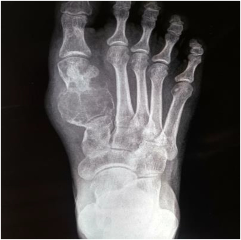
X-ray image of tumour
Fig. 2.

MRI image of tumour
Patient was taken for upfront surgery, resection of tumour-bearing first metatarsal, and reconstruction with vascularised fibular graft was done.
Technique of Reconstruction
- Wide excision of metatarsal tumour with surrounding soft tissue done (Fig. 3)
Fig. 3.
 Intra-op picture
 Osteotomy done through body of first cuneiform bone and distal to base of proximal phalanx.
Entire metatarsal bone-bearing tumour and two joints proximal and distal removed.
- Vascularised fibular graft taken from opposite leg (Fig. 4)
Fig. 4.
 Marking for fibular graft
 Microvascular anastomosis of fibular graft done, anastomosis of peroneal artery was done with dorsalis pedis artery.

Discussion
Osteosarcoma is one of the most common primary malignancy of bone. It is extremely rare in foot with calcaneum being most commonly involved. A review of cases at the Mayo Clinic showed that mean age was one decade older than conventional osteosarcoma and was more common in females. One explanation for older age of presentation was that osteosarcomas of small tubular bones arise secondarily to another process [3].
Vascularised bone grafts, by definition, are placed with their vascularity intact, and thus are immediately viable. Bone graft and bone graft substitutes have a number of inherent properties which allow them to initiate, stimulate, and facilitate bony healing [4, 5].
Biomechanically the fibula bears only 15% of the axial load across the ankle, allowing for its use as an autogenous bone graft with minimal biomechanical consequences on the weight bearing status of lower limb [6].
The endosteal blood supply to fibula is provided by a nutrient artery which typically enters the posterior fibular cortex at the junction of proximal one third and distal two thirds. This nutrient artery is a branch of peroneal artery. Fibula also receives additional vascularity from musculo-periosteal vessels which also emanate from peroneal artery [7].
Give the length of fibular diaphysis that may be harvested, free fibular grafts are well suited for the reconstruction of segmental defects of long bones, providing both mechanical strength and biological stimulus for healing. Based upon the fasciocutaneous arterial branches of peroneal artery, skin, fascia, and muscle may be harvested concomitantly with fibula to allow for more complex reconstruction. Apart from its advantages vascularised free fibular grafting is technically challenging. Donor site morbidity is seen in 10% of patients. Patients may also subsequently develop ankle pain, instability, and valgus deformity [8].
Preoperative planning should begin with exclusion of patients with peripheral vascular disease and deep venous thrombosis. Around 8% of population have hypoplasia or the absence of one or both of anterior and posterior tibial arteries, a condition called peronea arteria magna [9]. Absence of vessels at donor and recipient site is contraindication for this technique.
With current multidisciplinary approach for osteosarcoma limb salvage has become standard of care. Reconstruction with FVFGs are technically challenging but these have a very high bony union rates and can improve regional circulation, particularly when surrounding tissues have been damaged by chemotherapy and irradiation [10].
Conclusion
Vascularised free fibular grafting though technically challenging that to its complex reconstruction in foot has shown better results and should be used whenever its feasible. It provides immediate structural support and vascularity. Careful case selection and proper surgical technique results in better outcomes.
Footnotes
Publisher’s Note
Springer Nature remains neutral with regard to jurisdictional claims in published maps and institutional affiliations.
Contributor Information
Bibhuti Bhusan Borthakur, Email: borthakur.bb@gmail.com.
Srinivas Bannoth, Email: srinivasbannoth@gmail.com.
Sumanjit Boro, Email: Sumanjit.boro@yahoo.in.
Joydeep Purkayastha, Email: drjoydeeppurkayastha@gmail.com.
Abhijit Talukdar, Email: drabhijittalukdar881@gmail.com.
Deepjyoti Kalita, Email: kalita.deepjyoti@gmail.com.
References
- 1.Berlin S. A laboratory review of 67,000 foot tumours and lesion. J Am podiatric Assoc. 1984;74:341–347. doi: 10.7547/87507315-74-7-341. [DOI] [PubMed] [Google Scholar]
- 2.Green DP, Hotchkiss RN, Pederson WC, Wolfe S, editors. Green’s operative hand surgery. 5. Philadelphia: Churchill Livingston; 2005. [Google Scholar]
- 3.Peter FM. Choong, Abid A Qureshi, Franklin H Sim, K Krishnan Unni. A review of 52 patients at mayo clinic. Acta Orthop Scand. 1999;70(4):361–364. doi: 10.3109/17453679908997825. [DOI] [PubMed] [Google Scholar]
- 4.Buckwalter JA, Einhorn TA, Simon SR, editors. Orthopaedic basic science. Chicago: American Academy of Orthopaedic Surgeons; 2000. [Google Scholar]
- 5.Khan SN, Cammisa FP, Sandhu HS, Diwan AD, Firadi FP, Lane JM. The biology of bone grafting. J Am Acad Orthop Surg. 2005;13:77–86. doi: 10.5435/00124635-200501000-00010. [DOI] [PubMed] [Google Scholar]
- 6.Lambert KL. The weight bearing function of fibula. A strain gauge study. J Bone Joint Surg Am. 1996;78:204–211. doi: 10.2106/00004623-199602000-00006. [DOI] [PubMed] [Google Scholar]
- 7.Taylor GI, Miller GDH, Ham FJ. The free vascularized bone graft. A clinical extension of microsurgical technique. Plast Reconstr Surg. 1975;55:553. doi: 10.1097/00006534-197505000-00002. [DOI] [PubMed] [Google Scholar]
- 8.Tsai TM, Ludwig L, Tonkin M. Vascularized fibular epiphyseal transfer. A clinical study. Clin Othop Relat Res. 1986;210:228–234. [PubMed] [Google Scholar]
- 9.Heitmann C, Khan FN, Levin LS. Vasculature of peroneal artery: an anatomic study focused on the perforator vessels. J Reconstr Microsurg. 2003;19(3):157–162. doi: 10.1055/s-2003-39828. [DOI] [PubMed] [Google Scholar]
- 10.Malizos KN, Nunley JA, Goldner RD, Urbaniak JR, Harrelson JM. Free vascularized fibula in traumatic long bone defects and in limb salvaging following tumour resection: comparative study. Microsurgery. 1993;14(6):368–374. doi: 10.1002/micr.1920140603. [DOI] [PubMed] [Google Scholar]