

Abstract
Background
There is a controversy regarding whether ulnar styloid fractures affect the efficacy of treatment of distal radial fractures. The purpose of this study was to investigate whether ulnar styloid fractures impact wrist joint function in patients without distal radioulnar joint instability, after distal radial fracture fixation using a volar plate.
Materials and Methods
Seventy-five patients with a distal radial fracture were treated using a volar plate between February 2010 and February 2016 (33 men and 42 women; mean age 40.9 ± 9.4 years). Forty-four patients had ulnar styloid fractures (Group A) and 31 patients did not (Group B). There were no differences between the two groups with regard to sex, age, course of the disease and fracture type (P > 0.05).
Results
The mean follow-up time was 21 months. There were no differences between the groups in palmar tilt, radial inclination and radial length when the distal radial fractures had healed (P > 0.05). At the final follow-up visit, the two groups were similar in terms of wrist motion range, and grip and pinch strength (P > 0.05). The Gartland–Werley scores were 13 for excellent, 26 for good, 3 for fair and 2 for poor (excellence rate 89%) for Group A, and 10 for excellent, 17 for good, 2 for fair and 2 for poor (excellence rate 87%) for Group B. The difference between the two groups was not significant (Z = − 0.097, P = 0.922).
Conclusion
After open reduction and plate fixation of distal radial fractures, if stability of the distal radioulnar joint is achieved, untreated ulnar styloid fractures have no impact on wrist joint function.
Keywords: Distal radial fracture, Ulnar styloid fracture, Volar plate fixation, Wrist function, Treatment, Case–control study
Introduction
Distal radial fracture is common, accounting for 10% of bone injuries and ~ 75% of forearm fractures [1]. Approximately 50–65% of distal radial fractures are associated with ulnar styloid fractures [2]. Nevertheless, there is a controversy regarding whether an ulnar styloid fracture affects the efficacy of treatment of a distal radial fracture. Some previous studies have suggested that without treatment of the ulnar styloid fracture, there will be a poor clinical outcome, including distal radioulnar joint instability, ulnar-sided wrist pain, limited mobility and hand force reduction, which affect wrist joint function [3–5]. However, other studies have generated opposing radiographic and clinical results [6–9].
In many cases of distal radial fracture, triangular fibrocartilage complex traction leads to ulnar styloid fracture, which tends to cause distal radioulnar joint instability. To stabilise the distal radioulnar joint, open reduction and internal fixation is necessary for an ulnar styloid fracture [4, 10]. However, it is unclear whether the presence of an ulnar styloid fracture affects the efficacy of treatment of a distal radial fracture in patients without instability of the distal radioulnar joint. Therefore, the aim of this study was to investigate whether an ulnar styloid fracture affects wrist joint function in patients, in whom the distal radioulnar joint has been stabilised by screw fixation of a volar plate.
Materials and Methods
Study Population
Seventy-five patients with a distal radial fracture were treated using a volar plate between February 2005 and February 2011 (33 men and 42 women; mean age 40.9 ± 9.4 years). The inclusion criteria were: unilateral closed distal radial fracture, with or without ulnar styloid fracture; age ≥ 18 years; and treatment by open reduction and screw fixation of a volar plate. The exclusion criteria were: open fracture; old fracture; previous bilateral lesions or fracture of other parts of the radius and/or ulna; and intra-operative ballottement test showing distal radioulnar joint instability [11].
A prospective case–control study was conducted of patients with a distal radial fracture, with or without an ulnar styloid fracture. All of the patients satisfying the above criteria who were admitted to the hospital between February 2005 and February 2011 were enrolled. All of them underwent closed reduction by cast fixation in the emergency department. They were recommended to undergo open reduction and screw fixation of a volar plate, if the radiograph showed: a palmar tilt angle > 20°; ulnar inclination angle of < 15°; radial length reduction of > 5 mm; or a joint surface shift of > 2 mm. Of the 75 patients who underwent volar plate screw fixation and attended a follow-up appointment, 44 had an ulnar styloid fracture (Group A), of whom the ulnar styloid fracture shift was < 2 mm in 19 and ≥ 2 mm in 25. Thirty-one of the patients did not have an ulnar styloid fracture (Group B). The demographic characteristics of the patients are shown in Table 1. There were no statistically significant differences between the groups in the demographic characteristics of the patients (P > 0.05).
Table 1.
Demographic characteristics of the participants
| Group | Number of cases | Gender | Age | AO Type | Period from injury to surgery | Palmar tilt (°) | Radial inclination (°) | Radius length (mm) | |||
|---|---|---|---|---|---|---|---|---|---|---|---|
| Male | Female | A | B | C | |||||||
| A | 44 | 19 | 25 | 40.5 ± 15.3 | 17 | 13 | 14 | 3.4 ± 1.8 | 1.2 ± 1.6 | 12.3 ± 6.7 | 4.1 ± 1.2 |
| B | 31 | 14 | 17 | 41.3 ± 14.7 | 7 | 11 | 13 | 3.5 ± 1.5 | 1.5 ± 1.2 | 13.3 ± 5.3 | 4.5 ± 1.5 |
| Statistic | t = 0.029 P = 0.865 | t = 0.227 P = 0.821 | t = 4.839 P = 0.680 | t = 0.253 P = 0.801 | t = 0.882 P = 0.380 | t = 0.692 P = 0.491 | t = 1.281 P = 0.204 | ||||
Surgery and Post-operative Care
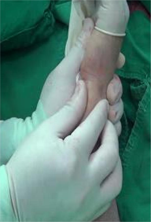
The surgical procedures were completed by the same group of surgeons. The ballottement test was used to determine the stability of the uninjured distal radioulnar joint (Figs. 1 and 2), and the same test was used to determine the stability of the injured joint after distal radial fracture reduction and fixation, for the purposes of comparison. The procedure used involved the Henry approach to reveal the fracture. Traction was achieved under direct visualisation, and reduction was achieved using reduction forceps, and verified using a C-arm X-ray machine. If the articular surface was still depressed, the joint capsule was opened and reduction was performed under direct visualisation to restore the articular surface, radial length, palmar tilt and radial inclination. Then, a smooth Kirschner wire was passed through the fracture from the radial styloid along an oblique line for temporary fixation, and a locking plate system (PuWei, Shanghai China) was used to fix the fracture. After fixation, wrist joint flexion and extension, radial deviation, pronation and supination range, and fracture stability were assessed, and the ballottement test was used to assess distal radioulnar joint stability. If the injured distal radioulnar joint was found to be less stable than the uninjured joint, the cause was identified and further treatment provided. This involved open reduction and internal fixation of any ulnar styloid fracture and repair of the triangular fibrocartilage complex if necessary, or plaster external fixation of the forearm at a 30° supination angle for 4 weeks. Such patients were not included in this study.
Fig. 1.

Ballottement test was used to determine the stability of the uninjured distal radioulnar joint before surgery
Fig. 2.
Ballottement test was used to compare the stability of the affected wrist joint after volar plate fixation for distal radial fracture
X-ray films were obtained as soon as the surgery was complete, and no subsequent external fixation was applied. The affected limb was raised to reduce post-surgical swelling. During the 1st day after the procedure, positive motion was applied to the wrist and all the more distal joints. After the pain of the incision had disappeared, the patients were instructed to undergo active functional training. Before and after the operation, antibiotics were administered, and post-operative analgesia was provided when necessary. Subsequently, X-ray images were periodically reviewed. After the fracture(s) had healed, the internal fixation was removed.
Evaluation of the Outcomes
X-ray films were used to evaluate the healing of the distal radial fractures, and to measure palmar tilt, radial inclination and radial length during healing. At the final follow-up visit, the Gartland–Werley score [12], wrist motion range, and hand grip and pinch strength were used to evaluate wrist joint function. The Gartland–Werley wrist joint rating was categorised as excellent, good, fair or poor, and comparisons of joint motion range and hand strength on the injured and uninjured sides were expressed using percentages.
Statistical Analysis
SPSS v.13.0 statistical software (SPSS Inc., Chicago, IL) was used for statistical analysis. Continuous data are presented as mean ± SD, normally distributed data were compared using Student’s t test, non-normally distributed data were compared using the rank sum test, and categorical data were analysed using the χ2 test. P < 0.05 was considered to represent statistical significance.
Results
The patients were followed up for a period of 12–39 months (mean 21 months). All experienced first intention healing of their incision, and there was no plate or screw breakage or loosening (Figs. 3, 4 and 5). The plate and screws were removed 12–18 months after the surgery. One patient in Group A experienced symptoms of median nerve entrapment 14 months after their procedure, and underwent carpal canal release surgery, during which bone entrapment and neurodegeneration were identified. However, the patient’s symptoms resolved following osteophyte removal and neurolysis. At the final follow-up visit, three patients in Group A and two patients in Group B showed ulnar-sided wrist pain. There were no statistically significant differences in the incidence of ulnar-sided wrist pain between the two groups (χ2 = 0.166, P = 0.684).
Fig. 3.
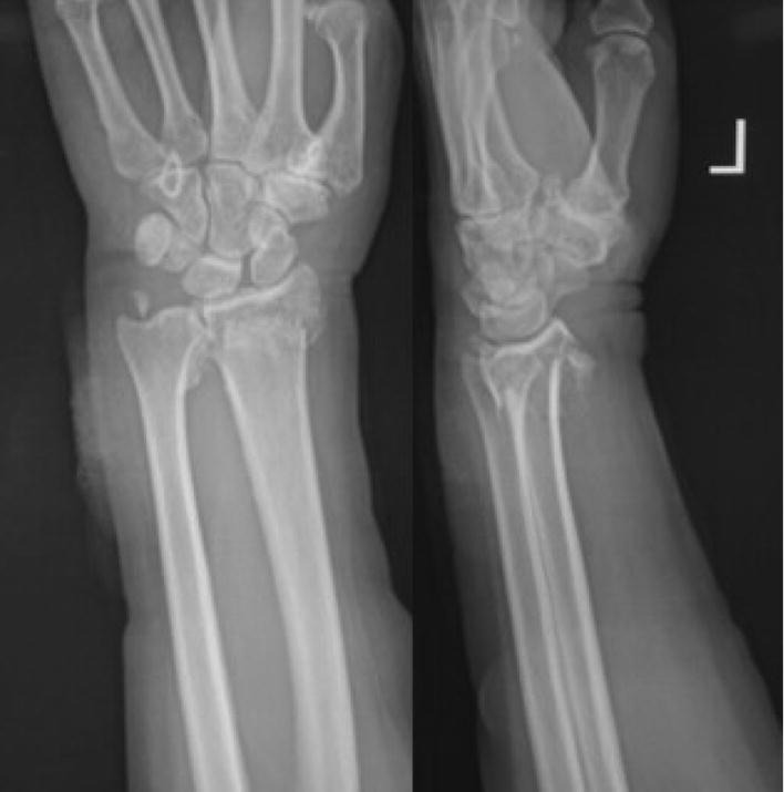
Preoperative radiographs of a 64-year-old woman, showing the distal radial fracture and non-union of the ulnar styloid, with > 2 mm styloid displacement
Fig. 4.
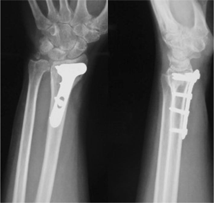
Postoperative radiographs were obtained immediately after surgery
Fig. 5.

Radiographs obtained at the follow-up appointment 13 weeks after surgery, showing union of the distal radial fracture and non-union of the ulnar styloid fracture
Radiographic Assessment
Radiography showed that all the distal radial fractures healed. The healing times for Groups A and B were similar (Group A 12.3 ± 2.5 weeks and Group B 11.4 ± 2.8 weeks; t = 1.461, P = 0.148). Twenty patients in Group A demonstrated healing of their ulnar styloid fracture (46%), with a mean healing time of 11.2 ± 1.6 weeks. There were no statistically significant differences in palmar tilt, radial inclination or radial length between the two groups (P > 0.05) (Table 2).
Table 2.
Palmar tilt, radial inclination and radius length for healed fractures in Groups A and B, determined radiographically
| Group | Number of cases | Palmar tilt (°) | Radial inclination (°) | Radius length (mm) |
|---|---|---|---|---|
| A | 44 | 8.9 ± 1.7 | 21.7 ± 1.9 | 9.8 ± 1.6 |
| B | 31 | 9.1 ± 1.4 | 22.5 ± 2.2 | 10.5 ± 1.5 |
| Statistic | t = 0.539 P = 0.592 | t = 1.681 P = 0.097 | t = 1.990 P = 0.051 | |
Evaluation of Function
At the last follow-up visit, the Gartland–Werley scores for Group A were 13 for excellent, 26 for good, 3 for fair and 2 for poor, giving an excellence rate of 89%. In Group B, the scores were 10 for excellent, 17 for good, 2 for fair and 2 for poor, giving an excellence rate of 87%. Thus, there were no statistically significant differences in Gartland–Werley score between the two groups (Z = − 0.097, P = 0.922). There were also no differences in wrist motion range, or grip or pinch strength between the two groups (P > 0.05) (Table 3). At the same time, the Gartland–Werley scores for participants in Group A with an ulnar styloid fracture shift of < 2 mm were 6 for excellent, 10 for good, 2 for fair and 1 for poor, giving an excellence rate of 84%, and the scores for participants with an ulnar styloid fracture shift of ≥ 2 mm were 7 for excellent, 16 for good, 1 for fair and 1 for poor, giving an excellence rate of 92%. Thus, there were no statistically significant differences in Gartland–Werley score between the two subgroups (Z = − 0.135, P = 0.892). In addition, for participants with ulnar styloid fracture shifts of < 2 mm and ≥ 2 mm, there were no statistically significant differences in wrist motion range, or grip or pinch strength (P > 0.05) (Table 4).The Gartland–Werley scores for participants in Group A in whom the ulnar styloid fracture healed were 7 for excellent, 11 for good, 2 for fair and 0 for poor, giving an excellence rate of 90%, and the scores for participants in whom the ulnar styloid fracture did not heal were 6 for excellent, 15 for good, 1 for fair and 2 for poor, giving an excellence rate of 88%. Thus, there were no statistically significant differences in the score between these two subgroups either (Z = − 0.740, P = 0.459). There were also no statistically significant differences in wrist motion range, or grip or pinch strength between the two subgroups (Figs. 6 and 7) (P > 0.05) (Table 5).
Table 3.
Wrist motion range, and grip and pinch strength in Groups A and B
| Group | Number of cases | Flexion and extension | Radioulnar partial | Pronation and supination | Grip strength | Pinch strength |
|---|---|---|---|---|---|---|
| A | 44 | 84.5 ± 5.8 | 84.4 ± 4.6 | 89.8 ± 3.9 | 82.6 ± 5.9 | 85.8 ± 7.1 |
| B | 31 | 85.3 ± 5.4 | 85.5 ± 5.7 | 90.6 ± 4.3 | 83.6 ± 6.4 | 86.6 ± 6.5 |
| Statistic | t = 0.605 P = 0.547 | t = 0.923 P = 0.360 | t = 0.838 P = 0.405 | t = 0.698 P = 0.487 | t = 0.497 P = 0.620 | |
Table 4.
Wrist motion range, and grip and pinch strength in participants with a styloid fracture shift of < 2 mm or ≥ 2 mm
| Group | Number of cases | Flexion and extension | Radioulnar partial | Pronation and supination | Grip strength | Pinch strength |
|---|---|---|---|---|---|---|
| < 2 mm | 19 | 84.8 ± 6.1 | 85.2 ± 4.5 | 90.2 ± 6.6 | 82.8 ± 4.5 | 86.4 ± 5.6 |
| ≥ 2 mm | 25 | 83.2 ± 5.3 | 84.2 ± 5.1 | 89.9 ± 5.8 | 81.7 ± 3.9 | 85.6 ± 5.2 |
| Statistic | t = 0.929 P = 0.358 | t = 0.677 P = 0.502 | t = 0.160 P = 0.874 | t = 0.867 P = 0.391 | t = 0.489 P = 0.627 | |
Fig. 6.

Wrist rotation being demonstrated at the final follow-up appointment 14 months after surgery
Fig. 7.
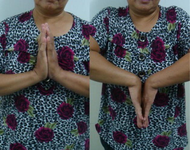
Wrist flexion and extension being demonstrated at the final follow-up appointment 14 months after surgery
Table 5.
Wrist motion range, and grip and pinch strength in participants with healed or non-healing fractures
| Group | Number of cases | Flexion and extension | Radioulnar partial | Pronation and supination | Grip strength | Pinch strength |
|---|---|---|---|---|---|---|
| United | 20 | 84.8 ± 6.4 | 85.2 ± 4.6 | 90.2 ± 4.8 | 82.3 ± 5.4 | 86.3 ± 5.8 |
| Ununited | 24 | 83.7 ± 5.4 | 84.2 ± 5.3 | 89.3 ± 3.7 | 82.9 ± 4.5 | 85.1 ± 4.6 |
| Statistic | t = 0.619 P = 0.540 | t = 0.661 P = 0.512 | t = 0.702 P = 0.486 | t = 0.402 P = 0.690 | t = 0.766 P = 0.448 | |
Discussion
The ulnar styloid plays an important role in wrist joint biomechanics. The distal radioulnar dorsal ligament is an important part of the triangular fibrocartilage complex, and its shallow and deep ligaments attach to the ulnar styloid base and ulnar nest. The deep ligament is the main stabilising structure of the distal radioulnar joint [13], and several studies have shown that ulnar styloid fractures are responsible for distal radioulnar joint instability. In 1996, ulnar styloid fractures were classified into two types by Hauck et al. [14]. The first type is a fracture below the distal radioulnar ligament attachment, which does not affect distal radioulnar joint stability, and the fracture fragments are small. In contrast, the second type is a fracture at the distal radioulnar ligament attachment site, which is associated with larger fracture fragments and distal radioulnar joint instability. Therefore, the authors recommended that patients with a distal radial fracture and an ulnar styloid fracture should undergo open reduction of their ulnar styloid fracture to prevent instability of the distal radioulnar joint.
Shaw et al. [10] found that patients with ulnar styloid fractures and avulsion of the triangular fibrocartilage complex can have their distal radioulnar joint stabilised by open reduction and internal fixation of the ulnar styloid fracture. Therefore, they recommended that open reduction and internal fixation should be the treatment of choice for ulnar styloid fractures involving an obvious shift, to stabilise the distal radioulnar joint. Of 272 patients with a distal radial fracture, Stoffelen et al. [4] reported 13 patients with distal radioulnar joint instability that was associated with ulnar styloid fracture. Finally, May et al. [5] conducted a retrospective study of 166 patients with a distal radial fracture, and found that 14 had distal radioulnar joint instability, 11 of whom had ulnar styloid fractures. They inferred that ulnar styloid fracture is a risk factor for distal radioulnar joint instability. Thus, our earlier observations and the findings of previous studies are consistent in implying that the presence of ulnar styloid fracture renders fracture treatment less effective.
However, other researchers have shown that a distal radial fracture in combination with an ulnar styloid fracture does not affect the stability of the distal radioulnar joint, but does lead to poor wrist joint function. Kim et al. [7] used volar plate fixation for 138 patients with an unstable distal radial fracture, 76 of whom also had an ulnar styloid fracture, and found that concomitant ulnar styloid fracture did not adversely affect distal radioulnar joint stability after internal fixation. The study by Lindau et al. [6, 15] generated consistent results. Theirs was a retrospective analysis of 76 patients with distal radial fracture, which found that patients with distal radioulnar joint instability demonstrated poor wrist joint function using both subjective and objective indicators, but distal radioulnar joint instability was not associated with ulnar styloid fracture. Therefore, it remains controversial whether distal radioulnar joint stability is promoted by the presence of an ulnar styloid fracture after the treatment of a distal radial fracture. However, few patients demonstrate instability of the distal radioulnar joint after the treatment of a distal radial fracture, and therefore lack of fixation of an ulnar styloid fracture would be unlikely to affect wrist joint function. For patients with a distal radial fracture accompanied by an ulnar styloid fracture, the present study has shown that if distal radial fracture reduction and fixation is sufficient to stabilise the distal radioulnar joint, an ulnar styloid fracture does not affect wrist joint function, regardless of whether the ulnar styloid fracture has healed, or its degree of separation.
Distal radioulnar joint stability can be evaluated using the ballottement test during surgery, and we found that if the distal radioulnar joint is stable, it is not necessary to treat an ulnar styloid fracture. However, if the distal radioulnar joint is unstable, it is necessary to determine the reasons for this: whether it is the result of the ulnar styloid fracture, or of distal radioulnar dorsal metacarpal ligament injury; and whether it is necessary to reduce and fix the ulnar styloid fracture, or repair the distal radioulnar dorsal ligament to stabilise the distal radioulnar joint.
In the present study, most patients achieved distal radioulnar joint stability through conservative treatment using forearm supination cast immobilization, although some did require repeated ligament reconstruction to stabilise the joint. Kim et al. [7] have reported that of 138 distal radial fracture patients that underwent volar plate fixation, 76 had an ulnar styloid fracture. An examination conducted during surgery showed that 32 patients had an unstable distal radioulnar joint, and after subsequent plywood forearm supination fixation for 4 weeks, two patients still demonstrated joint instability. Therefore, when treating such patients conservatively, it is necessary to inform them that distal radioulnar joint instability may remain in the long term that can affect joint function. If the patients are unwilling to accept this possibility, intra-operative assessment revealing distal radioulnar joint instability should be followed by immediate ulnar styloid reduction and fixation.
Wijffels et al. [16] and Yuan et al. [17] found that there was no relationship between the non-union of the ulnar styloid process and function in patients with a distal radial fracture in meta-analyses. Furthermore, Turan et al. [18] did not consider ulnar styloid fracture in the initial treatment of distal radial fracture in their study of 56 patients, but found that when the triangular fibrocartilage complex was also injured, surgical treatment of the ulnar fracture should be considered. Pidgeon et al. [19] found that an ulnar styloid fracture that disrupts the fovea causes instability of the distal radioulnar joint during 3 Nm torque pronation and supination in cadavers, and they therefore recommended that screw fixation of an ulnar styloid base fracture should be performed to achieve anatomic reduction. However, this only partially restores rotational stability to the joint, and only during pronation. Finally, Mam et al. [20] found that there was no significant difference in the outcomes of patients that did or did not have an ulnar styloid process fracture 1 year after surgery.
May et al. [5] reported that the presence of an ulnar styloid fracture is associated with post-surgical wrist pain after the repair of a distal radial fracture. The present study found that three patients in Group A had non-union of the ulnar styloid fracture and two patients in Group B had ulnar-sided wrist pain at the final follow-up appointment, and thus there was no difference between the prevalence of ulnar-sided wrist pain between the two groups. Careful analysis of the X-ray films found that ulnar-sided wrist pain was associated with poor distal radial fracture reduction and insufficient radial length, leading to ulnar positive variance. Indeed, wrist rotation, ulnar deviation and pressure are known to cause ulnar-sided wrist pain and wrist joint ulnar impaction syndrome. Thus, although ulnar styloid fracture non-union is the likely explanation for ulnar-sided wrist pain, as long as the radius length is restored, the risk of ulnar pain is likely to be significantly lower. Three patients in Group A had their ununited ulnar styloid removed when the distal radial plate was removed, which led to the disappearance of their post-surgical ulnar-sided wrist pain. In contrast, when Gogna et al. [21] conducted a clinical study of 47 patients, of whom 28 had an associated ulnar styloid fracture and 19 did not, they found that ulnar styloid fracture or its non-union does not affect the outcome of an adequately fixed distal radial fracture. They therefore urged caution in choosing surgical treatment for an ununited fracture of the ulnar styloid until a more comprehensive study of the treatments for pain associated with these fractures has been completed. Thus, in cases of non-healing ulnar styloid fracture that demonstrates ulnar side pain after surgery, MRI/CT should be used to evaluate in detail whether secondary surgery is indicated for ulnar styloid process fixation. If this is not attended to, wrist function is subsequently likely to be negatively impacted.
The principal limitation of this study was that the participants were limited to distal radial fracture patients who were treated using a volar plate, which prevented evaluation of the efficacy of the treatment method and its impact on the evaluation of the significance of ulnar styloid fracture for wrist joint function.
In summary, we have shown that if distal radioulnar joint stability is achieved by open reduction and fixation of a distal radial fracture, untreated ulnar styloid fracture has no impact on subsequent wrist joint function.
Author Contributions
Conceived the design of the study: SL and BS. Performed the research: JH and HW. Analyzed the data: HL and KS. Contributed new methods or models: HW and BL. Wrote the paper: SL. Contributed equally to the study: SL and DY. Read and approved the content of the final manuscript: all authors.
Availability of Supporting Data
The data sets supporting the results of this article are included within the article and its additional files.
Compliance with Ethical Standards
Conflict of interest
The author declare that they have no coflict of interest.
Declaration of Patient Consent
The authors certify that they have obtained all appropriate patient consent forms. In the form, the patient(s) has/have given his/her/their consent for his/her/their images and other clinical information to be reported in the journal. The patients understand that their names and initials will not be published and due efforts will be made to conceal their identity, but anonymity cannot be guaranteed. The study was approved by the Hospital Ethics Committee.
Consent for Publication
Written informed consent for publication of their clinical details and/or clinical images was obtained from the patient. A copy of the consent form is available for review by the Editor of this journal.
Footnotes
Publisher's Note
Springer Nature remains neutral with regard to jurisdictional claims in published maps and institutional affiliations.
Contributor Information
Shuzhen Li, Email: lishuzhenjoint@163.com.
Hao Wang, Email: 825977211@qq.com, Email: wanghaojoint@163.com.
Bo Su, Email: 15877126379@qq.com.
Jie Han, Email: Jham@163.com.
Haibo Liang, Email: 1023582191@qq.com.
Ke Sun, Email: SKgutou@163.com.
Dong Yin, Email: tangin2002@163.com.
References
- 1.Fujitani R, Omokawa S, Akahane M, Iida A, Ono H, Tanaka Y. Predictors of distal radioulnar joint instability in distal radius fractures. J Hand Surg Am. 2011;36:1919–1925. doi: 10.1016/j.jhsa.2011.09.004. [DOI] [PubMed] [Google Scholar]
- 2.Sammer DM, Shah HM, Shauver MJ, Chung KC. The effect of ulnar styloid fractures on patient-rated outcomes after volar locking plating of distal radius fractures. J Hand Surg Am. 2009;34:1595–1602. doi: 10.1016/j.jhsa.2009.05.017. [DOI] [PMC free article] [PubMed] [Google Scholar]
- 3.Mikic ZD, Yugoslavia NS. Treatment of acute injuries of the triangular fibrocartilage complex associated with distal radioulnar joint instability. J Hand Surg Am. 1995;20:319–323. doi: 10.1016/S0363-5023(05)80033-4. [DOI] [PubMed] [Google Scholar]
- 4.Stoffelen D, De Smet L, Broos P. The importance of the distal radioulnar joint in distal radial fractures. J Hand Surg Br. 1998;23:507–511. doi: 10.1016/S0266-7681(98)80134-4. [DOI] [PubMed] [Google Scholar]
- 5.May MM, Lawton JN, Blazar PE. Ulnar styloid fractures associated with distal radius fractures: incidence and implications for distal radioulnar joint instability. J Hand Surg Am. 2002;27:965–971. doi: 10.1053/jhsu.2002.36525. [DOI] [PubMed] [Google Scholar]
- 6.Lindau T, Hagberg L, Adlercreutz C, Jonsson K, Aspenberg P. Distal radioulnar instability is an independent worsening factor in distal radial fractures. Clin Orthop Relat Res. 2000;376:229–235. doi: 10.1097/00003086-200007000-00031. [DOI] [PubMed] [Google Scholar]
- 7.Kim JK, Koh YD, Do NH. Should an ulnar styloid fracture be fixed following volar plate fixation of a distal radial fracture? J Bone Joint Surg Am. 2010;92:1–6. doi: 10.2106/JBJS.H.01738. [DOI] [PubMed] [Google Scholar]
- 8.Souer JS, Ring D, Matschke S, Audige L, et al. Effect of an unrepaired fracture of the ulnar styloid base on outcome after plate-and-screw fixation of a distal radial fracture. J Bone Joint Surg Am. 2009;91:830–838. doi: 10.2106/JBJS.H.00345. [DOI] [PubMed] [Google Scholar]
- 9.Kazemian GH, Bakhshi H, Lilley M, et al. DRUJ instability after distal radius fracture: a comparison between cases with and without ulnar styloid fracture. Int J Surg. 2011;9:648–651. doi: 10.1016/j.ijsu.2011.08.005. [DOI] [PubMed] [Google Scholar]
- 10.Hauck RM, Skahen J, 3rd, Palmer AK. Classification and treatment of ulnar styloid nonunion. J Hand Surg Am. 1996;21:418–422. doi: 10.1016/S0363-5023(96)80355-8. [DOI] [PubMed] [Google Scholar]
- 11.Moriya T, Aoki M, Iba K, Ozasa Y, Wada T, Yamashita T. Effect of triangular ligament tears on distal radioulnar joint instability and evaluation of three clinical tests: a biomechanical study. J Hand Surg Eur. 2009;34:219–223. doi: 10.1177/1753193408098482. [DOI] [PubMed] [Google Scholar]
- 12.Gartland JJ, Jr, Werley CW. Evaluation of healed Colles’fractures. J Bone Joint Surg Am. 1951;33:895–907. doi: 10.2106/00004623-195133040-00009. [DOI] [PubMed] [Google Scholar]
- 13.Kleinman WB. Stability of the distal radioulna joint: biomechanics, pathophysiology, physical diagnosis, and restoration of function what we have learned in 25 years. J Hand Surg Am. 2007;32:1086–1106. doi: 10.1016/j.jhsa.2007.06.014. [DOI] [PubMed] [Google Scholar]
- 14.Shaw JA, Bruno A, Paul EM. Ulnar styloid fixation in the treatment of posttraumatic instability of the radioulnar joint: a biomechanical study with clinical correlation. J Hand Surg Am. 1990;15:712–720. doi: 10.1016/0363-5023(90)90142-E. [DOI] [PubMed] [Google Scholar]
- 15.Lindau T, Adlercreutz C, Aspenberg P. Peripheral tears of the triangular fibrocartilage complex cause distal radioulnar joint instability after distal radial fractures. J Hand Surg Am. 2000;25:464–468. doi: 10.1053/jhsu.2000.6467. [DOI] [PubMed] [Google Scholar]
- 16.Wijffels MME, Keizer J, Buijze GA, et al. Ulnar styloid process nonunion and outcome in patients with a distal radius fracture: a meta-analysis of comparative clinical trials[J] Injury. 2014;45(12):1889–1895. doi: 10.1016/j.injury.2014.08.007. [DOI] [PubMed] [Google Scholar]
- 17.Yuan C, Zhang H, Liu H, et al. Does concomitant ulnar styloid fracture and distal radius fracture portend poorer outcomes? a meta-analysis of comparative studies. Injury. 2017;48:2575–2581. doi: 10.1016/j.injury.2017.08.061. [DOI] [PubMed] [Google Scholar]
- 18.Turan S, Çankaya D, Yinolmaz S, et al. Effect of ulnar styloid fracture on outcomes after conservative treatment of distal radius fracture. Eklem Hastalık Cerrahisi. 2016;27(2):87. doi: 10.5606/ehc.2016.19. [DOI] [PubMed] [Google Scholar]
- 19.Pidgeon TS, Crisco JJ, Waryasz GR, et al. Ulnar styloid base fractures cause distal radioulnar joint instability in a cadaveric model. Hand. 2017;13:65–73. doi: 10.1177/1558944716685830. [DOI] [PMC free article] [PubMed] [Google Scholar]
- 20.Mam M, Fuhri Snethlage LJ, Ro DMK, et al. Functional outcomes of distal radius fractures with and without ulnar styloid fractures: a meta-analysis. J Hand Surg Eur. 2018;43(2):150–157. doi: 10.1177/1753193417730323. [DOI] [PMC free article] [PubMed] [Google Scholar]
- 21.Gogna P, Selhi HS, Mohindra M, et al. Ulnar styloid fracture in distal radius fractures managed with volar locking plates: to fix or not? J Hand Microsurg. 2014;6(2):53–58. doi: 10.1007/s12593-014-0133-7. [DOI] [PMC free article] [PubMed] [Google Scholar]
Associated Data
This section collects any data citations, data availability statements, or supplementary materials included in this article.
Data Availability Statement
The data sets supporting the results of this article are included within the article and its additional files.