PLOS ONE logoLink to PLOS ONE
. 2020 Mar 11;15(3):e0230083. doi: 10.1371/journal.pone.0230083

Characterization of brain dystrophins absence and impact in dystrophin-deficient Dmdmdx rat model

Dorian Caudal 1,*, Virginie François 2, Aude Lafoux 1, Mireille Ledevin 3, Ignacio Anegon 4, Caroline Le Guiner 2, Thibaut Larcher 3, Corinne Huchet 1,2
Editor: Gerhard Wiche5
PMCID: PMC7065776  PMID: 32160266

Abstract

Duchenne Muscular Dystrophy (DMD) is a severe muscle-wasting disease caused by mutations in the DMD gene encoding dystrophin, expressed mainly in muscles but also in other tissues like retina and brain. Non-progressing cognitive dysfunction occurs in 20 to 50% of DMD patients. Furthermore, loss of expression of the Dp427 dystrophin isoform in the brain of mdx mice, the most used animal model of DMD, leads to behavioral deficits thought to be linked to insufficiencies in synaptogenesis and channel clustering at synapses. Mdx mice where the locomotor phenotype is mild also display a high and maladaptive response to stress. Recently, we generated Dmdmdx rats carrying an out-of frame mutation in exon 23 of the DMD gene and exhibiting a skeletal and cardiac muscle phenotype similar to DMD patients. In order to evaluate the impact of dystrophin loss on behavior, we explored locomotion parameters as well as anhedonia, anxiety and response to stress, in Dmdmdx rats aged from 1.5 to 7 months, in comparison to wild-type (WT) littermates. Pattern of dystrophin expression in the brain of WT and Dmdmdx rats was characterized by western-blot analyses and immunohistochemistry. We showed that dystrophin-deficient Dmdmdx rats displayed motor deficits in the beam test, without association with depressive or anxiety-like phenotype. However, Dmdmdx rats exhibited a strong response to restraint-induced stress, with a large increase in freezings frequency and duration, suggesting an alteration in a functional circuit including the amygdala. In brain, large dystrophin isoform Dp427 was not expressed in mutant animals. Dmdmdx rat is therefore a good animal model for preclinical evaluations of new treatments for DMD but care must be taken with their responses to mild stress.

Introduction

Duchenne muscular dystrophy (DMD) is a X-linked neuromuscular disorder caused by mutations in the DMD gene, leading to a lack of dystrophin expression, a cytoskeletal protein mainly expressed in muscles, but also in other tissues like retina and brain. This disease is characterized by skeletal muscle pathology, but also cognitive and behavioral issues for around 20–50% of patients. Indeed, in addition to cognitive impairments [1], a subset of DMD patients suffer from attention-deficit/hyperactivity, anxiety, autism spectrum disorders, epilepsy and obsessive-compulsive disorders [25]. The reasons explaining these impairments rely on the variable location of mutations in the DMD gene, affecting shorter brain dystrophin isoforms normally produced from independent promoters. The more severe cognitive impairments in patients are, the more distal part of this gene is affected with mutations [6]. As opposed to muscular symptoms, cognitive disabilities are not progressive, and not a consequence of muscle alterations. Cognitive functioning in DMD also includes deficits in linguistic functions [7], short- and long-term memories [79]. Impairments in different types of memories have been underlined in DMD patients, even with a normal IQ, suggesting a link with the full-length brain dystrophin commonly lost in all patients [10, 11]. In the brain, it is expressed in areas involved in cognition and emotional behavior, such as hippocampus, amygdala, cerebellum and sensory cortices. More precisely, those impairments seem to be related to the absence of dystrophin in hippocampal, cerebellar and prefrontal cortex synapses. In neurons, dystrophin selectively localizes to the postsynaptic membrane in inhibitory synapses and acts as an actin-binding postsynaptic scaffold in GABAergic synapses [1215].

In the classical DMD model mdx mouse, the absence of the full-length brain dystrophin deficiency induces molecular, structural and physiological alterations in central inhibitory synapses, like an abnormal synaptic clustering and density of GABAA receptors in CA1 hippocampal dendritic layer [13, 1618], thus facilitating NMDA receptor-dependent synaptic plasticity and also inducing an abnormally increased hippocampal LTP [19]. We have to note, as an aside, that t the clinical level, an abnormal distribution of GABAA receptors has also been found in brain of Duchenne patients [20]. In mdx mouse, long term object recognition memory is altered [21, 22], as well as the acquisition and long-term retention of fear memories, depending on the amygdala, and hippocampal-dependent learning strategy in the water maze [23]. However, no deficits are encountered in this model for spatial working memory, flexibility, perception and sensorimotor gating of auditory inputs [23]. This model of mdx mice is also known for their enhanced fearfulness [24]. Indeed, they display elevated levels of freezing behavior in response to mild behavioral stress or electric shock, in an independent way from skeletal muscle impairment, but dependent on brain dystrophin, as fear responses can be reduced by rescuing brain dystrophin expression [24, 25].

The role of dystrophin in the brain is still not fully understood. It is thought to have a role in executive functions, perception and information processing, but has not yet been extensively studied. In this study, we used the Dmdmdx rat model, which was recently generated [26] in order to counteract the minor clinical dysfunction of mdx mouse [27] and the fact that their small size imposes limitations in the analysis of several aspects of the disease. Moreover, rats display complex social traits and have a convenient size since they are 10 times larger than mice, allowing the possibility to collect large quantities of biological tissues compared to mice. But rats remain a small laboratory animal model and allow studies with high statistical power. In this model, muscular function has been investigated. We previously showed that at 3 months, forelimb, hindlimb, diaphragm and cardiac muscles displayed severe fiber necrosis. At 7 months, in skeletal muscles regeneration activity was decreased with muscle showing abundant peri- and endomysial fibrosis with some adipose tissue infiltration as in skeletal muscles from DMD patients. Furthermore, in Dmdmdx rats muscle, strength and spontaneous activity were decreased and fatigue was a prominent finding of muscle function analysis [26]. The purpose of this work is to characterize in detail locomotion parameters, anxiety behavior and freezing response of Dmdmdx rats emerging in response to a short restraint stress. Indeed, quantitative evaluation of fearfulness in animal models may provide a relevant readout in preclinical assessment of therapies targeting the skeletal but also central nervous system [28].

Material and methods

Animals

This study was approved by the Ethics Committee on Animal Experimentation of the Pays de la Loire Region, France, in accordance with the guidelines from the French National Research Council for the Care and Use of Laboratory Animals (Permit Numbers: APAFIS#10792–2017061316021120). All efforts were made to minimize suffering. Sprague-Dawley (SD/Crl) rats were obtained from Charles River (L’Arbresle, France) and Dmdmdx (KO) littermates animals of different ages were generated as previously described [26]. The rats were housed in a controlled environment (ventilated racks, ambient temperature of 21°C, ambient hygrometry of 55%, 12 h light/dark cycle (dark at 8 pm, light at 8 am)), with several animals per cage, all males. Diet consisted of a standard diet (SAFE A04, Safe, Augy, France) given ad libitum, sterilized and filtrated water also given ad libitum. All behavioral tests were performed blind to animal identities.

Ledged beam-walking test

Dmdmdx and WT animals aged 7 months were trained on a tapered/ledged beam-walking test, adapted from the procedure previously described [29]. This test is sensitive to dopaminergic function [3032]. Rats walked along a 165 cm-long, progressively narrowing (6.5 cm wide at the wide end, 1.5 cm at the narrow end) Plexiglas beam, elevated above the floor on an incline of 15°, to reach their home cage. Two cm below the beam was a 2.5 cm-wide Plexiglas ledge that provided a platform to step on when there was a motor deficit. This ledge allowed rats to express their motor deficit, and removed the need for postural compensation to prevent falling off the beam. Taking a step with only one or two toes on the main surface of the beam (and the other four or three toes overhanging the ledge) was scored as a half foot-fault, whereas stepping with the entire foot on the ledge rather than on the main surface of the beam was scored as a full foot-fault. Before testing, each animal was allowed one refresher trial, which was not videotaped. One test consisted of 3 consecutive trials videotaped from the rear to allow a clear observation of the hindlimbs.

Elevated plus-maze

The elevated plus-maze is used to measure the level of anxiety-like behavior. The maze was made of 4 arms, two with high walls and two without walls and was elevated 1 meter above the floor, with an light intensity of 300 Lux in open arms. The behavioral model is based on the general aversion of rodents to open spaces. This aversion leads to thigmotaxis, a preference for remaining in enclosed spaces or close to the edges of a bounded space. In the elevated plus maze, this translates into the animals limiting their movement to the enclosed arms. At the start of each test, the rat was placed in the center of the maze, nose facing an open arm, and was allowed to explore it for 10 minutes. Each trial was videotaped, and the number of arm entries and the time spent in the opened and the closed arms were measured. The maze was cleaned with 70% ethanol between trials. Time spent in open arms was calculated, equal to (time spent in open arms) / (time spent in closed arms + time spent in open arms). This value is proportional to the anxiety level of the animal. Also, total number of entries into each arms, which is linked to global locomotion, was recorded.

Sucrose preference

Sucrose preference is a locomotor-independent test in which the relative preference for a sucrose-sweetened solution (vs. water) gives a measure related to the anhedonia observed in depressive patients [33]. Rats were given a choice between two bottles containing either tap water or 2% sucrose solution in their home cages during 48 h. The position of the two bottles was switched after 24 h in order to avoid a side preference. Water and sucrose consumptions were measured each day at the end of the afternoon by weighing the bottles. Sucrose preference (sucrose solution consumption (g) / water consumption (g) + sucrose solution consumption (g)) was calculated over the 48 h period and compared between both groups.

Restraint-stress and open-field

6 weeks-old Dmdmdx and WT rats were weighed and then restrained for 10 seconds by grasping the scruff and back skin between thumb and other fingers, and tilting the animal upside-down in order that the ventral part of its body faced the experimenter. Immediately after they were placed in the center of the open-field arena (Bioseb) and recorded from above during 5 minutes, at a light intensity of 100 Lux. Non-stressed rats were removed from cage, weighed, and directly placed in the arena center.

Unconditioned fear responses induced by this short restraint stress were characterized by periods of tonic immobility (freezing) and quantified during the 5 min recording period. Complete immobilization of the rat, except for respiration, was regarded as a freezing response [34]. The percent time spent freezing was calculated.

Central nervous system samples preparation for western blot studies

After euthanasia (deep anaesthesia and analgesia following i.p. injection of a mixture of ketamine (100 mg/kg, Imalgene, Merial, Lyon, France) and xylazine (10 mg/kg, Rompun, Bayer, Leverkusen, Germany) and then decapitation), brains from 6 weeks old animals from each genotype were extracted from the skull. For western blot analysis, samples from different brain regions were manually dissected, rapidly frozen in liquid nitrogen and stored at <-70°C: medial prefrontal cortex (P), amygdala (A), dorsal hippocampus (D), ventral hippocampus (V), cerebellum (C). Finally, spinal cord (SC) was dissected, sampled as previously described [35], rapidly frozen in liquid nitrogen and stored at <-70°C. Additionally, total brain (TB) samples consisting of a single brain hemisphere were prepared for each animal, and consisted of a whole brain preparation, with no distinction between brain areas.

Central nervous system preparation for immunohistochemistry

For immunohistochemistry, whole brain from one to 3 months old animals was immediately cut into 5 slices at the level of respectively (i) the frontal pole, (ii) the optic chiasm, (iii) the oculomotor nerve, (iv) the midbrain and (v) the occipital pole. Each slice was immediately snap frozen in liquid nitrogen cooled isopentane and stored at <-70°C. Brain slices were cut with a cryostat. Mouse monoclonal antibodies NCL-DYSB (1:25, Novocastra Laboratories (NL), Newcastle, UK), MANEX5556 (1:50, Developmental Studies Hybridoma Bank, University of Iowa, Iowa City, IA) and NCL-DYS2 (1:50, NL) were respectively used for the detection of Dp427, Dp427/Dp140 and Dp427/Dp140/Dp71 isoforms. For DYSB only, transverse cryosections were placed in 0.01 M citric acid, 0.05% Tween 20 (pH 6) and placed in a water bath for 15 min at 98°C. For all immunolabellings, sections were pre-incubated in PBS with 5% normal goat serum (Dako) for 30 min at room temperature (RT) and then incubated with primary antibody in 5% rat serum overnight at 4°C. After washing, primary antibody was revealed using a biotinylated secondary antibodies (1:300, Dako) in PBS with 5% rat serum for 1 hour at RT. Bound antibodies were detected with streptavidin (Dako) and DAB Liquid Substrate (Dako) for immunoperoxidase. Slides were counterstained with Gill’s hematoxylin and mounted. All slides evaluations were performed by a skilled pathologist certified by the European College of Veterinary Pathology.

Western blotting

Total proteins from different brain areas were extracted using 400 μL of RIPA extraction buffer containing protease inhibitors (Roche) and ground with TissueLyser II (Qiagen). 80 μg (for MANEX 1011C antibody) or 30 μg (for NCL-DYS2 antibody) of protein extracts were loaded on a 3–8% Tris-Acetate Precast polyacrylamide gel of NuPAGE Large Protein blotting kit (Invitrogen). Additionally, controls were loaded on each gel: muscle protein extract (biceps femoris) from a WT rat for detection of Dp427 and total brain protein extract for Dp71 and Dp140. In order to compare dystrophin levels between brain areas, these samples were loaded on one unique gel for the same animal. After Red Ponceau staining, membranes were incubated with two different mouse anti-Dystrophin antibodies: NCL-DYS2 (1:100, Novocastra), for the detection of Dp71 and Dp140 isoforms, MANEX 1011C (1:250, MDA Monoclonal Antibody Resource) for the detection of Dp427 isoform. An anti-GAPDH antibody (1:10000, Imgenex) was also used as a loading control. Detection was performed using either a secondary anti-mouse IgG HRP-conjugated antibody P0447 (1:5000, Dako) for dystrophin primary antibodies or secondary anti-goat IgG HRP-conjugated antibody P0449 (1:2000, Dako) for GAPDH primary antibody. Immunoblots were revealed with ECL Western blotting substrate (Pierce) and exposed to ECL-Hyperfilm (Amersham). For the semi-quantification analysis of the data, levels of dystrophin isoforms in each central nervous system region (n = 4 per genotype) were first normalized to GAPDH levels using ImageJ software, and then values were normalized to dystrophin Dp427 control muscle levels or to total brain Dp71 and Dp140 levels.

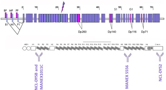
A schematic representation of the dystrophin epitopes recognized by all antibodies used in this study is depicted in Fig 1 (Fig 1).

Fig 1. Dystrophin epitopes recognized by different antibodies used in the study.

Fig 1

Western-blot antibodies (NCL-DYS2 and MANEX1011C) and immunolabelling antibodies (MANEX5556 and NCL-DYSB) are represented. Letters indicate the various promoters: brain (B), muscle (M), and Purkinje (P) promoters, and different isoforms Dp260, Dp140, Dp116 and Dp71 are represented. The arrow indicates the mutation position in Dmdmdx rat model.

Data analysis

All values are expressed as mean ± SEM, with a significance level set at p < 0.05. Statistical evaluation has been performed by using GraphPad Prism 5 (GraphPad Software, Inc., La Jolla, CA, USA). Data distribution was first evaluated with a D’Agostino & Pearson omnibus normality test, for each group, at each tested timepoint, for each measured parameter. We then used either Kruskal Wallis test or ordinary two-ANOVA analysis and if a significant interaction was found, we further performed multiple comparison tests.

Results

Dmdmdx rats display neuromotor alterations in the ledged beam-walking test

In order to complete the assessment of locomotor functions of Dmdmdx animals, as we did in our previous study [26], we used the ledged beam-walking test at 7 months, a time point where the muscular phenotype is strongly affected in Dmdmdx rats. Time spent to cross the beam was compared between WT and Dmdmdx rats. The latter animals spent more time crossing the apparatus compared to WT controls (p = 0.0339, Fig 2A), and this was correlated with a non-significant increase in the total number of steps to cross the beam (p = 0.1229, Fig 2B). However, no impairment was found for Dmdmdx rats in terms of stepping errors when performing the task (front limbs: p = 0.2333, hind limbs: p = 0.6378, Fig 2C).

Fig 2. Motor functions impairment in the ledged-beam walking test in Dmdmdx rats at 7 months.

Fig 2

For WT (black dots, n = 8) and Dmdmdx rats (grey dots, n = 10), (A) Time spent to cross the beam was significantly higher in mutant animals, (B) Number of steps was also higher, but without reaching significance, and (C) Number of errors per step was unaffected. Data are expressed as mean ± SEM. *p < 0.05.

Anhedonia in the sucrose preference test and anxiety levels in the elevated plus maze are not affected in Dmdmdx rats

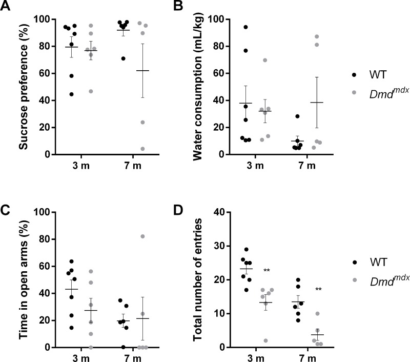
The sucrose preference test is usually used to evaluate anhedonia, indicated by a decrease in sucrose consumption, a typical depression-like behavior in rats and mice. Here evaluated this parameter at 2 different ages in terms of disease progression, 3 and 7 months. At 3 months and 7 months, no age nor genotype effect on sucrose preference was found, indicating that Dmdmdx rats had the same level of preference that WT rats over time (age: F(1,20) = 0.0125, p = 0.912; genotype: F(1,20) = 2.537, p = 0.127, Fig 3A), despite a large variability for older Dmdmdx animals, with some of them having a very low sucrose preference. In parallel, water consumption was evaluated and was shown to be constant over age and between both genotypes (age: F(1,20) = 0.825, p = 0.374; genotype: F(1,20) = 0.894, p = 0.356, Fig 3B). In order to study whether anxiety behavior is affected in Dmdmdx rats, we used the elevated plus-maze test. The percentage of time spent in the open arms, indicative of the anxiety level of the animal, was not significantly different between WT and Dmdmdx rats (age: F(1,20) = 2.474, p = 0.131; genotype: F(1,20) = 0.568, p = 0.460, Fig 3C), despite the fact that the total number of entries into either open or closed arms was significantly lower for Dmdmdx rats at both ages, due to locomotor impairment (age: F(1,20) = 25.49, p < 0.0001; genotype: F(1,20) = 26.38, p < 0.0001; Sidak’s multiple comparison test, p < 0.01 at 3 months and 7 months, Fig 3D). These results suggest that in this test, even if locomotion is affected, Dmdmdx animals are not more anxious than WT animals.

Fig 3. Anhedonia and anxiety are not affected in Dmdmdx rats at 3 and 7 months.

Fig 3

For WT (black dots, n = 7) and Dmdmdx rats (grey dots, n = 6), (A) Sucrose preference and (B) Water consumption are not affected in dystrophin-deficient rats, neither at 3 months, nor at 7 months, (C) Time spent in the open arm of the elevated plus maze (% of time spent on open and closed arms) is also stable over time in both genotypes, (D) Total number of entries into open and closed arms is significantly lower in Dmdmdx rats. Data are expressed as mean ± SEM. **p < 0.01.

Behavioral response to restraint-induced mild stress

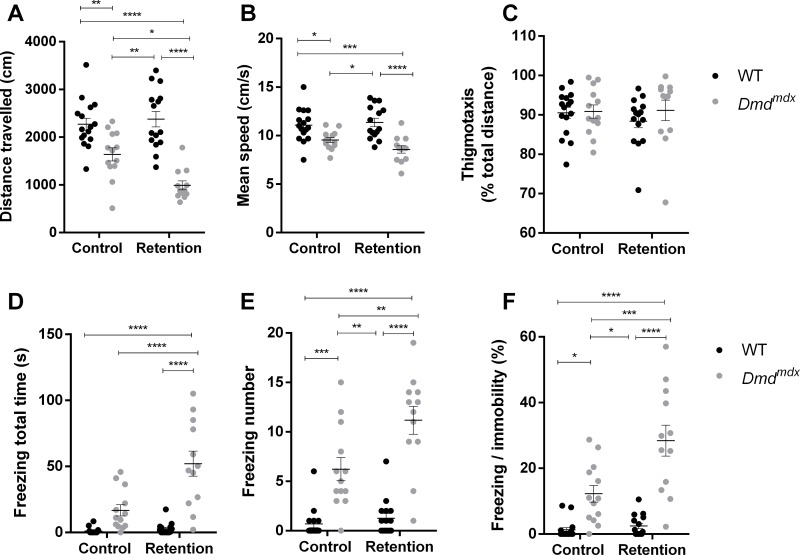
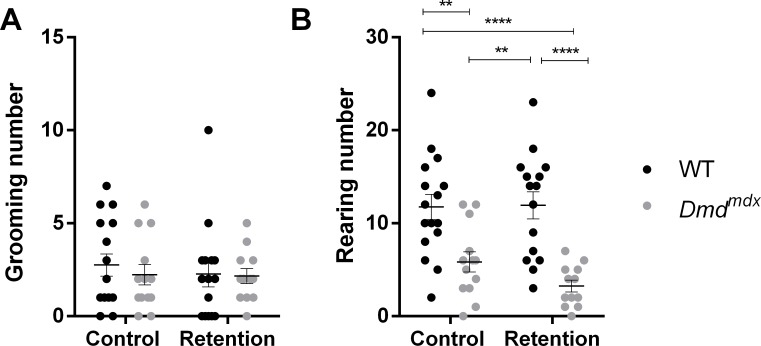
Locomotion was studied in an open-field arena for younger (6 weeks old) Dmdmdx mutants and WT rats. We used this age in order to assess if the response to stress is affected at an early timepoint. Duration of stress-induced tonic immobility, considered as a measure of unconditioned fearfulness [24], was also analyzed. For the retention group, animals were gently scruff-restrained for 10 seconds, in a way similar to the one used for the immobilization of rats for standard examination or intraperitoneal injections, whereas control animals were directly placed inside the open-field arena. Dmdmdx rats walked a shorter distance that WT littermates, and when looking at the effect of retention stress, we found that it only had an effect on Dmdmdx animals, because mutants from the retention group were less mobile than the unstressed Dmdmdx and WT animals (stress: F(1,52) = 3.9, p = 0.0536; genotype: F(1,52) = 54.38, p < 0.0001, Tukey’s multiple comparison test, p < 0.05 for Dmdmdx control vs. Dmdmdx retention, Fig 4A). However no impact of retention was found on mean speed during the test, a genotype effect was detected (stress: F(1,52) = 0.742, p = 0.393; genotype: F(1,52) = 29.3, p < 0.0001, Fig 4B). All animals from each group had the same levels of anxiety, as indicated by similar levels of thigmotaxis, with no effect of genotype nor stress (stress: F(1,52) = 0.259, p = 0.613; genotype: F(1,52) = 0.746, p = 0.392, Fig 4C). Interestingly, we show that restraint induced a large increase in tonic immobility in Dmdmdx rats characterized by a lasting freezing like behavior in terms of freezing duration (+83% between both Dmdmdx groups, stress: F(1,52) = 16.22, p = 0.0002; genotype: F(1,52) = 49.83, p < 0.0001, Tukey’s multiple comparison test, p < 0.0001 for Dmdmdx control vs. Dmdmdx retention, Fig 4D) and number of freezings (+80% between both Dmdmdx groups, stress: F(1,52) = 9.75, p = 0.0029; genotype: F(1,52) = 76.47, p < 0.0001, Tukey’s multiple comparison test, p < 0.01 for Dmdmdx control vs. Dmdmdx retention, Fig 4E). When freezing behavior was normalized to basal immobility, the same significant effect of stress was found throughout the 5-min testing period (stress: F(1,52) = 12.82, p = 0.0008; genotype: F(1,52) = 58.24, p < 0.0001, Tukey’s multiple comparison test, p < 0.001 for Dmdmdx control vs. Dmdmdx retention, Fig 4F). These results indicate that Dmdmdx animals, as opposed to WT littermates, display a maladaptive response to moderate stress, without being more anxious. These performances in the open-field cannot be attributed to overt changes in the behavioral expression of the fear response, as excessive grooming behavior was not observed during the sessions (stress: F(1,52) = 0.215, p = 0.645; genotype: F(1,52) = 0.276, p = 0.602, Fig 5A). Finally, vertical activity was only impacted by genotype, confirming the motor impairment, but not impacted by stress retention (stress: F(1,52) = 0.919, p = 0.342; genotype: F(1,52) = 33.60, p < 0.0001, Fig 5B).

Fig 4. Dmdmdx rats are characterized by early locomotion deficits (6 weeks old) and a maladaptive response to mild stress.

Fig 4

Compared to WT littermate controls (black dots, n = 16 unstressed and n = 15 stressed), Dmdmdx rats (grey dots, n = 13 unstressed and n = 12 stressed), stressed and non-stressed were characterized by a lower travelled distance (A) and mean speed (B) in the open-field, without effect on thigmotaxis (C). Specifically, stress significantly induced higher total freezing time (D) and number (E) and degree of freezing compared to immobility (F) in Dmdmdx animals. Data are expressed as mean ± SEM. *p < 0.05, **p < 0.01, ***p < 0.001, ****p < 0.0001.

Fig 5. Mild stress has no specific effect on ethological parameters grooming and rearings in young Dmdmdx rats (6 weeks old).

Fig 5

For WT (black dots, n = 16 unstressed and n = 15 stressed) and Dmdmdx rats (grey dots, n = 13 unstressed and n = 12 stressed), (A) Grooming number is stable in both genotypes, with or without prior stress, (B) Vertical activity, measured by rearing number is also unaffected by stress, but confirms the locomotor deficit in mutant rats. Data are expressed as mean ± SEM. **p < 0.01, ****p < 0.0001.

Dp71, Dp 140 and Dp427 levels in different regions of central nervous system

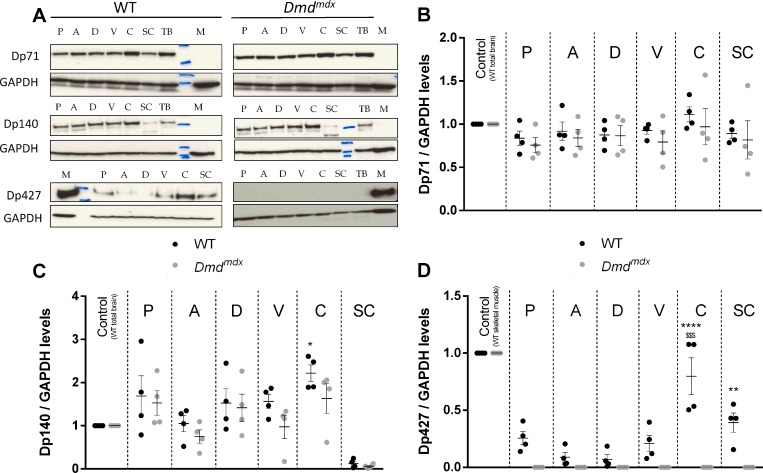
Western blot analysis of dystrophin levels and immunolabelling of dystrophin in different areas of central nervous system were performed using two antibodies directed against epitopes located at the C-terminus (for Dp71 and Dp140 detection) and within exons 10–11 (for full dystrophin Dp427 detection). A normalization step was added by using total WT rat brain for Dp71 and Dp140, because these isoforms are absent from WT muscles, and by using WT skeletal muscle extract for Dp427 quantification, this isoform being highly expressed in muscle.

Using western blot in WT animals, Dp71isoform, implicated in transmembrane receptor binding and vascular development, was found equally distributed throughout brain areas and spinal cord, as expected. Due to the point mutation in exon 23, Dmdmdx and WT animals displayed the same absence of regional specificity of Dp71 expression in central nervous system (genotype: F(1,42) = 1.526, p = 0.224; region: F(6,42) = 1.218, p = 0.316, Fig 6A and 6B). Concerning Dp140 isoform, which participates to early development via regulation of neuron differentiation, neuron projection morphogenesis and chromatin modification, we found in WT animals the highest levels in cerebellum, which happen to be significantly higher than amygdala levels. Dp140 levels in the same areas from Dmdmdx animals were not different than WT littermates (genotype: F(1,42) = 3.944, p = 0.054; region: F(6,42) = 11.99, p < 0.0001, Fig 6A and 6C). Interestingly, Dp140 was found at very low levels in spinal cord of animals from both groups, compared to other brain areas. Finally, we found that Dp427 isoform, which is found to the synaptic membrane of neurons with a function revolving around transmembrane transport and signal transmission, was unequally distributed throughout central nervous system areas of WT rats, with higher levels in cerebellum and spinal cord (genotype: F(1,42) = 73.90, p < 0.0001; region: F(6,42) = 74.24, p < 0.0001, Fig 6A and 6D). This is in line with previous mouse studies [8], where this isoform was found to be expressed in the cerebellum. As anticipated, using western blot no Dp427 was found for Dmdmdx rats, confirming the effectiveness of the generated mutation.

Fig 6. Specific lack of Dp427 dystrophin isoform in Dmdmdx rats central nervous system, but no modification of Dp140 and Dp71 levels.

Fig 6

WT controls (black dots, n = 4) and Dmdmdx animals (grey dots, n = 4) were sacrificed and samples from brain areas (P: prefrontal cortex, A: amygdala, D: dorsal hippocampus, V: ventral hippocampus, C: cerebellum), spinal cord (SC), total brain (TB) with no specific targeted area and biceps femoris muscle from a WT rat (M), were harvested. Western-blot of total proteins was incubated with either monoclonal antibodies NCL-DYS2 and 30 μg protein loading for Dp71 and Dp140) or Manex1011C and 80 μg protein loading for Dp427 (A). Quantification on western-blot of dystrophin isoforms in each central nervous system region (n = 4 per genotype) were normalized to GAPDH levels, and then to dystrophin Dp427 control muscle levels (B, C). This revealed identical levels of 71 kDa (A, B) and 140 kDa (A, C) dystrophin isoforms in all studied central nervous system areas from WT and Dmdmdx animals. Only Dp140 cerebellar levels were significantly higher compared to amygdalar levels in WT rats. Dp427 isoform was not detected in Dmdmdx animals (A, D), and had variable levels in WT rats, with higher levels in cerebellum and spinal cord. Total WT brain extracts were used as positive controls for Dp71 and Dp140 detections, and biceps femoris muscle from a WT rat was used as positive control for Dp427 detection. Staining with an anti-GAPDH polyclonal antibody validated equal protein loadings. Data are expressed as mean ± SEM. In Fig 6C, *p < 0.05 vs. A. In Fig 6D, ****p < 0.0001 vs. SC, $ $ $p < 0.001 vs P, A, D and V and **p<0.01 vs. A and D.

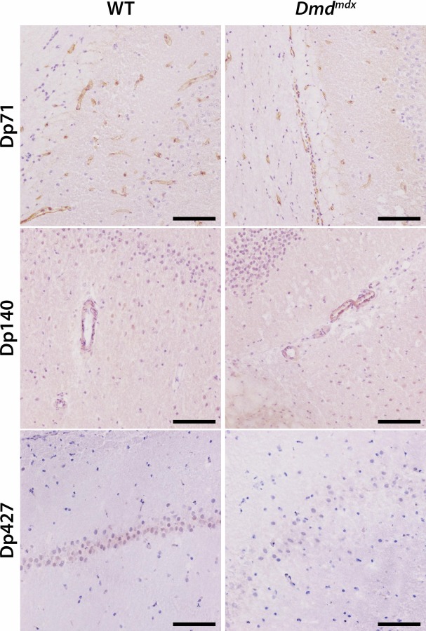
After immunohistolabelling using DYSB antibody, Dp427 was observed in WT animals with a faint intensity in large neuronal bodies of the hippocampal and cerebellar areas of the brain (Fig 7). This isoform was absent from the Dmdmdx littermates. Immunolabelling of Dp427/Dp140 isoforms, using MANEX5556 antibody, was similar in WT and Dmdmdx animals, therefore indicating that this marking is probably Dp140 specific, as we showed before that Dp427 is absent from mutant animals. This isoform was found with a light intensity in smooth muscle layer of blood vessels present in the whole brain area, but we cannot exclude the presence of a glial marking around vessels. Lastly, immunolabelling of Dp427/Dp140/Dp71 isoforms was found with similar intensities in WT and Dmdmdx, therefore indicating that this marking is probably Dp71 specific, as we showed before that Dp427 is absent from mutant animals and that Dp140 is only visible smooth muscle layers. Dp71 isoform was found exclusively in endothelial cells of blood capillaries throughout the entire brain area. No associated histologic lesions were identified.

Fig 7. Lack of Dp427 dystrophin isoform in Dmdmdx rats brain and no modification in distribution of Dp140 and Dp71 isoforms.

Fig 7

Males WT controls (n = 3) and Dmdmdx rats (n = 3) were sacrificed and slides from brain areas (at the levels of the frontal pole, the optic chiasm, the oculomotor nerve, the midbrain and the occipital pole) were processed for immunolabelling of Dp71, Dp140 and Dp427 dystrophin isoforms using respectively monoclonal antibodies NCL-DYS2, MANEX5556 and NCL-DYSB. Isoforms Dp71 and Dp140 were detected with similar intensities in WT littermate controls and Dmdmdx rats and were respectively localized in endothelial cells of blood capillaries and in smooth muscle layer of blood vessels throughout brain parenchyma. Isoform Dp427 was mostly detected in the cytoplasm of some large neurons of the hippocampus in WT controls but was absent in Dmdmdx littermate rats.

Discussion

This study describes the neuromotor, anxiety and anhedonia characterization of the recently described dystrophin deficient rat model, Dmdmdx rats [26]. It was previously demonstrated that cardiac and skeletal phenotypic properties of this model are very close to the human DMD pathology. Duchenne muscular dystrophy is the most common neuromuscular disorder, representing about 30% of muscular dystrophies [6, 36, 37], without any curative treatment to date. The present results demonstrate that Dmdmdx rats display neuromotor alterations at 7 months, as shown with the transversal beam test, in which mutant animals spent more time to cross the beam. However, no significant changes were found in the number of errors, neither for frontlimbs nor hindlimbs. This function mainly relies on the nigrostriatal dopaminergic pathway, which is known to control the dexterity of movement [38], correlated with the number of errors performed upon crossing the beam. Locomotor measurements are important parameters allowing to define phenotypes of animal models with muscular dystrophies. Motor deficit underlined here may therefore be a consequence of dystrophin absence in muscles from the limbs, but we cannot exclude they may also result from reduced levels of cerebellar dystrophin, as demonstrated by western blot.

In DMD patients, clinically significant internalizing disorders including anxiety and depression have been demonstrated, so that even a Duchenne muscular dystrophy neuropsychiatric syndrome has been suggested [39]. Indeed, a specific study on DMD patients demonstrated that 24% and 19% of them displayed anxiety and depression disorders, respectively [40]. Therefore, in order to measure anhedonia in mutant Dmdmdx animals, we used the sucrose preference test, reflecting the hedonic drive in a locomotor-independent manner [41]. The preference for sucrose solution was found to be unmodified in 3 months animals and not significantly reduced in 7 months old animals lacking dystrophin. This further supports the idea that this model does not manifest any depressive-like phenotype. In Dmdmdx rats, sucrose preference appears to decrease in a non-significant manner over time, this could be linked to the behavior of some animals that did not show any interest for the sucrose solution. Moreover, Dmdmdx rats performances in the elevated plus maze display a high variability indicating that anxiety-like behavior is absent in this model, as confirmed by the same level of thigmotaxis and grooming behavior in the open-field test between both groups. In this study, we have to report that motor impairment revealed in the beam test might have biased the assessment of emotional behaviors, which in many tests with a high-motor demand is dependent on mobility. Indeed, total number of entries into the elevated plus maze arms, an indicator of global locomotion, is significantly reduced at each studied timepoint. However, time in open arms did not differ between groups, so no misleading interpretation could be made. Contrasting results have been found on anxiety in the classical model of mdx mice. Indeed, in 2009 a group showed that this model did not display an anxiety-like phenotype in the elevated plus maze [24]. However, other recent studies indicate a deficit in the light-dark choice anxiety test [25, 42]. Moreover, another group demonstrated anxiety-like and depression-like behaviors in mdx mice, associated with decreased BDNF (Brain derived neurotrophic factor) levels [43]. It may therefore be useful to evaluate BDNF levels and perform light-dark choice anxiety tests to fully characterise the anxiety phenotype in Dmdmdx rats.

The open-field test was used to assess horizontal (distance and speed) and vertical (rearing) activities, which were shown to be reduced in 6 weeks-old Dmdmdx rats compared to WT rats. This reduced locomotion in the open-field arena has been previously reported for Dmdmdx animals [26], as opposed to studies using mdx mice, which do not display any motor deficit before 6 months of age [24, 44], thus confirming the early locomotor deficit in this rat DMD model. It was previously shown that the rodent normal defensive behavior in response to danger or a threat is enhanced in mdx mice, showing potent defensive freezing responses to a short stress restraint, as opposed to WT animals, in a way independent from hypothalamic–pituitary–adrenal axis activation [24]. Here we show for the first time that a 10 seconds restraint stress on young Dmdmdx rats has an effect on exploratory behavior. Indeed, freezing, defined as a lasting tonic immobility involving a brain circuit including the amygdala [24], is increased by retention for Dmdmdx rats in terms of freezing time and number of freezing events, while this effect is absent on WT littermates. It is important to notice that this effect is also found at a lower level in non-stressed Dmdmdx animals compared to WT. Therefore we have an exacerbation of freezing behavior following a brief stress event. This maladaptive response to stress confirms freezing behavior as an important and reliable study parameter in Dmdmdx rat. These analyses were performed on young animals at the age of 6 weeks, and we cannot exclude that this phenotype will be exacerbated at later timepoints, which has to be taken into account in the design and follow-up of preclinical studies using this model. In mdx mice, this behavior has been shown to increase with age and is thought to be underlined by an alteration of amygdala GABAergic circuits in dystrophin deficient animals [25]. We may hypothesize the same thing is occurring in our model, as well as alterations of central serotonin [44, 45] and cholinergic functions [46]. Reduced mobility of dystrophin deficient animals is often attributed to muscle wasting inducing fatigue [47]. Here we show that for Dmdmdx rats, this should be interpreted with care because this reduced mobility may indeed be a result of higher stress reactivity. For instance, when performing an intra-peritoneal injection, scruff restraint might appear to be a relatively mild stress, but in fact leading to confounding effects on locomotion parameters often used to evaluate treatments efficacies in preclinical studies. Indeed, this stress response is characterized by a strong motor inhibition which may be critical in the interpretation of functional measures based on quantification of mobility. One might hypothesize that this higher stress response in Dmdmdx rats is due a higher pain sensitivity, but during the evaluation animals did not show any paw rigidity while being in a tonic immobility state, and they reacted to sudden noises occurring in the animal house or touching, therefore interrupting their freezing behavior.

To understand the molecular mechanisms underlying these behavioral deficits, we performed studies of dystrophin isoforms expression level and localization in different brain areas. As demonstrated with the immunohistolabelling study, Dp427 isoform was present in large neuronal bodies in WT rats, and we found an absence of Dp427 isoform in all studied regions from Dmdmdx rats, which is consistent with the mutation generated on exon 23, inducing a complete loss of this large isoform and indicates that Dmdmdx rats are indeed dystrophin deficient animals, in muscles as well as in central nervous system. In WT animals, the highest levels of Dp427 isoforms were detected in the cerebellum and spinal cord, which is in agreement with studies demonstrating strong dystrophin expression in cerebellum, but also cortex and hippocampus in WT mice and rats [48, 49]. As prefrontal cortex and amygdala are known to communicate and be both involved in stress response [50, 51], dystrophin levels in these brain areas might participate in a typical adaptive stress response for WT rats. It was previously shown that brain dystrophin Dp427 is associated with a subpopulation of GABAA receptors at inhibitory synapses, and that in mdx mice, an impaired clustering of GABAA receptors in hippocampus, amygdala and cerebellum [12, 13, 16, 24, 26] has been associated with altered synaptic plasticity [22, 24, 52, 53]. Therefore, brain dystrophin deficiency in Dmdmdx rats may also affect synaptic transmission and GABAergic communication between cortex and amygdala, and more generally, the integrity of several brain areas might be compromised. Concerning the hippocampus, spatial memory needs to be evaluated in this model in order to assess potential specific short-term memory deficits. The known functional segmentation between dorsal hippocampus and ventral hippocampus, with the ventral part being involved with emotion and the dorsal part regulating information processing [54, 55] is also found here with dystrophin levels that seem to be higher in the ventral hippocampus. Behavioral and physiological consequences of this observation still need to be evaluated. Other structures expressing Dp427 in WT animals are implicated in stress response, like entorhinal and perirhinal cortices, hippocampus or cerebellum [56], so the dystrophin deficiency induced in these regions in our model may contribute to the abnormal contention response and to the locomotor deficits detailed in this study. Additionally, levels of other dystrophin isoforms Dp140, expressed in smooth muscle layer of blood vessels and Dp71, exclusively expressed in endothelial cells of blood capillaries from both groups, are not impacted by the dystrophin-deficient phenotype in comparison to control animals.

Taken together, these findings provide insights into the relevance of using the Dmdmdx rat model, with freezing confirmed as a good readout to assess dynamics of therapeutics targeting brain functions in DMD models. Moreover, given the robust clinical relationship between muscular and locomotion deficits already demonstrated in this model and human Duchenne pathology, present data further emphasize a potential beneficial role of Dmdmdx rat model to better understand human central nervous system symptoms to stress conditions in a Duchenne disease context.

Supporting information

S1 Raw images

(PDF)

Acknowledgments

We thank people from technical team of the animal house IRS2, who took care of all animals throughout these studies. We also thank the MDA Monoclonal Antibody Resource for providing the MANEX 1011C antibody.

Data Availability

All relevant data are within the manuscript and its Supporting Information files.

Funding Statement

The author(s) received no specific funding for this work.

References

  • 1.Cotton S, Voudouris NJ, Greenwood KM. Intelligence and Duchenne muscular dystrophy: full-scale, verbal, and performance intelligence quotients. Dev Med Child Neurol. 2001;43(7):497–501. 10.1017/s0012162201000913 [DOI] [PubMed] [Google Scholar]
  • 2.Ricotti V, Mandy WP, Scoto M, Pane M, Deconinck N, Messina S, et al. Neurodevelopmental, emotional, and behavioral problems in Duchenne muscular dystrophy in relation to underlying dystrophin gene mutations. Dev Med Child Neurol. 2016;58(1):77–84. 10.1111/dmcn.12922 [DOI] [PubMed] [Google Scholar]
  • 3.Pane M, Scalise R, Berardinelli A, D'Angelo G, Ricotti V, Alfieri P, et al. Early neurodevelopmental assessment in Duchenne muscular dystrophy. Neuromuscul Disord 2013;23(6):451–5. 10.1016/j.nmd.2013.02.012 [DOI] [PubMed] [Google Scholar]
  • 4.Hendriksen JG, Vles JS. Neuropsychiatric disorders in males with duchenne muscular dystrophy: frequency rate of attention-deficit hyperactivity disorder (ADHD), autism spectrum disorder, and obsessive—compulsive disorder. J Child Neurol. 2008;23(5):477–81. 10.1177/0883073807309775 [DOI] [PubMed] [Google Scholar]
  • 5.Banihani R, Smile S, Yoon G, Dupuis A, Mosleh M, Snider A, et al. Cognitive and Neurobehavioral Profile in Boys With Duchenne Muscular Dystrophy. J Child Neurol. 2015;30(11):1472–82. 10.1177/0883073815570154 [DOI] [PubMed] [Google Scholar]
  • 6.Muntoni F, Torelli S, Ferlini A. Dystrophin and mutations: one gene, several proteins, multiple phenotypes. Lancet Neurol 2003;12:731–40. [DOI] [PubMed] [Google Scholar]
  • 7.Billard C, Gillet P, Signoret JL, Uicaut E, Bertrand P, Fardeau M, et al. Cognitive functions in Duchenne muscular dystrophy: a reappraisal and comparison with spinal muscular atrophy. Neuromuscul Disord. 1992;2(5–6):371–8. 10.1016/s0960-8966(06)80008-8 [DOI] [PubMed] [Google Scholar]
  • 8.Snow WM, Fry M, Anderson JE. Increased density of dystrophin protein in the lateral versus the vermal mouse cerebellum. Cell Mol Neurobiol 2013;33(4):513–20. 10.1007/s10571-013-9917-8 [DOI] [PMC free article] [PubMed] [Google Scholar]
  • 9.Piccini G, Gazzellini S, D'Amico A, Pane M, Castelli E, Vicari S, et al. Developmental lag of visuospatial attention in Duchenne muscular dystrophy. Res Dev Disabil. 2014;36C:55–61. 10.1016/j.ridd.2014.09.021 [DOI] [PubMed] [Google Scholar]
  • 10.Hinton VJ, De Vivo DC, Nereo NE, Goldstein E, Stern Y. Selective deficits in verbal working memory associated with a known genetic etiology: The neuropsychological profile of duchenne muscular dystrophy. Journal of the International Neuropsychological. 2001;7:45–54. [DOI] [PMC free article] [PubMed] [Google Scholar]
  • 11.Hinton VJ, De Vivo DC, Nereo NE, Goldstein E, Stern Y. Poor verbal working memory across intellectual level in boys with Duchenne dystrophy. Neurology. 2000;54:2127–32. 10.1212/wnl.54.11.2127 [DOI] [PMC free article] [PubMed] [Google Scholar]
  • 12.Levi S, Grady RM, Henry MD, Campbell KP, Sanes JR, Craig AM. Dystroglycan is selectively associated with inhibitory GABAergic synapses but is dispensable for their differentiation. J Neurosci 2002;22:4274–85. 10.1523/JNEUROSCI.22-11-04274.2002 [DOI] [PMC free article] [PubMed] [Google Scholar]
  • 13.Knuesel I, Mastrocola M, Zuellig RA, Bornhauser B, Schaub MC, Fritschy JM. Short communication: Altered synaptic clustering of GABAA receptors in mice lacking dystrophin (mdx mice). European Journal of Neuroscience. 1999;11:4457–62. 10.1046/j.1460-9568.1999.00887.x [DOI] [PubMed] [Google Scholar]
  • 14.Graf ER, Zhang X-Z, Jin S-X, Linhoff MW, Craig AM. Neurexins induce differentiation of GABA and glutamate postsynaptic specializations via neuroligins. Cell. 2004;119:1013–26. 10.1016/j.cell.2004.11.035 [DOI] [PMC free article] [PubMed] [Google Scholar]
  • 15.Brunig I, Suter A, Knuesel I, Luscher B, Fritschy JM. GABAergic terminals are required for postsynaptic clustering of dystrophin but not of GABAA receptors and gephyrin. J Neurosci. 2002;22:4805–13. 10.1523/JNEUROSCI.22-12-04805.2002 [DOI] [PMC free article] [PubMed] [Google Scholar]
  • 16.Vaillend C, Perronnet C, Ros C, Gruszczynski C, Goyenvalle A, Laroche S, et al. Rescue of a dystrophin-like protein by exon skipping in vivo restores GABAA-receptor clustering in the hippocampus of the mdx mouse. Mol Ther. 2010;18:1683–8. 10.1038/mt.2010.134 [DOI] [PMC free article] [PubMed] [Google Scholar]
  • 17.Miranda R, Sébrié C, Degrouard J, Gillet B, Jaillard D, Laroche S, et al. Reorganization of inhibitory synapses and increased PSD length of perforated excitatory synapses in hippocampal area CA1 of dystrophindeficient mdx mice. Cereb Cortex. 2009;19:876–88. 10.1093/cercor/bhn135 [DOI] [PubMed] [Google Scholar]
  • 18.Krasowska E, Zabłocki K, Gorecki DC, Swinny JD. Aberrant location of inhibitory synaptic marker proteins in the hippocampus of dystrophin-deficient mice: implications for cognitive impairment in Duchenne muscular dystrophy. PLoS One. 2014;9. Epub e108364. [DOI] [PMC free article] [PubMed] [Google Scholar]
  • 19.Vaillend C, Billard JM. Facilitated CA1 hippocampal synaptic plasticity in dystrophin-deficient mice: role for GABAA receptors? Hippocampus. 2002;12(6):713–7. 10.1002/hipo.10068 [DOI] [PubMed] [Google Scholar]
  • 20.Suzuki H, Aoki Y, Kameyama T, Saito T, Masuda S, Tanihata J, et al. Endogenous Multiple Exon Skipping and Back-Splicing at the DMD Mutation Hotspot. Int J Mol Sci. 2016;17(10). [DOI] [PMC free article] [PubMed] [Google Scholar]
  • 21.Vaillend C, Rendon A, Misslin R, Ungerer A. Influence of dystrophin-gene mutation on mdx mouse behavior. I. Retention deficits at long delays in spontaneous alternation and bar-pressing tasks. Behav Genet. 1995;25(6):569–79. 10.1007/bf02327580 [DOI] [PubMed] [Google Scholar]
  • 22.Vaillend C, Billard JM, Laroche S. Impaired long-term spatial and recognition memory and enhanced CA1 hippocampal LTP in the dystrophin-deficient Dmd(mdx) mouse. Neurobiol Dis. 2004;17(1):10–20. 10.1016/j.nbd.2004.05.004 [DOI] [PubMed] [Google Scholar]
  • 23.Chaussenot R, Edeline JM, Le Bec B, El Massioui N, Laroche S, Vaillend C. Cognitive dysfunction in the dystrophin-deficient mouse model of Duchenne muscular dystrophy: A reappraisal from sensory to executive processes. Neurobiol Learn Mem. 2015;124:111–22. 10.1016/j.nlm.2015.07.006 [DOI] [PubMed] [Google Scholar]
  • 24.Sekiguchi M, Zushida K, Yoshida M, Maekawa M, Kamichi S, Yoshida M, et al. A deficit of brain dystrophin impairs specific amygdala GABAergic transmission and enhances defensive behavior in mice. Brain 2009;132:124–35. 10.1093/brain/awn253 [DOI] [PubMed] [Google Scholar]
  • 25.Vaillend C, Chaussenot R. Relationships linking emotional, motor, cognitive and GABAergic dysfunctions in dystrophin-deficient mdx mice. Hum Mol Genet. 2017;26(6):1041–55. 10.1093/hmg/ddx013 [DOI] [PubMed] [Google Scholar]
  • 26.Larcher T, Lafoux A, Tesson L, Remy S, Thepenier V, François V, et al. Characterization of dystrophin deficient rats: a new model for Duchenne muscular dystrophy. PLoS One. 2014;9(10). 10.1371/journal.pone.0110371 [DOI] [PMC free article] [PubMed] [Google Scholar]
  • 27.Partridge TA. The mdx mouse model as a surrogate for Duchenne muscular dystrophy. FEBS J. 2013;280(17):4177–86. 10.1111/febs.12267 [DOI] [PMC free article] [PubMed] [Google Scholar]
  • 28.Goyenvalle A, Griffith G, Babbs A, El Andaloussi S, Ezzat K, Avril A, et al. Functional correction in mouse models of muscular dystrophy using exon-skipping tricyclo-DNA oligomers. Nat Med. 2015;21:270–5. 10.1038/nm.3765 [DOI] [PubMed] [Google Scholar]
  • 29.Zhao CS, Puurunen K, Schallert T, Sivenius J, Jolkkonen J. Behavioral and histological effects of chronic antipsychotic and antidepressant drug treatment in aged rats with focal ischemic brain injury. Behav Brain Res. 2005;158(2):211–20. 10.1016/j.bbr.2004.09.001 [DOI] [PubMed] [Google Scholar]
  • 30.Walsh SL, Wagner GC. Motor impairments after methamphetamine-induced neurotoxicity in the rat. J Pharmacol Exp Ther. 1992;263:617–26. [PubMed] [Google Scholar]
  • 31.Feeney DM, Gonzalez A, Law WA. Amphetamine, haloperidol, and experience interact to affect rate of recovery after motor cortex injury. Science. 1982;217:855–7. 10.1126/science.7100929 [DOI] [PubMed] [Google Scholar]
  • 32.Bowenkamp KE, Ujhelyi L, Cline EJ, Bickford PC. Effects of intra-striatal GDNF on motor coordination and striatal electrophysiology in aged F344 rats. Neurobiol Aging 2000;21:117–24. 10.1016/s0197-4580(99)00112-8 [DOI] [PubMed] [Google Scholar]
  • 33.Willner P, Towell A, Sampson D, Sophokleous S, Muscat R. Reduction of sucrose preference by chronic unpredictable mild stress, and its restoration by a tricyclic antidepressant. Psychopharmacology (Berl) 1987;93:358–64. [DOI] [PubMed] [Google Scholar]
  • 34.Paylor R, Zhao Y, Libbey M, Westphal H, Crawley JN. Learning impairments and motor dysfunctions in adult Lhx5-deficient mice displaying hippocampal disorganization. Physiol Behav. 2001;73:781–92. 10.1016/s0031-9384(01)00515-7 [DOI] [PubMed] [Google Scholar]
  • 35.Richner M, Jager SB, Siupka P, Vaegter CB. Hydraulic Extrusion of the Spinal Cord and Isolation of Dorsal Root Ganglia in Rodents. J Vis Exp. 2017;119 10.3791/55226 [DOI] [PMC free article] [PubMed] [Google Scholar]
  • 36.Koenig M, Monaco AP, Kunkel LM. The complete sequence of dystrophin predicts a rod-shaped cytoskeletal protein. Cell. 1988;53:219–28. 10.1016/0092-8674(88)90383-2 [DOI] [PubMed] [Google Scholar]
  • 37.Hoffman EP, Brown RH Jr, Kunkel LM. Dystrophin: the protein product of the Duchenne muscular dystrophy locus. Cell. 1987;51:919–28. 10.1016/0092-8674(87)90579-4 [DOI] [PubMed] [Google Scholar]
  • 38.Takakusaki K. Neurophysiology of gait: from the spinal cord to the frontal lobe. Mov Disord. 2013;28(11). 10.1002/mds.25669 [DOI] [PubMed] [Google Scholar]
  • 39.Lee AJ, Buckingham ET, Kauer AJ, Mathews KD. Descriptive Phenotype of Obsessive Compulsive Symptoms in Males With Duchenne Muscular Dystrophy. J Child Neurol 2018;33(9):572–9. 10.1177/0883073818774439 [DOI] [PMC free article] [PubMed] [Google Scholar]
  • 40.Pangalila RF, van den Bos GA, Bartels B, Bergen M, Stam HJ, Roebroeck ME. Prevalence of fatigue, pain, and affective disorders in adults with Duchenne muscular dystrophy and their associations with quality of life. Arch Phys Med Rehabil 2015;96:1242–7. 10.1016/j.apmr.2015.02.012 [DOI] [PubMed] [Google Scholar]
  • 41.Cryan JF, Holmes A. The ascent of mouse: advances in modelling human depression and anxiety. Nat Rev Drug Discov. 2005;4(9):775–90. 10.1038/nrd1825 [DOI] [PubMed] [Google Scholar]
  • 42.Remmelink E, Aartsma-Rus A, Smit AB, Verhage M, Loos M, van Putten M. Cognitive flexibility deficits in a mouse model for the absence of full-length dystrophin. Genes Brain Behav. 2016. July;15(6):558–67. 10.1111/gbb.12301 [DOI] [PubMed] [Google Scholar]
  • 43.Comim CM, Ventura L, Freiberger V, Dias P, Bragagnolo D, Dutra ML, et al. Neurocognitive Impairment in mdx Mice. Mol Neurobiol 2019. 10.1007/s12035-019-1573-7 [DOI] [PubMed] [Google Scholar]
  • 44.Manning J, Kulbida R, Rai P, Jensen L, Bouma J, Singh SP, et al. Amitriptyline is efficacious in ameliorating muscle inflammation and depressive symptoms in the mdx mouse model of Duchenne muscular dystrophy. Exp Physiol. 2014;99:1370–86. 10.1113/expphysiol.2014.079475 [DOI] [PubMed] [Google Scholar]
  • 45.Sogos V, Reali C, Fanni V, Curto M, Gremo F. Dystrophin antisense oligonucleotides decrease expression of nNOS in human neurons. Brain ResMol Brain Res. 2003;118:52–9. [DOI] [PubMed] [Google Scholar]
  • 46.Cohen EJ, Quarta E, Fulgenzi G, Minciacchi D. Acetylcholine, GABA and neuronal networks: a working hypothesis for compensations in the dystrophic brain. Brain Res Bull. 2015;110(1–13). [DOI] [PubMed] [Google Scholar]
  • 47.Rafael JA, Nitta Y, Peters J, Davies KE. Testing of SHIRPA, a mouse phenotypic assessment protocol, on Dmd(mdx) and Dmd(mdx3cv) dystrophin-deficient mice. Mamm Genome. 2000;11:725–8. 10.1007/s003350010149 [DOI] [PubMed] [Google Scholar]
  • 48.Snow WM, Anderson JE, Jakobson LS. Neuropsychological and neurobehavioral functioning in Duchenne muscular dystrophy: A review. Neuroscience and Biobehavioral Reviews. 2013;37:743–52. 10.1016/j.neubiorev.2013.03.016 [DOI] [PubMed] [Google Scholar]
  • 49.Gorecki D, Geng Y, Thomas K, Hunt SP, Barnard EA, Barnard PJ. Expression of the dystrophin gene in mouse and rat brain. Neuroreport. 1991;2(12):773–6. 10.1097/00001756-199112000-00011 [DOI] [PubMed] [Google Scholar]
  • 50.Maroun M, Richter-Levin G. Exposure to acute stress blocks the induction of long-term potentiation of the amygdala-prefrontal cortex pathway in vivo. J Neurosci. 2003;23:4406–9. 10.1523/JNEUROSCI.23-11-04406.2003 [DOI] [PMC free article] [PubMed] [Google Scholar]
  • 51.Caudal D, Godsil BP, Mailliet F, Bergerot D, Jay TM. Acute stress induces contrasting changes in AMPA receptor subunit phosphorylation within the prefrontal cortex, amygdala and hippocampus. PLoS One. 2010;5(12). 10.1371/journal.pone.0015282 [DOI] [PMC free article] [PubMed] [Google Scholar]
  • 52.Dallérac G, Perronnet C, Chagneau C, Leblanc-Veyrac P, Samson-Desvignes N, Peltekian E, et al. Rescue of a dystrophin-like protein by exon skipping normalizes synaptic plasticity in the hippocampus of the mdx mouse. Neurobiology of Disease. 2011;43:635–41. 10.1016/j.nbd.2011.05.012 [DOI] [PubMed] [Google Scholar]
  • 53.Anderson JL, Morley JW, Head SI. Enhanced homosynaptic LTD in cerebellar Purkinje cells of the dystrophic MDX mouse. Muscle and Nerve. 2010;41(3):329–34. 10.1002/mus.21467 [DOI] [PubMed] [Google Scholar]
  • 54.Moser M, Moser E. Functional differentiation in the hippocampus. Hippocampus. 1998;8:608–19. [DOI] [PubMed] [Google Scholar]
  • 55.Bannerman DM, Grubb M, Deacon RM, Yee BK, Feldon J, Rawlins JN. Ventral hippocampal lesions affect anxiety but not spatial learning. Behav Brain Res. 2003;139(1–2). [DOI] [PubMed] [Google Scholar]
  • 56.LeDoux JE. Emotion circuits in the brain. Annual Review of Neuroscience. 2000;23:155–84. 10.1146/annurev.neuro.23.1.155 [DOI] [PubMed] [Google Scholar]

Decision Letter 0

Gerhard Wiche

2 Jan 2020

PONE-D-19-28211

Dystrophin-deficient Dmdmdx rat model displays an increased behavioral response to restraint-induced mild stress

PLOS ONE

Dear Dr. Caudal,

Thank you for submitting your manuscript to PLOS ONE. After careful consideration, we feel that it has merit but does not fully meet PLOS ONE’s publication criteria as it currently stands. Therefore, we invite you to submit a revised version of the manuscript that addresses the points raised during the review process.

Your manuscript has been evaluated by 2 reviewers. One of them raised several major and some minor concerns that need to be addressed and consequently a major revision is suggested. A few more technical points were  raised by the second referee. However, there was also some concern about one of the assays being underpowered.

We would appreciate receiving your revised manuscript within the next 6 weeks. When you are ready to submit your revision, log on to https://www.editorialmanager.com/pone/ and select the 'Submissions Needing Revision' folder to locate your manuscript file.

If you would like to make changes to your financial disclosure, please include your updated statement in your cover letter.

To enhance the reproducibility of your results, we recommend that if applicable you deposit your laboratory protocols in protocols.io, where a protocol can be assigned its own identifier (DOI) such that it can be cited independently in the future. For instructions see: http://journals.plos.org/plosone/s/submission-guidelines#loc-laboratory-protocols

Please include the following items when submitting your revised manuscript:

  • A rebuttal letter that responds to each point raised by the academic editor and reviewer(s). This letter should be uploaded as separate file and labeled 'Response to Reviewers'.

  • A marked-up copy of your manuscript that highlights changes made to the original version. This file should be uploaded as separate file and labeled 'Revised Manuscript with Track Changes'.

  • An unmarked version of your revised paper without tracked changes. This file should be uploaded as separate file and labeled 'Manuscript'.

Please note while forming your response, if your article is accepted, you may have the opportunity to make the peer review history publicly available. The record will include editor decision letters (with reviews) and your responses to reviewer comments. If eligible, we will contact you to opt in or out.

We look forward to receiving your revised manuscript.

Kind regards,

Gerhard Wiche, Ph.D.

Academic Editor

PLOS ONE

Journal Requirements:

When submitting your revision, we need you to address these additional requirements.

1. Please ensure that your manuscript meets PLOS ONE's style requirements, including those for file naming. The PLOS ONE style templates can be found at

http://www.journals.plos.org/plosone/s/file?id=wjVg/PLOSOne_formatting_sample_main_body.pdf and http://www.journals.plos.org/plosone/s/file?id=ba62/PLOSOne_formatting_sample_title_authors_affiliations.pdf

2. Please amend the methods section of your manuscript to include the method of euthanasia

3. PLOS ONE now requires that authors provide the original uncropped and unadjusted images underlying all blot or gel results reported in a submission’s figures or Supporting Information files. This policy and the journal’s other requirements for blot/gel reporting and figure preparation are described in detail at https://journals.plos.org/plosone/s/figures#loc-blot-and-gel-reporting-requirements and https://journals.plos.org/plosone/s/figures#loc-preparing-figures-from-image-files. When you submit your revised manuscript, please ensure that your figures adhere fully to these guidelines and provide the original underlying images for all blot or gel data reported in your submission. See the following link for instructions on providing the original image data: https://journals.plos.org/plosone/s/figures#loc-original-images-for-blots-and-gels.

In your cover letter, please note whether your blot/gel image data are in Supporting Information or posted at a public data repository, provide the repository URL if relevant, and provide specific details as to which raw blot/gel images, if any, are not available. Email us at plosone@plos.org if you have any questions.

4. We note that you have included the phrase “data not shown” in your manuscript. Unfortunately, this does not meet our data sharing requirements. PLOS does not permit references to inaccessible data. We require that authors provide all relevant data within the paper, Supporting Information files, or in an acceptable, public repository. Please add a citation to support this phrase or upload the data that corresponds with these findings to a stable repository (such as Figshare or Dryad) and provide and URLs, DOIs, or accession numbers that may be used to access these data. Or, if the data are not a core part of the research being presented in your study, we ask that you remove the phrase that refers to these data.

[Note: HTML markup is below. Please do not edit.]

Reviewers' comments:

Reviewer's Responses to Questions

Comments to the Author

1. Is the manuscript technically sound, and do the data support the conclusions?

The manuscript must describe a technically sound piece of scientific research with data that supports the conclusions. Experiments must have been conducted rigorously, with appropriate controls, replication, and sample sizes. The conclusions must be drawn appropriately based on the data presented.

Reviewer #1: Partly

Reviewer #2: Yes

**********

2. Has the statistical analysis been performed appropriately and rigorously?

Reviewer #1: Yes

Reviewer #2: Yes

**********

3. Have the authors made all data underlying the findings in their manuscript fully available?

The PLOS Data policy requires authors to make all data underlying the findings described in their manuscript fully available without restriction, with rare exception (please refer to the Data Availability Statement in the manuscript PDF file). The data should be provided as part of the manuscript or its supporting information, or deposited to a public repository. For example, in addition to summary statistics, the data points behind means, medians and variance measures should be available. If there are restrictions on publicly sharing data—e.g. participant privacy or use of data from a third party—those must be specified.

Reviewer #1: Yes

Reviewer #2: Yes

**********

4. Is the manuscript presented in an intelligible fashion and written in standard English?

PLOS ONE does not copyedit accepted manuscripts, so the language in submitted articles must be clear, correct, and unambiguous. Any typographical or grammatical errors should be corrected at revision, so please note any specific errors here.

Reviewer #1: Yes

Reviewer #2: Yes

**********

5. Review Comments to the Author

Please use the space provided to explain your answers to the questions above. You may also include additional comments for the author, including concerns about dual publication, research ethics, or publication ethics. (Please upload your review as an attachment if it exceeds 20,000 characters)

Reviewer #1: This work by Caudal et al focuses on the behavioral characterization of the dystrophin deficient DMD rat model previously described by some of the authors.

The manuscript is relatively well written, with experiments and results well designed and easy to follow. The figures are also very clear. Overall, this is an interesting study which confirms a behavioral phenotype observed in another animal model of DMD, the mdx mouse and is therefore of interest to the research community in the DMD field.

However, the authors draw conclusions that may be premature based on the presented results. Some experiments do not strongly support the conclusions drawn by the authors and I have several concerns that should be addressed before this work could be considered acceptable for publication.

Major comments

1- The main behavioral characterization of this work is the enhanced response to restraint-induced mild stress (similar to the one described in the mdx mouse). Unfortunately, this test was only performed in 6-wk old animals. While the authors explain that they “used this age in order to assess if the response to stress is affected at an early timepoint”, it is also necessary to include later time points. Especially considering that other tests were performed in older animals, it is extremely surprising to not show this test in 3 month- and 7 month-old animals. These data should absolutely be included since 1) it is known that the response to mild stress increases with age in mdx mice and 2) if this model of rat is used for therapeutic purpose, it is very likely that the tests may be used at much later time points than 6 weeks.

2- The conclusion of the authors that the DMD rat model does not show major cognitive deficit is not supported by their experimental data. Learning and memory tasks need to be performed to document this assumption. Tests previously used in the mdx mouse, which holds comparable genetic defect, should be used for comparison. Moreover, the underlying mechanisms of the enhanced fear response to mild stress are not characterized, but studies in mdx mice suggest they may involve altered cognitive processes.

3- Anxiety: Using only the elevated plus-maze test is inadequate to conclude that the DMD rats do not exhibit enhanced anxiety. Indeed, although mdx mice show normal behaviour in plus maze, other studies indicate a deficit in the light-dark choice anxiety test (Remmelink et al., 2016; Chaussenot and Vaillend, 2017). As the DMD rat shows enhanced fear responses comparable with mdx mice, one may expect similar phenotypes in other tests not included in the present study. At least the light-dark test should be performed before concluding on anxiety in this model. Moreover, please indicate the intensity of the light which participates as an aversive stimulus to induce anxiety, and refer to publication using same conditions and demonstrating the protocol induces anxiety in rats.

4- The authors insist on the effect observed in the ledged beam-walking test but the experimental data do not support this conclusion that strongly, and the conclusions are therefore misleading. For example: the authors write: “this was correlated with a non-significant increase in the total number of steps to cross the beam (p = 0.1229, Fig. 1B)” and just after they conclude “This indicates that mutant animals spend more time to perform this task, with more and slower steps.” However, here there is no significant difference in the number of steps and on the figure the difference is minimal from a quantitative point of view (16 steps versus 17 steps). The conclusions should therefore be toned down.

5- Western blots and figure 5: there is, in addition to a normalization with the GAPDH, a normalization with a positive control named “Control” in fig5B, C and D. Hovere this is not the same control for Dp71, Dp140 and DP427 (as stated in the figre legend). For more clarity, it should be written on the figure what the positive control is (WT total brain for B and C, and WT skeletal muscle for D) and probably explained in the results section why this normalization is being carried out and what it provides.

Moreover, the authors affirm that there is no dp140 in the spinal cord, yet a small band is visible in the western blot, so this shows that there seems to be a slight expression and not an absence of expression of dp140 in the spinal cord.

6- Regarding motor deficit, the authors say: "Hence, motor deficit underlined here only seems to be a consequence of dystrophin absence in muscles from the limbs, and not in the brain”. This conclusion is false, indeed the cerebellum is involved in motor coordination and dystrophin is expressed in the cerebellum, so it is wrong to say that this is due only to muscular dystrophin. Moreover, the authors themselves show that the brain structure in which dp427 seems to be most expressed is the cerebellum. It is therefore inappropriate to conclude that cerebral dystrophin is not involved, especially since one of the tests used (the ledged beam-walking test) involves motor coordination and therefore potentially the cerebellum.

7- Introduction: The author should explain why the rat model could be more suitable than mouse models for preclinical studies

8- The number of tested animals appears different depending on the test (n=8 and 10 in figure 1, then n=7 and 6 in fig2, etc). N numbers should be clearly indicated in all figure legends.

Minor:

1- Introduction : « abnormal distribution of GABAA receptors has also been found in brain of Duchenne patients [19], thus facilitating NMDA receptor-dependent synaptic plasticity and also inducing an abnormally increased hippocampal LTP [20] » ; here the authors mix data from patients and from mdx mice to give a general conclusion on patients. This is awkward as no LTP not NMDA studies have been done in patients.

2- Discussion: “The highest levels found in prefrontal cortex compared to amygdala, although non-significant, still tells us a possible explanation for the highest stress sensitivity for Dmdmdx rats”; this is a highly speculative assumption. Also, “levels of other dystrophin isoforms Dp140, expressed in smooth muscle layer of blood vessels and Dp71, exclusively expressed in endothelial cells of blood capillaries from both groups”: It has not been demonstrated that Dp140 is expressed in smooth muscles nor that Dp71 is expressed in endothelial cells (rather, many publications indicate localization in glial endfeet contacting blood vessels).

3- Figure 3 and 4 were inversed in the manuscript

Reviewer #2: The manuscript describes a series of experiments designed to assess the behavioural phenotype of the Dmdmdx rat model of Duchenne muscular dystrophy (DMD). The results show a clear locomoter deficit but no clear behavioural defects, other than a marked freezing response following mild handling stress, similar to that described in the mdx mouse model of DMD. The experiments appear competently performed and sufficiently powered (in most cases), although the 7 month old Dmdmdx rats showed a great degree of variation in the sucrose and water consumption assays and consequently these specific studies were underpowered.

The data presented in this paper support assays for future studies of therapeutics targeting muscle and brain function in this rat model.

Specific questions for the authors:

1. What sex of rats was used for the experiments? If both sexes were used, please show the number of each sex in each experiment.

2. Please give details of the diet for the rats (as suggested by the ARRIVE guidelines).

3. Were the video analyses of behaviour analysed blind to animal identity?

**********

6. PLOS authors have the option to publish the peer review history of their article (what does this mean?). If published, this will include your full peer review and any attached files.

If you choose “no”, your identity will remain anonymous but your review may still be made public.

Do you want your identity to be public for this peer review? For information about this choice, including consent withdrawal, please see our Privacy Policy.

Reviewer #1: No

Reviewer #2: No

[NOTE: If reviewer comments were submitted as an attachment file, they will be attached to this email and accessible via the submission site. Please log into your account, locate the manuscript record, and check for the action link "View Attachments". If this link does not appear, there are no attachment files to be viewed.]

While revising your submission, please upload your figure files to the Preflight Analysis and Conversion Engine (PACE) digital diagnostic tool, https://pacev2.apexcovantage.com/. PACE helps ensure that figures meet PLOS requirements. To use PACE, you must first register as a user. Registration is free. Then, login and navigate to the UPLOAD tab, where you will find detailed instructions on how to use the tool. If you encounter any issues or have any questions when using PACE, please email us at figures@plos.org. Please note that Supporting Information files do not need this step.

PLoS One. 2020 Mar 11;15(3):e0230083. doi: 10.1371/journal.pone.0230083.r002

Author response to Decision Letter 0


28 Jan 2020

We thank the reviewer for his positive feedback and his/her constructive comments. We understand his concerns and hope that the answers that we give below will satisfy him.

Major comments

1- The main behavioral characterization of this work is the enhanced response to restraint-induced mild stress (similar to the one described in the mdx mouse). Unfortunately, this test was only performed in 6-wk old animals. While the authors explain that they “used this age in order to assess if the response to stress is affected at an early timepoint”, it is also necessary to include later time points. Especially considering that other tests were performed in older animals, it is extremely surprising to not show this test in 3 month- and 7 month-old animals. These data should absolutely be included since 1) it is known that the response to mild stress increases with age in mdx mice and 2) if this model of rat is used for therapeutic purpose, it is very likely that the tests may be used at much later time points than 6 weeks.

Indeed, restraint-induced mild stress is only performed in 6 weeks young animals, as it was the objective of this study. The other tests included in this study, performed in older animals, were done on a previous cohort of animals, more than a year before thinking about performing restraint stress on these animals. This explains why we did not study response to stress in older animals. We deeply agree that this should be done in the future on other cohorts of rats (3 and 7 months). The objective of this work is to offer answers to people asking questions about the quantification and evaluation of brain dystrophin. Moreover, we found an early hyper responsivity to stress during the behavioral evaluation, so we thought it was important to quickly communicate about this finding to teams already using this model. This is a first step characterizing neuronal aspects on this model that was only previously studied for muscle and heart disease aspects, and we think that this is an important message to share, without waiting for data at more advanced ages, that will be long to obtain. We have added a few words about this aspect in the discussion, from lines 420 to 422:

These analyses were performed on young animals at the age of 6 weeks, and we cannot exclude that this phenotype will be exacerbated at later timepoints, which has to be taken into account in the design and follow-up of preclinical studies using this model. In mdx mice, this behavior has been shown to increase with age and is thought to be underlined by an alteration of amygdala GABAergic circuits in dystrophin deficient animals [25].

2- The conclusion of the authors that the DMD rat model does not show major cognitive deficit is not supported by their experimental data. Learning and memory tasks need to be performed to document this assumption. Tests previously used in the mdx mouse, which holds comparable genetic defect, should be used for comparison. Moreover, the underlying mechanisms of the enhanced fear response to mild stress are not characterized, but studies in mdx mice suggest they may involve altered cognitive processes.

As previously explained, cognitive exploration was not extensively performed on these animals, but this is planned for a later study, especially in order to characterize fear response following stress. We have therefore removed mentions about cognition in the paper.

3- Anxiety: Using only the elevated plus-maze test is inadequate to conclude that the DMD rats do not exhibit enhanced anxiety. Indeed, although mdx mice show normal behaviour in plus maze, other studies indicate a deficit in the light-dark choice anxiety test (Remmelink et al., 2016; Chaussenot and Vaillend, 2017). As the DMD rat shows enhanced fear responses comparable with mdx mice, one may expect similar phenotypes in other tests not included in the present study. At least the light-dark test should be performed before concluding on anxiety in this model. Moreover, please indicate the intensity of the light which participates as an aversive stimulus to induce anxiety, and refer to publication using same conditions and demonstrating the protocol induces anxiety in rats.

We agree that EPM and dark-light box assess two different types of anxiety. EPM measures acrophobia-induced anxiety (fear of height) and light-dark test evaluates photophobia-induced anxiety (repulsion for light). Since rodents are naturally acrophobic and photophobic, these tests are good models to study anxiety in rats. As these tests focus on different kinds of anxiety, they can dissociate. One may find a pathological phenotype in one of the tests but not in the others.

In the light-dark box test and in the EPM, animals have a place where to hide, whereas in the open-field, the whole arena is exposed to light, so the open field may be considered as more stressful than the light-dark box of EPM, especially if the brightness is the same in the open field as in the light compartment of the light-dark box. In this study, for the open-field, light intensity was set at 100 Lux, as the main objective was not to study anxiety, but rather evaluate locomotion and response to restraint-stress. In the EPM, light intensity was higher (300 Lux). These details have been added in the manuscript on line 158 and 184.

4- The authors insist on the effect observed in the ledged beam-walking test but the experimental data do not support this conclusion that strongly, and the conclusions are therefore misleading. For example: the authors write: “this was correlated with a non-significant increase in the total number of steps to cross the beam (p = 0.1229, Fig. 1B)” and just after they conclude “This indicates that mutant animals spend more time to perform this task, with more and slower steps.” However, here there is no significant difference in the number of steps and on the figure the difference is minimal from a quantitative point of view (16 steps versus 17 steps). The conclusions should therefore be toned down.

We agree with this remark concerning a mitigated effect, therefore we have simply removed the last sentence in this result part, on lines 262 and 263.

5- Western blots and figure 5: there is, in addition to a normalization with the GAPDH, a normalization with a positive control named “Control” in fig5B, C and D. Hovere this is not the same control for Dp71, Dp140 and DP427 (as stated in the figure legend). For more clarity, it should be written on the figure what the positive control is (WT total brain for B and C, and WT skeletal muscle for D) and probably explained in the results section why this normalization is being carried out and what it provides.

Moreover, the authors affirm that there is no dp140 in the spinal cord, yet a small band is visible in the western blot, so this shows that there seems to be a slight expression and not an absence of expression of dp140 in the spinal cord.

We have modified Fig 5 by adding the type of control used for Dp71, Dp140 and Dp427 normalization: total muscle (Dp427) or total brain (Dp71 and Dp140) of WT animal. Also, in the Methods section (not the Results section as suggested by the reviewer), we have explained why we used either brain or muscle for this normalization, from lines 327 to 330:

A normalization step was added by using total WT rat brain for Dp71 and Dp140, because these isoforms are absent from WT muscles, and by using WT skeletal muscle extract for Dp427 quantification, this isoform being highly expressed in muscle.

Concerning Dp140 in spinal cord, this comment is true, so we have changed this point in the text on line 341.

6- Regarding motor deficit, the authors say: "Hence, motor deficit underlined here only seems to be a consequence of dystrophin absence in muscles from the limbs, and not in the brain”. This conclusion is false, indeed the cerebellum is involved in motor coordination and dystrophin is expressed in the cerebellum, so it is wrong to say that this is due only to muscular dystrophin. Moreover, the authors themselves show that the brain structure in which dp427 seems to be most expressed is the cerebellum. It is therefore inappropriate to conclude that cerebral dystrophin is not involved, especially since one of the tests used (the ledged beam-walking test) involves motor coordination and therefore potentially the cerebellum.

This is a true and very relevant comment. We have revised the text accordingly in the discussion part from lines 376 to 378:

Motor deficit underlined here may therefore be a consequence of dystrophin absence in muscles from the limbs, and / or of reduced levels of cerebellar dystrophin, as demonstrated by western blot.

7- Introduction: The author should explain why the rat model could be more suitable than mouse models for preclinical studies

An explanation has been added on lines 109 to 115:

In this study, we used the Dmdmdx rat model, which was recently generated [26] in order to counteract the minor clinical dysfunction of mdx mouse [27] and the fact that their small size imposes limitations in the analysis of several aspects of the disease. Moreover, one of the advantages of rat over mice in preclinical studies is that rat behavior is much better characterized. They have finer and more accurate motor coordination than mice and exhibit a richer behavioral display, including more complex social traits. Moreover, rats have a convenient size since they are 10 times larger than mice but are still a small laboratory animal model and allow studies with high statistical power.

8- The number of tested animals appears different depending on the test (n=8 and 10 in figure 1, then n=7 and 6 in fig2, etc). N numbers should be clearly indicated in all figure legends.

We have indeed added the N values in each figure legend.

Minor:

1- Introduction : « abnormal distribution of GABAA receptors has also been found in brain of Duchenne patients [19], thus facilitating NMDA receptor-dependent synaptic plasticity and also inducing an abnormally increased hippocampal LTP [20] » ; here the authors mix data from patients and from mdx mice to give a general conclusion on patients. This is awkward as no LTP not NMDA studies have been done in patients.

We totally agree with the reviewer, as it is a mistake from our side that slipped through our internal correction process. We have therefore modified this part from lines 92 to 94:

[…] like an abnormal synaptic clustering and density of GABAA receptors in CA1 hippocampal dendritic layer [13, 16-18], thus facilitating NMDA receptor-dependent synaptic plasticity and also inducing an abnormally increased hippocampal LTP [19]. We have to note, as an aside, that t the clinical level, an abnormal distribution of GABAA receptors has also been found in brain of Duchenne patients [20] […]

2- Discussion: “The highest levels found in prefrontal cortex compared to amygdala, although non-significant, still tells us a possible explanation for the highest stress sensitivity for Dmdmdx rats”; this is a highly speculative assumption. Also, “levels of other dystrophin isoforms Dp140, expressed in smooth muscle layer of blood vessels and Dp71, exclusively expressed in endothelial cells of blood capillaries from both groups”: It has not been demonstrated that Dp140 is expressed in smooth muscles nor that Dp71 is expressed in endothelial cells (rather, many publications indicate localization in glial endfeet contacting blood vessels).

We have toned down the first speculation concerning the comparison between prefrontal cortex and amygdala from lines 448 to 452:

Moreover, although quite speculative at the moment, the highest levels found in prefrontal cortex compared to amygdala, could tell us a possible explanation for the highest stress sensitivity for Dmdmdx rats. Indeed, as prefrontal cortex and amygdala are known to communicate and be both involved in stress response [49, 50],

Concerning the second comment, it is strictly based on our histology observations (Fig. 6), where the immunolabelling was very clear on the endothelium and the smooth muscles (including for the vessels outside the nervous tissue like the meninges, therefore showing that it is not associated with glial cells, at least for this localization). For the smallest vessels in the nervous tissue, glial cells and endothelial cells are very close, so it has been interpreted as endothelial tissue but we maybe cannot exclude the presence of a glial marking around the vessels. This remark has been added on line 355.

3- Figure 3 and 4 were inversed in the manuscript

Actually, the name of the figures submitted during the submission process was reversed. The nomenclature in the paper is indeed the correct one.

Reviewer #2: The manuscript describes a series of experiments designed to assess the behavioural phenotype of the Dmdmdx rat model of Duchenne muscular dystrophy (DMD). The results show a clear locomoter deficit but no clear behavioural defects, other than a marked freezing response following mild handling stress, similar to that described in the mdx mouse model of DMD. The experiments appear competently performed and sufficiently powered (in most cases), although the 7 month old Dmdmdx rats showed a great degree of variation in the sucrose and water consumption assays and consequently these specific studies were underpowered.

We agree with this point, a high variability is observed, as already mentioned in the text.

The data presented in this paper support assays for future studies of therapeutics targeting muscle and brain function in this rat model.

We thank the reviewer for this positive feedback.

Specific questions for the authors:

1. What sex of rats was used for the experiments? If both sexes were used, please show the number of each sex in each experiment.

All animals used were males. This has been added in the text on line 137:

The rats were housed in a controlled environment (ventilated racks, ambient temperature of 21°C, ambient hygrometry of 55%, 12 h light/dark cycle (dark at 8 pm, light at 8 am)), with several animals per cage, all males.

2. Please give details of the diet for the rats (as suggested by the ARRIVE guidelines).

Thanks for pointing us to these guidelines, that are indeed really useful when reporting work performed with animals. Details on the diet have been added in the Methods section from line 137 to 139:

Diet consisted of a standard diet (SAFE A04, Safe, Augy, France) given ad libitum, sterilized and filtrated water also given ad libitum.

3. Were the video analyses of behaviour analysed blind to animal identity?

Yes, all analyses were performed blind the animal identities. This has been added to the text on line 139:

All behavioral tests were performed blind to animal identities.

Attachment

Submitted filename: Response to Reviewers.docx

Decision Letter 1

Gerhard Wiche

10 Feb 2020

PONE-D-19-28211R1

Dystrophin-deficient Dmdmdx rat model displays an increased behavioral response to restraint-induced mild stress

PLOS ONE

Dear Dr. Caudal,

Thank you for submitting your revised manuscript to PLOS ONE. The revision has been evaluated again by the reviewer who originally suggested a major revision. As you can see, your responses have satisfactory addressed the major concerns that had been raised. However, a few suggestions for further improvement came up, which seem feasible. Therefore, we invite you to submit a re-revised version of the manuscript that addresses these additional points .

When you are ready to submit your revision, log on to https://www.editorialmanager.com/pone/ and select the 'Submissions Needing Revision' folder to locate your manuscript file.

If you would like to make changes to your financial disclosure, please include your updated statement in your cover letter.

To enhance the reproducibility of your results, we recommend that if applicable you deposit your laboratory protocols in protocols.io, where a protocol can be assigned its own identifier (DOI) such that it can be cited independently in the future. For instructions see: http://journals.plos.org/plosone/s/submission-guidelines#loc-laboratory-protocols

Please include the following items when submitting your revised manuscript:

  • A rebuttal letter that responds to each point raised by the academic editor and reviewer(s). This letter should be uploaded as separate file and labeled 'Response to Reviewers'.

  • A marked-up copy of your manuscript that highlights changes made to the original version. This file should be uploaded as separate file and labeled 'Revised Manuscript with Track Changes'.

  • An unmarked version of your revised paper without tracked changes. This file should be uploaded as separate file and labeled 'Manuscript'.

Please note while forming your response, if your article is accepted, you may have the opportunity to make the peer review history publicly available. The record will include editor decision letters (with reviews) and your responses to reviewer comments. If eligible, we will contact you to opt in or out.

We look forward to receiving your revised manuscript.

Kind regards,

Gerhard Wiche, Ph.D.

Academic Editor

PLOS ONE

[Note: HTML markup is below. Please do not edit.]

Reviewers' comments:

Reviewer's Responses to Questions

Comments to the Author

1. If the authors have adequately addressed your comments raised in a previous round of review and you feel that this manuscript is now acceptable for publication, you may indicate that here to bypass the “Comments to the Author” section, enter your conflict of interest statement in the “Confidential to Editor” section, and submit your "Accept" recommendation.

Reviewer #1: (No Response)

**********

2. Is the manuscript technically sound, and do the data support the conclusions?

The manuscript must describe a technically sound piece of scientific research with data that supports the conclusions. Experiments must have been conducted rigorously, with appropriate controls, replication, and sample sizes. The conclusions must be drawn appropriately based on the data presented.

Reviewer #1: Partly

**********

3. Has the statistical analysis been performed appropriately and rigorously?

Reviewer #1: Yes

**********

4. Have the authors made all data underlying the findings in their manuscript fully available?

The PLOS Data policy requires authors to make all data underlying the findings described in their manuscript fully available without restriction, with rare exception (please refer to the Data Availability Statement in the manuscript PDF file). The data should be provided as part of the manuscript or its supporting information, or deposited to a public repository. For example, in addition to summary statistics, the data points behind means, medians and variance measures should be available. If there are restrictions on publicly sharing data—e.g. participant privacy or use of data from a third party—those must be specified.

Reviewer #1: Yes

**********

5. Is the manuscript presented in an intelligible fashion and written in standard English?

PLOS ONE does not copyedit accepted manuscripts, so the language in submitted articles must be clear, correct, and unambiguous. Any typographical or grammatical errors should be corrected at revision, so please note any specific errors here.

Reviewer #1: Yes

**********

6. Review Comments to the Author

Please use the space provided to explain your answers to the questions above. You may also include additional comments for the author, including concerns about dual publication, research ethics, or publication ethics. (Please upload your review as an attachment if it exceeds 20,000 characters)

Reviewer #1: (No Response)

**********

7. PLOS authors have the option to publish the peer review history of their article (what does this mean?). If published, this will include your full peer review and any attached files.

If you choose “no”, your identity will remain anonymous but your review may still be made public.

Do you want your identity to be public for this peer review? For information about this choice, including consent withdrawal, please see our Privacy Policy.

Reviewer #1: No

[NOTE: If reviewer comments were submitted as an attachment file, they will be attached to this email and accessible via the submission site. Please log into your account, locate the manuscript record, and check for the action link "View Attachments". If this link does not appear, there are no attachment files to be viewed.]

While revising your submission, please upload your figure files to the Preflight Analysis and Conversion Engine (PACE) digital diagnostic tool, https://pacev2.apexcovantage.com/. PACE helps ensure that figures meet PLOS requirements. To use PACE, you must first register as a user. Registration is free. Then, login and navigate to the UPLOAD tab, where you will find detailed instructions on how to use the tool. If you encounter any issues or have any questions when using PACE, please email us at figures@plos.org. Please note that Supporting Information files do not need this step.

Attachment

Submitted filename: Reviewers comment on revised version.docx

PLoS One. 2020 Mar 11;15(3):e0230083. doi: 10.1371/journal.pone.0230083.r004

Author response to Decision Letter 1


19 Feb 2020

PONE-D-19-28211

Dystrophin-deficient Dmdmdx rat model displays an increased behavioral response to

restraint-induced mild stress

Reviewer #1: This work by Caudal et al focuses on the behavioral characterization of the

dystrophin deficient DMD rat model previously described by some of the authors.

The manuscript is relatively well written, with experiments and results well designed and easy

to follow. The figures are also very clear. Overall, this is an interesting study which confirms

a behavioral phenotype observed in another animal model of DMD, the mdx mouse and is

therefore of interest to the research community in the DMD field.

However, the authors draw conclusions that may be premature based on the presented results.

Some experiments do not strongly support the conclusions drawn by the authors and I have

several concerns that should be addressed before this work could be considered acceptable for

publication.

We thank the reviewer for his positive feedback and his/her constructive comments. We

understand his concerns and hope that the answers that we give below will satisfy him.

Major comments

1- The main behavioral characterization of this work is the enhanced response to restraint induced mild stress (similar to the one described in the mdx mouse). Unfortunately, this test

was only performed in 6-wk old animals. While the authors explain that they “used this age in

order to assess if the response to stress is affected at an early timepoint”, it is also necessary to

include later time points. Especially considering that other tests were performed in older

animals, it is extremely surprising to not show this test in 3 month- and 7 month-old animals.

These data should absolutely be included since 1) it is known that the response to mild stress

increases with age in mdx mice and 2) if this model of rat is used for therapeutic purpose, it is

very likely that the tests may be used at much later time points than 6 weeks.

Indeed, restraint-induced mild stress is only performed in 6 weeks young animals, as it was

the objective of this study. The other tests included in this study, performed in older animals,

were done on a previous cohort of animals, more than a year before thinking about

performing restraint stress on these animals. This explains why we did not study response to

stress in older animals. We deeply agree that this should be done in the future on other cohorts

of rats (3 and 7 months). The objective of this work is to offer answers to people asking

questions about the quantification and evaluation of brain dystrophin. Moreover, we found an

early hyper responsivity to stress during the behavioral evaluation, so we thought it was

important to quickly communicate about this finding to teams already using this model. This

is a first step characterizing neuronal aspects on this model that was only previously studied

for muscle and heart disease aspects, and we think that this is an important message to share,

without waiting for data at more advanced ages, that will be long to obtain. We have added a

few words about this aspect in the discussion, from lines 420 to 422:

The reviewer finds this answer acceptable however since 1) the authors confirm that “The objective of this work is to offer answers about the quantification and evaluation of brain dystrophin” and 2) agree that the restraint-induced mild stress should be done in older animals to fully characterize this phenotype, the title of the manuscript should be changed to a more general title (not only focusing on the behavioral response to restraint-induced mild stress which is only partially characterized here).

Maybe something like: “Characterization of brain dystrophins absence and its impact in Dystrophin-deficient Dmdmdx rat model” or

“A first characterization of the impact of brain Dp47 dystrophin deficiency in the dystrophin-deficient Dmdmdx rat model”. (these are just suggestions)

We agree with the reviewer and have therefore modified the title accordingly.

2- The conclusion of the authors that the DMD rat model does not show major cognitive

deficit is not supported by their experimental data. Learning and memory tasks need to be

performed to document this assumption. Tests previously used in the mdx mouse, which

holds comparable genetic defect, should be used for comparison. Moreover, the underlying

mechanisms of the enhanced fear response to mild stress are not characterized, but studies in

mdx mice suggest they may involve altered cognitive processes.

As previously explained, cognitive exploration was not extensively performed on these

animals, but this is planned for a later study, especially in order to characterize fear response

following stress. We have therefore removed mentions about cognition in the paper.

The authors took into consideration the criticisms concerning the cognitive part of the disease, they decided to nuance their remarks. OK.

3- Anxiety: Using only the elevated plus-maze test is inadequate to conclude that the DMD

rats do not exhibit enhanced anxiety. Indeed, although mdx mice show normal behaviour in

plus maze, other studies indicate a deficit in the light-dark choice anxiety test (Remmelink et

al., 2016; Chaussenot and Vaillend, 2017). As the DMD rat shows enhanced fear responses

comparable with mdx mice, one may expect similar phenotypes in other tests not included in

the present study. At least the light-dark test should be performed before concluding on

anxiety in this model. Moreover, please indicate the intensity of the light which participates as

an aversive stimulus to induce anxiety, and refer to publication using same conditions and

demonstrating the protocol induces anxiety in rats.

We agree that EPM and dark-light box assess two different types of anxiety. EPM measures

acrophobia-induced anxiety (fear of height) and light-dark test evaluates photophobia-induced

anxiety (repulsion for light). Since rodents are naturally acrophobic and photophobic, these

tests are good models to study anxiety in rats. As these tests focus on different kinds of

anxiety, they can dissociate. One may find a pathological phenotype in one of the tests but not

in the others.

In the light-dark box test and in the EPM, animals have a place where to hide, whereas in the

open-field, the whole arena is exposed to light, so the open field may be considered as more

stressful than the light-dark box of EPM, especially if the brightness is the same in the

open field as in the light compartment of the light-dark box. In this study, for the open-field,

light intensity was set at 100 Lux, as the main objective was not to study anxiety, but rather

evaluate locomotion and response to restraint-stress. In the EPM, light intensity was higher

(300 Lux). These details have been added in the manuscript on line 158 and 184.

The reviewer accepts the authors answer but a comment about previous studies in mdx mice should be added in the discussion where they mention:

“Contrasting results have been found on anxiety in the classical model of mdx mice. Indeed, in 2009 a group showed that this model did not display an anxiety-like phenotype in the elevated plus maze (24). However, other recent studies indicate a deficit in the light-dark choice anxiety test (Remmelink et al., 2016; Chaussenot and Vaillend, 2017). Moreover, another group demonstrated anxiety-like and depression-like behaviors in mdx mice, associated with decreased BDNF (Brain derived neurotrophic factor) levels [42]. It may therefore be useful to evaluate BDNF levels and perform light-dark choice anxiety tests to fully characterise the anxiety phenotype in Dmdmdx rats.”

Please amend accordingly.

We have amended the text according to reviewer’s comment.

4- The authors insist on the effect observed in the ledged beam-walking test but the

experimental data do not support this conclusion that strongly, and the conclusions are

therefore misleading. For example: the authors write: “this was correlated with a non significant increase in the total number of steps to cross the beam (p = 0.1229, Fig. 1B)” and just after they conclude “This indicates that mutant animals spend more time to perform this

task, with more and slower steps.” However, here there is no significant difference in the

number of steps and on the figure the difference is minimal from a quantitative point of view

(16 steps versus 17 steps). The conclusions should therefore be toned down.

We agree with this remark concerning a mitigated effect, therefore we have simply removed

the last sentence in this result part, on lines 262 and 263.

Ok, but please also remove the following sentence from the discussion: “The present results demonstrate that Dmdmdx rats display neuromotor alterations at 7 months, as shown with the transversal beam test, in which mutant animals spent more time to cross the beam, as they did slightly more steps to reach the other extremity.

We have amended the text according to reviewer’s comment.

5- Western blots and figure 5: there is, in addition to a normalization with the GAPDH, a

normalization with a positive control named “Control” in fig5B, C and D. However this is not

the same control for Dp71, Dp140 and DP427 (as stated in the figure legend). For more

clarity, it should be written on the figure what the positive control is (WT total brain for B and

C, and WT skeletal muscle for D) and probably explained in the results section why this

normalization is being carried out and what it provides.

Moreover, the authors affirm that there is no dp140 in the spinal cord, yet a small band is

visible in the western blot, so this shows that there seems to be a slight expression and not an

absence of expression of dp140 in the spinal cord.

We have modified Fig 5 by adding the type of control used for Dp71, Dp140 and Dp427

normalization: total muscle (Dp427) or total brain (Dp71 and Dp140) of WT animal. Also, in the Methods section (not the Results section as suggested by the reviewer), we have explained why we used either brain or muscle for this normalization, from lines 327 to 330:

A normalization step was added by using total WT rat brain for Dp71 and Dp140, because

these isoforms are absent from WT muscles, and by using WT skeletal muscle extract for Dp427 quantification, this isoform being highly expressed in muscle.

Concerning Dp140 in spinal cord, this comment is true, so we have changed this point in the

text on line 341.

Reviewer accepts this change, however still finds unfortunate that the authors do not have the same control tissues for the different tissues analyzed. Concerning cerebral Dp427, why did they use a WT control from muscle tissue since Dp427 is expressed in the CNS? This is what the reviewer implied when asking “explain in the results section why this normalization is being carried out and what it provides”. Did the author intend to comment on the difference of expression between muscle and brain? Because if no explanation is given, it seems inappropriate to use muscle as control (since DP427 is expressed in total brain).

This muscle control was used as an internal control for Dp427. Indeed, at the beginning of western blot experiments, we had no idea how Dp427 would appear on the gels, therefore we had to choose an internal control that was used routinely in our lab. This allowed us to validate our brain dystrophin extraction protocol, protein dosing, and also, and more importantly revelation by ECL. Indeed, as we did several gels, and therefore several films, the exposure time could vary from a few seconds betwwen films, so this well established internal control was important for us. The objective here is not to compare Dp427 levels between brain and muscles, but rather to remove an experimental bias that could occur before we knew anything about rat brain dystrophin western blotting study in our samples with our experimental conditions. Moreover, we have cautioulsy performed another normalization with GAPDH.

6- Regarding motor deficit, the authors say: "Hence, motor deficit underlined here only seems

to be a consequence of dystrophin absence in muscles from the limbs, and not in the brain”.

This conclusion is false, indeed the cerebellum is involved in motor coordination and

dystrophin is expressed in the cerebellum, so it is wrong to say that this is due only to

muscular dystrophin. Moreover, the authors themselves show that the brain structure in which

dp427 seems to be most expressed is the cerebellum. It is therefore inappropriate to conclude

that cerebral dystrophin is not involved, especially since one of the tests used (the ledged

beam-walking test) involves motor coordination and therefore potentially the cerebellum.

This is a true and very relevant comment. We have revised the text accordingly in the

discussion part from lines 376 to 378:

Motor deficit underlined here may therefore be a consequence of dystrophin absence in muscles from the limbs, and / or of reduced levels of cerebellar dystrophin, as demonstrated by western blot.

The authors have taken into consideration the remarks concerning the possible impact of cerebellum in the motor dysfonction and have revised the text. The reviewer suggests to rephrase as follow for more clarity:

“Motor deficit underlined here may be a consequence of dystrophin absence in muscles

from the limbs, but we cannot exclude they may also result from reduced levels of cerebellar dystrophin, as demonstrated by western blot.”

We have amended the text according to reviewer’s comment.

7- Introduction: The author should explain why the rat model could be more suitable than

mouse models for preclinical studies

An explanation has been added on lines 109 to 115:

In this study, we used the Dmdmdx rat model, which was recently generated [26] in order to

counteract the minor clinical dysfunction of mdx mouse [27] and the fact that their small size

imposes limitations in the analysis of several aspects of the disease. Moreover, one of the

advantages of rat over mice in preclinical studies is that rat behavior is much better

characterized. They have finer and more accurate motor coordination than mice and exhibit a

richer behavioral display, including more complex social traits. Moreover, rats have a

convenient size since they are 10 times larger than mice but are still a small laboratory animal model and allow studies with high statistical power.

Ok but Authors should supplement their arguments with bibliographical references. Specifically to argue that rat behavior is better characterized and has richer behavioral display, which may not be obvious regarding the major advances made in characterizing complex cognitive functions in transgenic mice since the 90’s. In addition, it would be appreciated if they could better detail the positive points of their model compared respectively to the murine model (how is being 10 times larger better?)

We have removed the sentence concerning rat behavior which is indeed a bit exaggerated. The nex text is now:

In this study, we used the Dmdmdx rat model, which was recently generated [26] in order to counteract the minor clinical dysfunction of mdx mouse [27] and the fact that their small size imposes limitations in the analysis of several aspects of the disease. Moreover, rats display complex social traits and have a convenient size since they are 10 times larger than mice, allowing the possibility to collect large quantities of biological tissues compared to mice. But rats remain a small laboratory animal model and allow studies with high statistical power.

8- The number of tested animals appears different depending on the test (n=8 and 10 in figure

1, then n=7 and 6 in fig2, etc). N numbers should be clearly indicated in all figure legends.

We have indeed added the N values in each figure legend.

OK

Minor:

1- Introduction : « abnormal distribution of GABAA receptors has also been found in brain of

Duchenne patients [19], thus facilitating NMDA receptor-dependent synaptic plasticity and

also inducing an abnormally increased hippocampal LTP [20] » ; here the authors mix data

from patients and from mdx mice to give a general conclusion on patients. This is awkward as

no LTP not NMDA studies have been done in patients.

We totally agree with the reviewer, as it is a mistake from our side that slipped through our

internal correction process. We have therefore modified this part from lines 92 to 94:

[…] like an abnormal synaptic clustering and density of GABAA receptors in CA1 hippocampal dendritic layer [13, 16-18], thus facilitating NMDA receptor-dependent synaptic plasticity and also inducing an abnormally increased hippocampal LTP [19]. We have to note, as an aside, that t the clinical level, an abnormal distribution of GABAA receptors has also been found in brain of Duchenne patients [20] […]

OK

2- Discussion: “The highest levels found in prefrontal cortex compared to amygdala, although

non-significant, still tells us a possible explanation for the highest stress sensitivity for

Dmdmdx rats”; this is a highly speculative assumption. Also, “levels of other dystrophin

isoforms Dp140, expressed in smooth muscle layer of blood vessels and Dp71, exclusively

expressed in endothelial cells of blood capillaries from both groups”: It has not been

demonstrated that Dp140 is expressed in smooth muscles nor that Dp71 is expressed in

endothelial cells (rather, many publications indicate localization in glial endfeet contacting

blood vessels).

We have toned down the first speculation concerning the comparison between prefrontal

cortex and amygdala from lines 448 to 452:

Moreover, although quite speculative at the moment, the highest levels found in prefrontal

cortex compared to amygdala, could tell us a possible explanation for the highest stress

sensitivity for Dmdmdx rats. Indeed, as prefrontal cortex and amygdala are known to

communicate and be both involved in stress response [49, 50],

Reviewer still feels that this is highly speculative and based on a non-significant result, and therefore advises to remove the entire sentence.

As suggested, we have removed the speculative part of the sentence, but not the part mentioning the relationship between both regions, which is still true:

In WT animals, the highest levels of Dp427 isoforms were detected in the cerebellum and spinal cord, which is in agreement with studies demonstrating strong dystrophin expression in cerebellum, but also cortex and hippocampus in WT mice and rats [48, 49]. As prefrontal cortex and amygdala are known to communicate and be both involved in stress response [50, 51], dystrophin levels in these brain areas might participate in a typical adaptive stress response for WT rats.

Concerning the second comment, it is strictly based on our histology observations (Fig. 6),

where the immunolabelling was very clear on the endothelium and the smooth muscles

(including for the vessels outside the nervous tissue like the meninges, therefore showing that

it is not associated with glial cells, at least for this localization). For the smallest vessels in the

nervous tissue, glial cells and endothelial cells are very close, so it has been interpreted as

endothelial tissue but we maybe cannot exclude the presence of a glial marking around the

vessels. This remark has been added on line 355.

The reviewer acknowledges that the authors find that the “immunolabelling was very clear on the endothelium and the smooth muscles (including for the vessels outside the nervous tissue like the meninges, therefore showing that it is not associated with glial cells, at least for this localization)”, however it cannot be stated without showing either the data mentioned by the authors, or co-staining data of endothelium and smooth muscles to conclude about the expression of dystrophin there.

As mentioned by the authors themselves, the main objective of this work is to characterize the expression and localization of dystrophin in the brain of this novel dmd-mdx rat model and the reviewer completely agrees that this work is very important and will likely serve as reference for future work in the field. As such, it is crucial to characterize properly the localization of the various isoforms using appropriate controls.

Therefore, in addition to adding co-staining, it is also necessary to better explain the choice of antibodies as this may be a little confusing for the reader:

-Dys2 was chosen to detect Dp71 but as a C-ter Ab, it will stain all isoforms of dystrophin (including Dp427 and Dp140 – even in the dmd-mdx model for the latter one). The authors should therefore provide controls that the staining shown for Dp71 is not Dp427 or Dp140 staining (for Dp427, this can be compared with dmd-mdx tissues and explained in the text but for Dp140, how can the authors be sure that this is not Dp140 staining for example?)

-Similar point for Mannex5556 which is specific of exon55-56 and which should stain both Dp140 and Dp427. The authors should better explain and justify why they consider the staining to be Dp140 specific.

Also, since it is the first time that Manex5556 is used in rats (at least to the reviewer’s knowledge), it would be better to provide at least one negative control without primary antibody as supplemental data.

Maybe this was not clear in the text, but the main objective of this immunolabelling with these 3 antibodies is of course not to be specific of a given isoform, we agree with the reviewer on this aspect, despite immunolabelling were different between three antibodies and between two genotypes, indicating that they recognize different epitopes. These antibodies have been chosen to cover three different regions of dystrophin protein, not to be specific of a given region. As such, we have modified the text in the Methods section accordingly to erase the idea that these antibodies are specific. Concerning co-staining, it is rather difficult to perform with our monoclonal antibodies, and we have to mention that all these analyses and distinction between endothelial cells and smooth muscles is very typical and was performed by a skilled anatomopatholgist. We have added this sentence at the end of Methods section:“All slides evaluations were performed by a skilled pathologist certified by the European College of Veterinary Pathology.”

We also add below, as requested by the reviewer, a table showing the negative controls (x20) that were performed for the 3 antibodies in parallel, but we don’t think this needs to be added into a supplemental figure. Light non-specific binding is detected on some collagen fibers.

Negative control for NCL-DYSB

Negative control for MANEX5556

Negative control for NCL-DYS2

The addition of a schematic representation of the dystrophin epitopes recognized by the different antibodies used would be very useful here.

We have added a figure in order to show this representation (Figure 7).

3- Figure 3 and 4 were inversed in the manuscript

Actually, the name of the figures submitted during the submission process was reversed. The

nomenclature in the paper is indeed the correct one.

OK

Attachment

Submitted filename: Response to reviewers.docx

Decision Letter 2

Gerhard Wiche

21 Feb 2020

Characterization of brain dystrophins absence and impact in dystrophin-deficient Dmdmdx rat model

PONE-D-19-28211R2

Dear Dr. Caudal,

We are pleased to inform you that your manuscript has been judged scientifically suitable for publication and will be formally accepted for publication once it complies with all outstanding technical requirements.

Within one week, you will receive an e-mail containing information on the amendments required prior to publication. When all required modifications have been addressed, you will receive a formal acceptance letter and your manuscript will proceed to our production department and be scheduled for publication.

Shortly after the formal acceptance letter is sent, an invoice for payment will follow. To ensure an efficient production and billing process, please log into Editorial Manager at https://www.editorialmanager.com/pone/, click the "Update My Information" link at the top of the page, and update your user information. If you have any billing related questions, please contact our Author Billing department directly at authorbilling@plos.org.

If your institution or institutions have a press office, please notify them about your upcoming paper to enable them to help maximize its impact. If they will be preparing press materials for this manuscript, you must inform our press team as soon as possible and no later than 48 hours after receiving the formal acceptance. Your manuscript will remain under strict press embargo until 2 pm Eastern Time on the date of publication. For more information, please contact onepress@plos.org.

With kind regards,

Gerhard Wiche, Ph.D.

Academic Editor

PLOS ONE

Additional Editor Comments (optional):

Reviewers' comments:

Acceptance letter

Gerhard Wiche

24 Feb 2020

PONE-D-19-28211R2

Characterization of brain dystrophins absence and impact in dystrophin-deficient Dmdmdx rat model

Dear Dr. Caudal:

I am pleased to inform you that your manuscript has been deemed suitable for publication in PLOS ONE. Congratulations! Your manuscript is now with our production department.

If your institution or institutions have a press office, please notify them about your upcoming paper at this point, to enable them to help maximize its impact. If they will be preparing press materials for this manuscript, please inform our press team within the next 48 hours. Your manuscript will remain under strict press embargo until 2 pm Eastern Time on the date of publication. For more information please contact onepress@plos.org.

For any other questions or concerns, please email plosone@plos.org.

Thank you for submitting your work to PLOS ONE.

With kind regards,

PLOS ONE Editorial Office Staff

on behalf of

Prof. Gerhard Wiche

Academic Editor

PLOS ONE

Associated Data

    This section collects any data citations, data availability statements, or supplementary materials included in this article.

    Supplementary Materials

    S1 Raw images

    (PDF)

    Attachment

    Submitted filename: Response to Reviewers.docx

    Attachment

    Submitted filename: Reviewers comment on revised version.docx

    Attachment

    Submitted filename: Response to reviewers.docx

    Data Availability Statement

    All relevant data are within the manuscript and its Supporting Information files.


    Articles from PLoS ONE are provided here courtesy of PLOS

    RESOURCES