PLOS ONE logoLink to PLOS ONE
. 2020 Mar 11;15(3):e0229681. doi: 10.1371/journal.pone.0229681

The relationship between environmental sources and the susceptibility of Acanthamoeba keratitis in the United Kingdom

Nicole A Carnt 1,2,3,*, Dinesh Subedi 1,4, Sophie Connor 5, Simon Kilvington 6
Editor: Manuel Garza León7
PMCID: PMC7065798  PMID: 32160218

Abstract

Purpose

To determine whether Acanthamoeba keratitis (AK) patients have higher rates of Acanthamoeba and free-living amoeba (FLA) colonising domestic sinks than control contact lens (CL) wearers, and whether these isolates are genetically similar to the corneal isolates from their CL associated AK.

Methods

129 AK patients from Moorefield Eye Hospital, London and 64 control CL wearers from the Institute of Optometry were included in this study. The participants self-collected home kitchen and bathroom samples from tap-spouts, overflows and drains using an instructional kit. The samples were cultured by inoculating onto a non-nutrient agar plate seeded with Escherichia coli, incubated at 32°C and examined for amoebae by microscopy for up to 2 weeks. Partial sequences of mitochondrial cytochrome oxidase genes (coxA) of Acanthamoeba isolates from four AK patients were compared to Acanthamoeba isolated from the patient’s home. The association between sampling sites was analysed with the chi-square test.

Results

A total of 513 samples from AK patients and 189 from CL controls were collected. The yield of FLA was significantly greater in patients’ bathrooms (72.1%) than CL controls’ bathrooms (53.4%) (p<0.05). Spouts (kitchen 6.7%, bathroom 11%) had the lowest rate of Acanthamoeba isolation compared to drains (kitchen 18.2%, bathroom 27.9%) and overflow (kitchen 39.1%, bathroom 25.9%) either in kitchens or bathrooms (p<0.05). There was no statistically significant difference between the average prevalence of Acanthamoeba in all three sample sites in kitchens (16.9%) compared to all three sample sites in bathrooms (21.5%) and no association for Acanthamoeba prevalence between AK patients and CL controls. All four corneal isolates had the same coxA sequence as at least one domestic water isolate from the patients’ sink of the kitchen and the bathroom.

Conclusion

The prevalence of Acanthamoeba and FLA was high in UK homes. FLA colonisation was higher in AK patients compared to controls but the prevalence of Acanthamoeba between AK patients and CL controls domestic sinks was similar. This study confirms that domestic water isolates are probably the source of AK infection. Advice about avoiding water contact when using CL’s should be mandatory.

Introduction

Free-living amoebae (FLA) are unicellular eukaryotic organisms that can grow independently in different environments, including natural and man-made bodies of water; lakes, ponds, swimming pools, and even treated water supplies [13]. Some genera of FLA such as Acanthamoeba, Vahlkampfia, Naegleria and Hartmannella are opportunistically pathogenic to humans [1, 4].

The term Acanthamoeba keratitis (AK) refers to infection of the cornea by Acanthamoeba. However, other FLA such as Vahlkampfia and Hartmannella are also known causative agents of keratitis [4, 5]. AK and other amoebal keratitis are increasingly being recognized as a severe ocular infection worldwide that occurs most often among contact lens (CL) wearers and can lead to blindness [610]. Water contamination has been recognized as the most important risk factor for CL-associated AK [1114].

Acanthamoeba keratitis was first reported in 1974 as an extremely rare disease [15]. With the increased population of CL wearers, the incidence of AK has significantly risen [16, 17]. The reported incidence of AK in developed countries is up to 149 cases per million per year for contact lens wearers but it is less than 2 per million per year for non-contact lens wearers [18, 19]. In an outbreak in England and Wales during 1997–1999, the annual incidence of AK was 1.13 (in general adults) to 21.14 per million (in CL wearers) [18]. The latter study also found that the incidence of AK was much greater in areas supplied with hard water, which enhances limescale formation on pipes and so increases colonisation of Acanthamoeba [18]. Furthermore, distance from water purification plants, use of stagnant water (for example cisterns), and warmer air temperature were found can be associated with higher incidence of AK [14, 1926]. In a more recent outbreak that started in the UK in 2010–2011 a three-fold increase in the incidence of AK was reported compared to the outbreak in 2004–2009 [27]. The increased number of AK cases in the UK has been linked to increased use of disposable contact lenses in case control studies [28, 29] and improper lens hygiene [29].

Kilvington et al. have found 89% of patients with culture-positive AK contained FLA including Acanthamoeba in tap water from their kitchens, or bathrooms, and water storage tanks were implicated as promoting this colonisation [11]. Acanthamoeba were cultured from 30% of all homes, and 75% of isolates from domestic water and isolates from the corneas of AK patients had identical mtDNA profiles [11]. However, that study did not examine water samples from CL wearers who were not AK patients. Such sampling may help the understanding of the CL-wearing population’s risk of developing AK. In the current study, samples from both AK patients and control CL wearers and from different areas of their kitchen and bathroom sinks were cultured to understand whether AK patients have higher rates of Acanthamoeba sink colonisation than control CL wearers. The current study also aimed to determine the differences in the prevalence of Acanthamoeba and FLA in spouts, overflows and drains of kitchens and bathrooms, whether Acanthamoeba colonisation of domestic water systems remained constant over time and whether domestic waterborne Acanthamoeba isolates were similar to those isolated from cases of AK.

Methods

Sample collection

A total of 129 AK patients from Moorfield’s Eye Hospital, London and 64 control CL wearers from Institute of Optometry, London were included in this study. The research protocol received approval from the National Research Ethics Service Committee London-Hampstead (REC reference 13/LO/0032) and the Moorfields Eye Hospital Research governance committee. Written informed consent was received from participants before initiation of the study. Each participant was provided with a sampling pack containing six sterile polyester-tipped applicators and sterile screw-cap test tubes, written instructions (Supplementary information S1 Data) and a questionnaire which included questions on the suburb, date and time of sample collection, and the date and time that samples were returned to researchers. Participants were requested to swab the inside of their bathroom and kitchen spouts, sink drains and overflows for 10 seconds with the applicator, place the swab into the test tube, fill the tube with 5mL cold tap-water, then fasten the test tube cap tightly. A total of 23 repeat samples at least one month apart were collected from patient’s kitchen and bathroom.

Culture and microscopy

Upon receipt at the laboratory, tubes were vortexed and 500 μL of water was inoculated onto 1.4% non-nutrient agar (NNA) plate pre-seeded with 100μL of viable Escherichia coli. Each agar plate was then incubated at 32°C in sealed polythene bags. After incubation for 3–4 days, plates were examined daily for up to 10 days using an inverted light microscope for the presence of FLA and Acanthamoeba. Isolates were identified by morphologic examination of the trophozoite and cyst forms [30]. Samples identified with Acanthamoeba were classified as Acanthamoeba positive and those with Acanthamoeba or FLA were classified as FLA in current analysis.

PCR assay, coxA sequencing and sequence homology analysis

Nucleotide sequence of the mitochondrial cytochrome oxidase subunit-1 and -2 (cox1/2) of corneal isolates were compared with isolates from the patients’ homes. DNA was extracted using Chelex resin (MB Chelex-100 resin; Bio-Rad Laboratories, Hercules, CA, USA) following the method described by Kilvington et al.[31]. Cox1/2 was amplified by PCR using previously established primer and cycle conditions [11]. The amplified products were sent for Sanger sequencing and DNA sequences were aligned using ClustalW and a phylogenetic tree constructed using MEGA 7 [32].

Statistical analysis

The Pearson Chi-square test was used to assess whether there was a statistically significant difference in the association between sampling sites. Odds ratios and their 95% confidence intervals (95% CIs) were calculated to measure association between AK cases and detection rate of Acanthamoeba and FLA in AK patients’ and CL controls’ homes.

Results

Acanthamoeba and free living amoeba colonisation

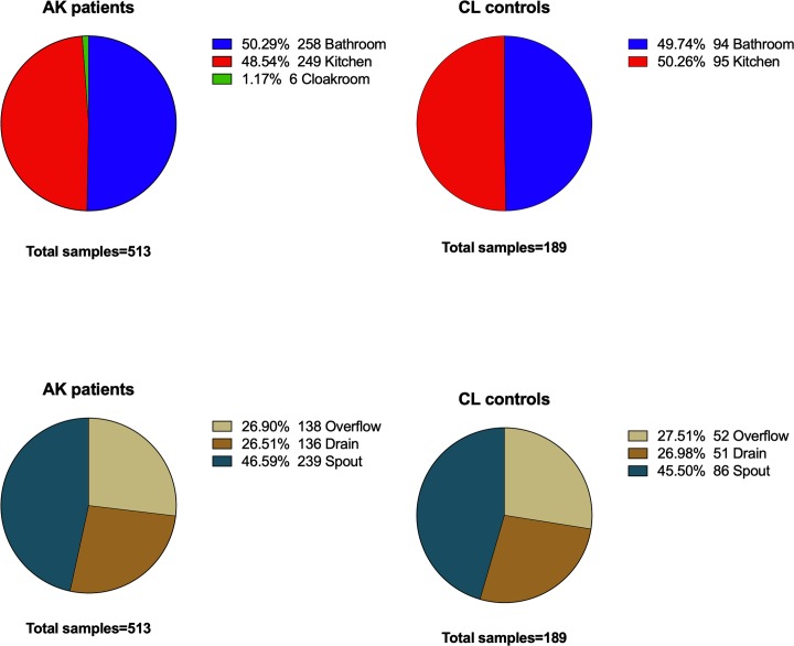
A total of 513 samples from 77 AK patients and 189 samples from 40 CL controls were retrieved and examined in this study. Samples were collected from water tap-spouts, sink overflows and drains from the kitchen and bathroom. The proportion of samples from kitchens and bathrooms were broadly similar (Fig 1). Samples collected from the cloakrooms of AK patients were excluded from the current analysis because the number of samples were small (1.7%) and there were no cloakrooms samples from CL controls group.

Fig 1. Distribution of samples analysed in current study.

Fig 1

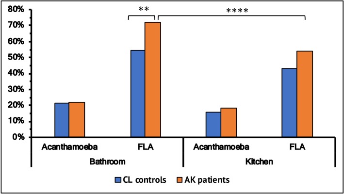
A slightly higher proportion of Acanthamoeba were cultured from bathrooms (average 21.5%) compared to kitchens (average16.9%) (Fig 2) and there was no difference in this proportion between the patient and control group. However, bathrooms yielded a higher proportion of FLA positive samples compared to kitchens. Bathrooms from the AK cohort had a higher proportion (72.1%) of FLA than samples from the control’s bathrooms (54.3%) (p<0.05) (OR 2.1; 95% CI 1.33–3.55). Furthermore, AK patient’s bathrooms had a significantly higher proportion of FLA than kitchens (p<0.05) (Fig 2). Controls had a similar higher proportion of FLA from bathrooms, but this did not reach statistical significance.

Fig 2. Overall colonisation of Acanthamoeba and free-living amoeba (FLA) in samples from bathroom and kitchen in patients and control groups.

Fig 2

* denotes level of significance (*p ≤ 0.05, **p ≤ 0.01, ***p ≤ 0.001, ****p≤ 0.0001).

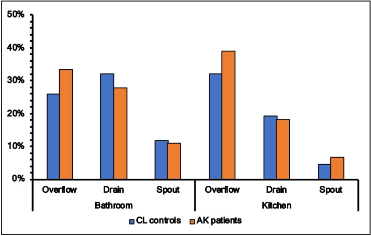
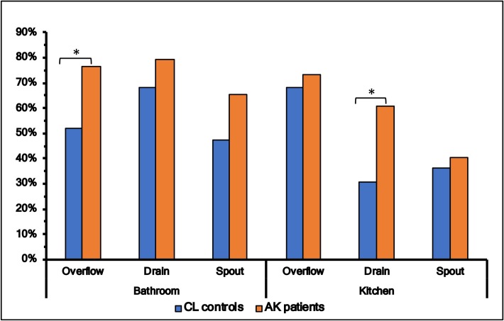
Colonisation of Acanthamoeba in tap-spouts was lower in both bathrooms and kitchens (p<0.05) than drains or overflows, but there was no statistical difference between patients and controls in the prevalence of Acanthamoeba in these sites (Fig 3). On the other hand, the rate of colonisation of FLA was higher in drains, overflows and tap-spouts of patients’ kitchens and bathrooms compared to these three sites from controls, and this reached statistical significance between bathroom’s overflow (controls 14/27 vs patients, 55/72, p = 0.02) and kitchen’s drain (controls 8/25 vs patients 40/66, p = 0.02) (Fig 4).

Fig 3. Colonisation of Acanthamoeba in samples for spout, drain and overflow from bathroom and kitchen in patients and control groups.

Fig 3

Fig 4. Colonisation of free-living amoeba (FLA) in samples for spout, drain and overflow from bathroom and kitchen in patients and control groups.

Fig 4

* denotes level of significance (*p ≤ 0.05, **p ≤ 0.01, ***p ≤ 0.001, ****p≤ 0.0001).

Twenty-three repeat samples at the two-sampling time-points for both bathrooms and kitchens spouts were examined to understand whether Acanthamoeba was consistently present at the same sites. Acanthamoeba isolation appeared to be sporadic from either bathrooms or kitchens (Table 1).

Table 1. Repeatability of samples.

Bathroom Kitchen
First Sample First Sample
Positive Negative Positive Negative
Second samples Positive 1 2 Second samples Positive 0 3
Negative 3 17 Negative 3 17

Patients and environmental Acanthamoeba colonisation

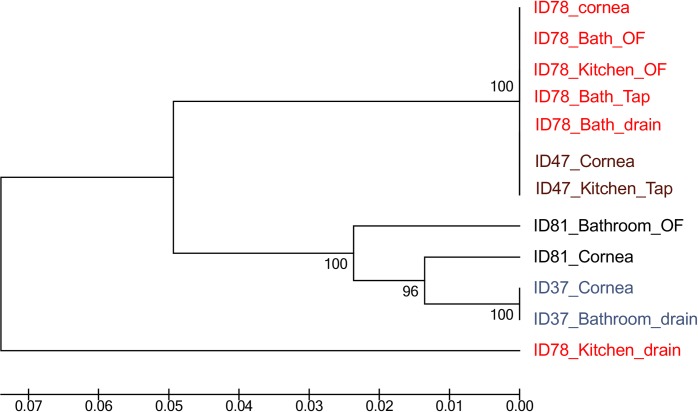
Four AK patients were selected to the study genetic relatedness between pathogenic and environmental isolates on the basis of association between case and detection of Acanthamoeba in water samples from their homes. Partial sequence of the mitochondrial cytochrome oxidase gene (coxA 1/2) was obtained from Acanthamoeba isolates of four different keratitis patients and those sequences were compared with isolates from the patient’s home. Fig 5 shows that each pathogenic isolate had the identical coxA 1/2 sequence to at least one domestic water isolate including isolates from both the kitchen and the bathroom. The Acanthamoeba isolate from patient ID78 was similar to isolates from the patient’s bathroom spout, drain and overflow and their kitchen’s overflow suggesting a possible source of keratitis isolate. This isolate was also similar to the corneal isolate from another patient ID47 and an isolate from that patient’s kitchen spout, suggesting possibility of common source of infections. For patients ID81 and ID37, their corneal isolates were similar to those from their bathrooms. Water contact was reported by three of these four patients in the days prior to suffering AK.

Fig 5. Cytochrome oxidase gene (coxA 1/2) sequence comparison of patient and environmental Acanthamoeba isolates.

Fig 5

Discussion

Domestic tap water is a major risk factor for contact-lens associated Acanthamoeba keratitis [11, 18, 20] and is a reservoir for Acanthamoeba and other FLA [11]. Based on coxA 1/2 gene sequence [33], this study identified that isolates from water sources were very similar to isolates from the corneas of AK patients, suggesting that domestic waterborne Acanthamoeba could be associated with AK. The current study examined water samples from CL wearers who were not AK patients and compared these to samples from AK patients. There was no variation in the rate of colonisation of Acanthamoeba in kitchens and bathrooms of AK patients compared to that of CL wearers controls, suggesting that all contact lens wearers are at risk of developing AK and it is not that AK patients have bathrooms or kitchens that are more frequently colonised. Patient’s bathrooms yielded a higher proportion of FLA compared to the bathrooms of control CL wearers or the kitchens of patients. Poorly maintained sinks may allow microbial biofilms to form, and this is believed to facilitate amoebal colonisation of water outlets and drains [11], not least because amoebae can graze of the bacteria in biofilms [34].

The prevalence of Acanthamoeba observed in this study (21.5%) was slightly lower than the prevalence reported by another UK based study, in which Kilvington et al. isolated Acanthamoeba from 30% of homes [11]. In the current study tap spouts tended to have filters on them making it difficult to swab the inside of spouts and this could be the reason for these differences [11]. In addition, spouts have the fastest water flow and large volume of water regularly flows through it, which self-flushes the spouts and may prevent colonisation of FLA. Furthermore, there is a wide variation in the rate of isolation of Acanthamoeba in previous reports. Studies based in Hong Kong have shown that household tap water yielded 10% [35], 8.3% [36] and 7.7% [36] Acanthamoeba positive samples. In addition, studies from Scotland (12% bathrooms, 2% kitchen) [37], Jamaica (36%, tap-water) [38] and Florida (2.8%, domestic water) [39] have reported various isolation rates in water samples. One of the reasons for these variations may be due to use of different methods for identification of Acanthamoeba, for example morphological [39] versus molecular [38] techniques. Given that PCR-based methods are more sensitive than culture for detection of Acanthamoeba [40], further studies based on molecular identification will be required to validate rate of isolation of Acanthamoeba observed in the current study.

The domestic water supply system of the UK, which uses water storage cisterns as well as often having hard water, is believed to be a cause for the higher prevalence of Acanthamoeba in the tap water in this region [11]. Acanthamoeba and other microbes proliferate in the cistern water. Also, there is a seasonal trend with the colonisation of Acanthamoeba increasing in warmer months [2224].

A limitation of this study is that the genus-level identification of FLA by molecular methods was not performed. FLA such as Hartmannella and Vahlkampfiid amoebae [4, 5], although rare compared to Acanthamoeba, are also causative agents of keratitis. In addition, the significantly higher FLA positive samples in the patient cohort suggests that their identification in the genus-level will help to better understand the risk of keratitis associated with domestic tap water. Further studies regarding the pathogenicity of FLA and Acanthamoeba will also require assessing the source of transmission as well as the severity of AK.

In conclusion, this study suggested that Acanthamoeba colonisation in UK water supply is high and occurs in CL-wearers and AK patients. Domestic water isolates were similar to isolates from the cornea of AK patient, confirming that domestic waterborne Acanthamoeba is still associated with keratitis. Advice about avoiding domestic water contact when using CL’s should be mandatory.

Supporting information

S1 Data. Water sample instruction.

(DOCX)

Acknowledgments

The authors would like to thank Ms Varshini Parayoganathan for database management, Ms Melanie Mason and Mr Scott Hau, Dr Judith Morris and Institute of Ophthalmology for subject recruitment. We would also like to thank Prof John Dart, Moorefields Eye Hospital for providing guidance and recruitments and Prof Mark Willcox, School of Optometry and Vision Science, UNSW Sydney for editing the draft.

Data Availability

All relevant data are within the manuscript and its Supporting Information files.

Funding Statement

Dr Carnt and Dr Kilvington received Dallos Award 2015 from British Contact Lens Association (BCLA) to conduct this study. Dr Carnt was supported by an NHMRC Early Career CJ Martin Fellowship (APP1036728), National Institute of Research (NIHR) Biomedical Research Centre (BRC) at Moorfields Eye Hospital NIHR BRC. The views expressed are those of the author(s) and not necessarily those of the BCLA, the NHS, the NIHR or the Department of Health. The funders provided support in the form of salaries for author NC and research materials but did not have any additional role in the study design, data collection and analysis, decision to publish, or preparation of the manuscript.

References

  • 1.Schuster FL, Visvesvara GS. Free-living amoebae as opportunistic and non-opportunistic pathogens of humans and animals. Int J Parasitol. 2004;34(9):1001–27. Epub 2004/08/18. 10.1016/j.ijpara.2004.06.004 . [DOI] [PubMed] [Google Scholar]
  • 2.Khan NA. Acanthamoeba: Biology and increasing importance in human health. FEMS Microbiol Rev. 2006;30(4):564–95. Epub 2006/06/16. 10.1111/j.1574-6976.2006.00023.x . [DOI] [PubMed] [Google Scholar]
  • 3.Schuster FL. Cultivation of pathogenic and opportunistic free-living amebas. Clinical microbiology reviews. 2002;15(3):342–54. 10.1128/CMR.15.3.342-354.2002 [DOI] [PMC free article] [PubMed] [Google Scholar]
  • 4.Pinna A, Porcu T, Boscia F, Cano A, Erre G, Mattana A. Free-Living Amoebae Keratitis. Cornea. 2017;36(7):785–90. Epub 2017/05/10. 10.1097/ICO.0000000000001226 . [DOI] [PubMed] [Google Scholar]
  • 5.Aitken D, Hay J, Kinnear FB, Kirkness CM, Lee WR, Seal DV. Amebic keratitis in a wearer of disposable contact lenses due to a mixed Vahlkampfia and Hartmannella infection. Ophthalmology. 1996;103(3):485–94. Epub 1996/03/01. 10.1016/s0161-6420(96)30667-2 . [DOI] [PubMed] [Google Scholar]
  • 6.Clarke DW, Niederkorn JY. The pathophysiology of Acanthamoeba keratitis. Trends Parasitol. 2006;22(4):175–80. 10.1016/j.pt.2006.02.004 . [DOI] [PubMed] [Google Scholar]
  • 7.Marciano-Cabral F, Cabral G. Acanthamoeba spp. as agents of disease in humans. Clinical microbiology reviews. 2003;16(2):273–307. Epub 2003/04/15. 10.1128/CMR.16.2.273-307.2003 [DOI] [PMC free article] [PubMed] [Google Scholar]
  • 8.Ku JY, Chan FM, Beckingsale P. Acanthamoeba keratitis cluster: an increase in Acanthamoeba keratitis in Australia. Clin Exp Ophthalmol. 2009;37(2):181–90. Epub 2009/09/03. 10.1111/j.1442-9071.2008.01910.x . [DOI] [PubMed] [Google Scholar]
  • 9.Stehr-Green JK, Bailey TM, Visvesvara GS. The epidemiology of Acanthamoeba keratitis in the United States. American journal of ophthalmology. 1989;107(4):331–6. 10.1016/0002-9394(89)90654-5 [DOI] [PubMed] [Google Scholar]
  • 10.Stehr-Green JK, Bailey TM, Brandt FH, Carr JH, Bond WW, Visvesvara GS. Acanthamoeba keratitis in soft contact lens wearers: A case-control study. Jama. 1987;258(1):57–60. 10.1001/jama.1987.03400010061028 [DOI] [PubMed] [Google Scholar]
  • 11.Kilvington S, Gray T, Dart J, Morlet N, Beeching JR, Frazer DG, et al. Acanthamoeba keratitis: the role of domestic tap water contamination in the United Kingdom. Investigative ophthalmology & visual science. 2004;45(1):165–9. Epub 2003/12/24. 10.1167/iovs.03-0559 . [DOI] [PubMed] [Google Scholar]
  • 12.Arshad M, Carnt N, Tan J, Ekkeshis I, Stapleton F. Water exposure and the risk of contact lens-related disease. Cornea. 2019;38(6):791–7. Epub 2019/02/23. 10.1097/ICO.0000000000001898 . [DOI] [PubMed] [Google Scholar]
  • 13.Fraser MN, Wong Q, Shah L, Holland SP, Morshed M, Isaac-Renton J, et al. Characteristics of an Acanthamoeba keratitis outbreak in British Columbia between 2003 and 2007. Ophthalmology. 2012;119(6):1120–5. Epub 2012/03/01. 10.1016/j.ophtha.2011.12.041 . [DOI] [PubMed] [Google Scholar]
  • 14.Mathers WD, Sutphin JE, Lane JA, Folberg R. Correlation between surface water contamination with amoeba and the onset of symptoms and diagnosis of amoeba-like keratitis. The British journal of ophthalmology. 1998;82(10):1143–6. Epub 1999/01/30. 10.1136/bjo.82.10.1143 [DOI] [PMC free article] [PubMed] [Google Scholar]
  • 15.Naginton J, Watson PG, Playfair TJ, McGill J, Jones BR, Steele AD. Amoebic infection of the eye. Lancet. 1974;2(7896):1537–40. Epub 1974/12/28. 10.1016/s0140-6736(74)90285-2 . [DOI] [PubMed] [Google Scholar]
  • 16.Schaumberg DA, Snow KK, Dana MR. The epidemic of Acanthamoeba keratitis: where do we stand? Cornea. 1998;17(1):3–10. Epub 1998/01/22. 10.1097/00003226-199801000-00001 . [DOI] [PubMed] [Google Scholar]
  • 17.Butler TK, Males JJ, Robinson LP, Wechsler AW, Sutton GL, Cheng J, et al. Six-year review of Acanthamoeba keratitis in New South Wales, Australia: 1997–2002. Clin Exp Ophthalmol. 2005;33(1):41–6. Epub 2005/01/27. 10.1111/j.1442-9071.2004.00911.x . [DOI] [PubMed] [Google Scholar]
  • 18.Radford CF, Minassian DC, Dart JK. Acanthamoeba keratitis in England and Wales: incidence, outcome, and risk factors. The British journal of ophthalmology. 2002;86(5):536–42. 10.1136/bjo.86.5.536 [DOI] [PMC free article] [PubMed] [Google Scholar]
  • 19.Ibrahim YW, Boase DL, Cree IA. Factors affecting the epidemiology of Acanthamoeba keratitis. Ophthalmic epidemiology. 2007;14(2):53–60. Epub 2007/04/28. 10.1080/09286580600920281 . [DOI] [PubMed] [Google Scholar]
  • 20.Joslin CE, Tu EY, McMahon TT, Passaro DJ, Stayner LT, Sugar J. Epidemiological characteristics of a Chicago-area Acanthamoeba keratitis outbreak. American journal of ophthalmology. 2006;142(2):212–7. Epub 2006/08/01. 10.1016/j.ajo.2006.04.034 . [DOI] [PubMed] [Google Scholar]
  • 21.Kao PM, Chou MY, Tao CW, Huang WC, Hsu BM, Shen SM, et al. Diversity and seasonal impact of Acanthamoeba species in a subtropical rivershed. BioMed research international. 2013;2013:405794 10.1155/2013/405794 [DOI] [PMC free article] [PubMed] [Google Scholar]
  • 22.Kyle DE, Noblet GP. Seasonal distribution of thermotolerant free-living amoebae. I. Willard's Pond. J Protozool. 1986;33(3):422–34. 10.1111/j.1550-7408.1986.tb05634.x . [DOI] [PubMed] [Google Scholar]
  • 23.Kyle DE, Noblet GP. Seasonal distribution of thermotolerant free-living amoebae. II. Lake Issaqueena. J Protozool. 1987;34(1):10–5. 10.1111/j.1550-7408.1987.tb03122.x . [DOI] [PubMed] [Google Scholar]
  • 24.McAllum P, Bahar I, Kaiserman I, Srinivasan S, Slomovic A, Rootman D. Temporal and seasonal trends in Acanthamoeba keratitis. Cornea. 2009;28(1):7–10. 10.1097/ICO.0b013e318181a863 . [DOI] [PubMed] [Google Scholar]
  • 25.Taher EE, Meabed EMH, Abdallah I, Abdel Wahed WY. Acanthamoeba keratitis in noncompliant soft contact lenses users: Genotyping and risk factors, a study from Cairo, Egypt. Journal of infection and public health. 2018;11(3):377–83. 10.1016/j.jiph.2017.09.013 . [DOI] [PubMed] [Google Scholar]
  • 26.Yoder JS, Verani J, Heidman N, Hoppe-Bauer J, Alfonso EC, Miller D, et al. Acanthamoeba keratitis: the persistence of cases following a multistate outbreak. Ophthalmic epidemiology. 2012;19(4):221–5. 10.3109/09286586.2012.681336 . [DOI] [PubMed] [Google Scholar]
  • 27.Carnt N, Hoffman JM, Verma S, Hau S, Radford CF, Minassian DC, et al. Acanthamoeba keratitis: Confirmation of the UK outbreak and a prospective case-control study identifying contributing risk factors. The British journal of ophthalmology. 2018;102(12):1621–8. Epub 2018/09/21. 10.1136/bjophthalmol-2018-312544 . [DOI] [PubMed] [Google Scholar]
  • 28.Bacon AS, Frazer DG, Dart JK, Matheson M, Ficker LA, Wright P. A review of 72 consecutive cases of Acanthamoeba keratitis, 1984–1992. Eye. 1993;7 (Pt 6):719–25. Epub 1993/01/01. 10.1038/eye.1993.168 . [DOI] [PubMed] [Google Scholar]
  • 29.Radford CF, Bacon AS, Dart JK, Minassian DC. Risk factors for acanthamoeba keratitis in contact lens users: a case-control study. Bmj. 1995;310(6994):1567–70. Epub 1995/06/17. 10.1136/bmj.310.6994.1567 [DOI] [PMC free article] [PubMed] [Google Scholar]
  • 30.Khan NA. Acanthamoeba: Biology and Pathogenesis. Norfolk, UK: Caister Academic Press; 2009. 289 p. [Google Scholar]
  • 31.Iovieno A, Miller D, Lonnen J, Kilvington S, Alfonso EC. Extraction of Acanthamoeba DNA by use of Chelex resin. Journal of clinical microbiology. 2011;49(1):476–7. Epub 2010/11/19. 10.1128/JCM.01795-10 [DOI] [PMC free article] [PubMed] [Google Scholar]
  • 32.Kumar S, Stecher G, Tamura K. MEGA7: Molecular evolutionary genetics analysis version 7.0 for bigger datasets. Mol Biol Evol. 2016;33(7):1870–4. 10.1093/molbev/msw054 . [DOI] [PMC free article] [PubMed] [Google Scholar]
  • 33.Burger G, Plante I, Lonergan KM, Gray MW. The mitochondrial DNA of the amoeboid protozoon, Acanthamoeba castellanii: Complete sequence, gene content and genome organization. Journal of molecular biology. 1995;245(5):522–37. Epub 1995/02/03. 10.1006/jmbi.1994.0043 . [DOI] [PubMed] [Google Scholar]
  • 34.Seiler C, van Velzen E, Neu TR, Gaedke U, Berendonk TU, Weitere M. Grazing resistance of bacterial biofilms: a matter of predators' feeding trait. FEMS Microbiol Ecol. 2017;93(9). Epub 2017/09/30. 10.1093/femsec/fix112 . [DOI] [PubMed] [Google Scholar]
  • 35.Boost M, Cho P, Lai S, Sun WM. Detection of Acanthamoeba in tap water and contact lens cases using polymerase chain reaction. Optometry and vision science: official publication of the American Academy of Optometry. 2008;85(7):526–30. Epub 2008/07/03. 10.1097/OPX.0b013e31817dba7c . [DOI] [PubMed] [Google Scholar]
  • 36.Houang E, Lam D, Fan D, Seal D. Microbial keratitis in Hong Kong: relationship to climate, environment and contact-lens disinfection. Transactions of the Royal Society of Tropical Medicine and Hygiene. 2001;95(4):361–7. Epub 2001/10/03. 10.1016/s0035-9203(01)90180-4 . [DOI] [PubMed] [Google Scholar]
  • 37.Seal D, Stapleton F, Dart J. Possible environmental sources of Acanthamoeba spp in contact lens wearers. The British journal of ophthalmology. 1992;76(7):424–7. Epub 1992/07/01. 10.1136/bjo.76.7.424 [DOI] [PMC free article] [PubMed] [Google Scholar]
  • 38.Lorenzo-Morales J, Lindo JF, Martinez E, Calder D, Figueruelo E, Valladares B, et al. Pathogenic Acanthamoeba strains from water sources in Jamaica, West Indies. Ann Trop Med Parasitol. 2005;99(8):751–8. Epub 2005/11/22. 10.1179/136485905X65215 . [DOI] [PubMed] [Google Scholar]
  • 39.Shoff ME, Rogerson A, Kessler K, Schatz S, Seal DV. Prevalence of Acanthamoeba and other naked amoebae in South Florida domestic water. J Water Health. 2008;6(1):99–104. Epub 2007/11/14. 10.2166/wh.2007.014 . [DOI] [PubMed] [Google Scholar]
  • 40.Khan NA, Jarroll EL, Paget TA. Acanthamoeba can be differentiated by the polymerase chain reaction and simple plating assays. Current microbiology. 2001;43(3):204–8. Epub 2001/06/16. 10.1007/s002840010288 . [DOI] [PubMed] [Google Scholar]

Decision Letter 0

Manuel Garza León

31 Dec 2019

PONE-D-19-26513

The relationship between environmental sources and the susceptibility of Acanthamoeba keratitis in the United Kingdom

PLOS ONE

Dear Dr Carnt,

Thank you for submitting your manuscript to PLOS ONE. After careful consideration, we feel that it has merit but does not fully meet PLOS ONE’s publication criteria as it currently stands. Therefore, we invite you to submit a revised version of the manuscript that addresses the points raised during the review process.

We would appreciate receiving your revised manuscript by Feb 14 2020 11:59PM. When you are ready to submit your revision, log on to https://www.editorialmanager.com/pone/ and select the 'Submissions Needing Revision' folder to locate your manuscript file.

If you would like to make changes to your financial disclosure, please include your updated statement in your cover letter.

To enhance the reproducibility of your results, we recommend that if applicable you deposit your laboratory protocols in protocols.io, where a protocol can be assigned its own identifier (DOI) such that it can be cited independently in the future. For instructions see: http://journals.plos.org/plosone/s/submission-guidelines#loc-laboratory-protocols

Please include the following items when submitting your revised manuscript:

  • A rebuttal letter that responds to each point raised by the academic editor and reviewer(s). This letter should be uploaded as separate file and labeled 'Response to Reviewers'.

  • A marked-up copy of your manuscript that highlights changes made to the original version. This file should be uploaded as separate file and labeled 'Revised Manuscript with Track Changes'.

  • An unmarked version of your revised paper without tracked changes. This file should be uploaded as separate file and labeled 'Manuscript'.

Please note while forming your response, if your article is accepted, you may have the opportunity to make the peer review history publicly available. The record will include editor decision letters (with reviews) and your responses to reviewer comments. If eligible, we will contact you to opt in or out.

We look forward to receiving your revised manuscript.

Kind regards,

Manuel Garza León

Academic Editor

PLOS ONE

Journal Requirements:

When submitting your revision, we need you to address these additional requirements.

1. Please ensure that your manuscript meets PLOS ONE's style requirements, including those for file naming. The PLOS ONE style templates can be found at

http://www.journals.plos.org/plosone/s/file?id=wjVg/PLOSOne_formatting_sample_main_body.pdf and http://www.journals.plos.org/plosone/s/file?id=ba62/PLOSOne_formatting_sample_title_authors_affiliations.pdf

2. Thank you for stating the following in the Financial Disclosure section:

'British Contact Lens Association, Dallos award, 2015 (Carnt and Kilvington). Dr

Carnt was supported by an NHMRC Early Career CJ Martin Fellowship (APP1036728).  The views expressed are those of the author(s) and not necessarily those of the NHS, the NIHR or the Department of Health'

We note that one or more of the authors are employed by a commercial company: 

Research Organisation (KC) Ltd, and Ophtecs Corporation.

a. Please provide an amended Funding Statement declaring this commercial affiliation, as well as a statement regarding the Role of Funders in your study. If the funding organization did not play a role in the study design, data collection and analysis, decision to publish, or preparation of the manuscript and only provided financial support in the form of authors' salaries and/or research materials, please review your statements relating to the author contributions, and ensure you have specifically and accurately indicated the role(s) that these authors had in your study. You can update author roles in the Author Contributions section of the online submission form.

Please also include the following statement within your amended Funding Statement.

“The funder provided support in the form of salaries for authors [insert relevant initials], but did not have any additional role in the study design, data collection and analysis, decision to publish, or preparation of the manuscript. The specific roles of these authors are articulated in the ‘author contributions’ section.”

If your commercial affiliation did play a role in your study, please state and explain this role within your updated Funding Statement.

b. Please also provide an updated Competing Interests Statement declaring this commercial affiliation along with any other relevant declarations relating to employment, consultancy, patents, products in development, or marketed products, etc.  

Within your Competing Interests Statement, please confirm that this commercial affiliation does not alter your adherence to all PLOS ONE policies on sharing data and materials by including the following statement: "This does not alter our adherence to  PLOS ONE policies on sharing data and materials.” (as detailed online in our guide for authors http://journals.plos.org/plosone/s/competing-interests) . If this adherence statement is not accurate and  there are restrictions on sharing of data and/or materials, please state these. Please note that we cannot proceed with consideration of your article until this information has been declared.

c. Please include both an updated Funding Statement and Competing Interests Statement in your cover letter. We will change the online submission form on your behalf.

Please know it is PLOS ONE policy for corresponding authors to declare, on behalf of all authors, all potential competing interests for the purposes of transparency. PLOS defines a competing interest as anything that interferes with, or could reasonably be perceived as interfering with, the full and objective presentation, peer review, editorial decision-making, or publication of research or non-research articles submitted to one of the journals. Competing interests can be financial or non-financial, professional, or personal. Competing interests can arise in relationship to an organization or another person. Please follow this link to our website for more details on competing interests: http://journals.plos.org/plosone/s/competing-interests

3. Please include captions for your Supporting Information files at the end of your manuscript, and update any in-text citations to match accordingly. Please see our Supporting Information guidelines for more information: http://journals.plos.org/plosone/s/supporting-information

[Note: HTML markup is below. Please do not edit.]

Reviewers' comments:

Reviewer's Responses to Questions

Comments to the Author

1. Is the manuscript technically sound, and do the data support the conclusions?

The manuscript must describe a technically sound piece of scientific research with data that supports the conclusions. Experiments must have been conducted rigorously, with appropriate controls, replication, and sample sizes. The conclusions must be drawn appropriately based on the data presented.

Reviewer #1: Yes

Reviewer #2: Yes

**********

2. Has the statistical analysis been performed appropriately and rigorously?

Reviewer #1: Yes

Reviewer #2: No

**********

3. Have the authors made all data underlying the findings in their manuscript fully available?

The PLOS Data policy requires authors to make all data underlying the findings described in their manuscript fully available without restriction, with rare exception (please refer to the Data Availability Statement in the manuscript PDF file). The data should be provided as part of the manuscript or its supporting information, or deposited to a public repository. For example, in addition to summary statistics, the data points behind means, medians and variance measures should be available. If there are restrictions on publicly sharing data—e.g. participant privacy or use of data from a third party—those must be specified.

Reviewer #1: Yes

Reviewer #2: Yes

**********

4. Is the manuscript presented in an intelligible fashion and written in standard English?

PLOS ONE does not copyedit accepted manuscripts, so the language in submitted articles must be clear, correct, and unambiguous. Any typographical or grammatical errors should be corrected at revision, so please note any specific errors here.

Reviewer #1: Yes

Reviewer #2: Yes

**********

5. Review Comments to the Author

Please use the space provided to explain your answers to the questions above. You may also include additional comments for the author, including concerns about dual publication, research ethics, or publication ethics. (Please upload your review as an attachment if it exceeds 20,000 characters)

Reviewer #1: Line 138-139 – Spouts have the fastest flowing and largest volume of water flowing through it with thousands of gallons per month, this may be the reason why you do not see as much bacteria/Acanthamoeba/FLA in that particular area for Fig 3. Should repeat samples be taken from the drain or overflows from these areas for future studies? Please comment/discuss this finding.

Do you think that paranoia of patients when sampling they didn’t want to see Acanthamoeba or FLA within their home that they may have stringently cleaned between sampling?

All figures/graphs are of poor quality please use another software program.

Were attempts to characterize the species/isolate causing the AK in these 4 patients?

Supplemental information is clear for samplers.

Molecular characterization of FLA should become mandatory in future studies.

Reviewer #2: Overall, it is a very well written case-control study on the association between environmental sources of contamination and Acanthamoeba keratitis in UK.

As recommended, please include the statistical analysis used for this study in the manuscript (as it is included only in the abstract). As a suggestion, including the odds ratio of exposure to tap water in the AK patient vs the corresponding ratio for control group, would be really interesting.

Great job!

**********

6. PLOS authors have the option to publish the peer review history of their article (what does this mean?). If published, this will include your full peer review and any attached files.

If you choose “no”, your identity will remain anonymous but your review may still be made public.

Do you want your identity to be public for this peer review? For information about this choice, including consent withdrawal, please see our Privacy Policy.

Reviewer #1: Yes: Christopher Aaron Rice

Reviewer #2: No

[NOTE: If reviewer comments were submitted as an attachment file, they will be attached to this email and accessible via the submission site. Please log into your account, locate the manuscript record, and check for the action link "View Attachments". If this link does not appear, there are no attachment files to be viewed.]

While revising your submission, please upload your figure files to the Preflight Analysis and Conversion Engine (PACE) digital diagnostic tool, https://pacev2.apexcovantage.com/. PACE helps ensure that figures meet PLOS requirements. To use PACE, you must first register as a user. Registration is free. Then, login and navigate to the UPLOAD tab, where you will find detailed instructions on how to use the tool. If you encounter any issues or have any questions when using PACE, please email us at figures@plos.org. Please note that Supporting Information files do not need this step.

Attachment

Submitted filename: The relationship between environmental sources and the susceptibility of Acanthamoeba keratitis in the United Kingdom.Carnt.2019.docx

PLoS One. 2020 Mar 11;15(3):e0229681. doi: 10.1371/journal.pone.0229681.r002

Author response to Decision Letter 0


15 Jan 2020

Major changes

• As your study was designed to search for Acanthamoeba and then record the presence of other FLA I would switch this throughout the text. “To determine whether Acanthamoeba keratitis (AK) patients have higher rates of Acanthamoeba or other free-living amoeba (FLA) colonising domestic sinks than control contact lens (CL) wearers, and whether these isolates are genetically similar to the corneal isolates identified from their CL associated AK.”

Answer: The sentence has been modified as suggested.

• There is no description of the other FLA identified, can you describe which others were investigated and found?

o Can this also be broken down within the bar graphs?

Answer: Classification of FLA were not attempted in this study and this was mentioned as a limitation of the study.

“A limitation of this study is that the genus-level identification of FLA by molecular methods was not performed. FLA such as Hartmannella and Vahlkampfiid amoebae [4, 5], although rare compared to Acanthamoeba, are also causative agents of keratitis. In addition, the significantly higher FLA positive samples in the patient cohort suggests that their identification in the genus-level will help to better understand the risk of keratitis associated with domestic tap water. Further studies regarding the pathogenicity of FLA and Acanthamoeba will also require assessing the source of transmission as well as the severity of AK”

• The quality of all of the figures need to be improved (use graph pad prism or another software for better images).

Answer: Figures have been reformatted as suggested

Figure 1.

• Do these graphs describe the full picture? Although the graphs tell the reader total numbers it doesn’t break down the exact number of samples obtained from the spout from the kitchen of an AK wearer or the drain from the bathroom of a CL control as examples. A reorganization and coloration to be more descriptive may be useful.

Answer: Figures have been reformatted to keep numbers of samples.

Figure 2.

• As you are specifically identifying different FLA, this graph could be a stacked bar graph with different colors representing the different genus of FLA found for both groups and areas swabbed.

Answer: Figures have been reformatted as suggested

As mentioned above genus level identification for FLA was not attempted.

Minor Changes

• Lines 19-21, 37-41, 79, 113 – switch Acanthamoeba and FLA order.

Answer: The order has been corrected as suggested throughout the manuscript.

• Don’t you think using E. coli and 32°C selects for Acanthamoeba over other FLA?

Answer: FLA could be thermophilic (Hartmannella vermiformis and Naegleria) as indicated in this paper https://www.ncbi.nlm.nih.gov/pmc/articles/PMC3469187/. Data of the current study showed that rate of isolation of FLA was higher than that of Acanthamoeba. Therefore, we believed that the incubation condition does not select one over the other.

• There was no discussion to where the patients were from within the UK (distribution, were these all local to a particular area? – This data should be easily accessible since collected during the questionnaire). Can you make a conclusion about geographic location, potential differences in water storage methods, sources that the water was from or treatment methods? You may find that there was a particular area that could have an increased risk or contamination. This could be useful for water regulatory and treatment of such bodies of water.

Answer: Yes, we agree that this information would be useful in many ways. However, we were able to collect this information from less than 1/4 of case and controls. Therefore, this analysis has not been added in the manuscript.

• Why were only 4 patients out of 129 compared for Ac coxA? Please discuss.

Answer: Due to time frame of the project, resources and funding availability, we have selected samples from only four patients for genetic characterization. These four patients appeared to have striking association with detection of Acanthamoeba in water sample from their homes. Our aim was to show relatedness between pathogenic and environmental isolates. We have clarified this in manuscript as

Four AK patients were selected to the study genetic relatedness between pathogenic and environmental isolates on the basis of association between case and detection of Acanthamoeba in water samples from their homes. Partial sequence of the mitochondrial cytochrome oxidase gene (coxA 1/2) was obtained from Acanthamoeba isolates of four different keratitis patients and those sequences were compared with isolates from the patient’s home.

Lines 24-26 – “The samples were cultured by inoculating onto a non-nutrient agar plate seeded with Escherichia coli and the plates were incubated at 32˚C and examined for amoebae by microscopy for up to 2 weeks.” Could read as “The samples were cultured by inoculating onto a non-nutrient agar plate seeded with Escherichia coli, incubated at 32˚C and examined for amoebae by microscopy for up to 2 weeks.”

Answer: The sentence has been corrected as suggested

• Lines 30-36 – Please add the %’s for spouts, drains, overflow in kitchens and bathrooms.

Answer: Percentage for sampling sites have been added

“Spouts (kitchen 6.7%, bathroom 11%) had the lowest rate of Acanthamoeba isolation compared to drains (kitchen 18.2%, bathroom 27.9%) and overflow (kitchen 39.1%, bathroom 25.9%) either in kitchens or bathrooms (p<0.05). There was no statistically significant difference between the prevalence of Acanthamoeba in all three sample sites in kitchens (16.9%) compared to all three sample sites in bathrooms (21.5%) and no association for Acanthamoeba prevalence between AK patients and CL controls.”

• Line 36 – Was this the kitchen or bathroom sink?

Answer: It was from both the kitchen and the bathroom. The sentence has been modified as

“All four corneal isolates had the same coxA sequence as at least one domestic water isolate from the patients’ sink of the kitchen and the bathroom.”

• Line 46 – Change “potentially” to opportunistically. Where these the different types of FLA identified within this study? Please make reference to which ones?

Answer: This has been changed as suggested.

• Line 71 – Reference to ref 11 not needed here since it is referenced at the end of the sentence.

Answer: The reference has been deleted.

• Line 95-96 – If 23x repeat samples were taken from 129 patients from 6 locations within the home there would be 17,802 samples? I would remove this sentence. Or please show the succession of each patient over the 23 month period as a supplemental figure.

Answer: The sentence has been rewritten to avoid this confusion.

“A total of 23 repeat samples at least one month apart were collected from patient’s kitchen and bathroom”

• Line 114 – 513 total samples were obtained from AK patients = 6.6 samples per patient? 189 samples from 40 CL controls = 4.7 samples per patient?

Answer: As mentioned in methods, each patient was provided with six sample collecting pack. However, some of them had not returned all six tubes with sample plus we collected 23 repeat samples from AK patents. This is the reason; the sample numbers are not exact multiple of patient number. To avoid any confusion, the sentence has been modified as

“A total of 513 samples from 77 AK patients and 189 samples from 40 CL controls were retrieved and examined in this study.”

Furthermore, the distribution of sample in figure 1 shows that proportion of samples collected from different sites are different.

Figure 1.

• Please add in total numbers for each of the described %’s within the pie chart.

Answer: This has been updated in figure 1.

• Please add discussion about why the 1.17% (6 cases) from the Cloakroom of AK patients was removed.

Answer: This has been added as

“Samples collected from the cloakrooms of AK patients were excluded from the current analysis because the number of samples were small (1.7%) and there were no cloakrooms samples from CL controls group.”

• Line 121 – (The Acanthamoeba comparison should be Figure 2 not “Fig 1”. Also spell out figure to keep consistent throughout the MS.

Answer: Corrections made as suggested.

• Line 123-124 – Do both groups (AK patients and CL control) FLA total include the Acanthamoeba data as well? If so, shouldn’t this be removed/normalized?

Answer: In the current analysis, FLA group includes Acanthamoeba or FLA. The rate of presence of Acanthamoeba and FLA was analysed separately for each sampled area. Therefore, there were no duplicate isolates in the analysis. To clarify this, following modifications have been added

“Samples identified with Acanthamoeba were classified as Acanthamoeba positive and those with Acanthamoeba or FLA were classified as FLA in the current analysis.”

Figure 2.

• Please shorten the significance bar for the ** between the FLA found in patients and controls.

Answer: Figure has been reformatted as suggested

• Please keep consistency between titles of groups for all figures, “AK patients” and “CL controls”.

Answer: Corrections have been made throughout

• Line 131-137 – Please include the * for level of significance.

Answer: Correction has been made

• Line 133-137 – Please spell out figure to keep consistency throughout the MS.

Answer: Corrections have been made throughout

• Line 142 – Please Italicize Acanthamoeba.

Answer: Corrections have been made throughout

Figure 3&4.

• Please keep consistency between titles of groups for all figures, “AK patients” and “CL controls”.

Answer: Corrections have been made throughout

• Please shorten the significance bar and center the * for kitchen drain and bathroom overflow.

Answer: Figure has been reformatted as suggested

Table 1.

• Please centers align and middle aligns text into the center of all boxes of the table.

Answer: Table has been formatted as suggested.

• References font changes.

Answer: Font has been changed to match with manuscript’s font.

Reviewer # 1

Line 138-139 – Spouts have the fastest flowing and largest volume of water flowing through it with thousands of gallons per month, this may be the reason why you do not see as much bacteria/Acanthamoeba/FLA in that particular area for Fig 3. Should repeat samples be taken from the drain or overflows from these areas for future studies? Please comment/discuss this finding.

Answer: Discussion has been added (line 191 -194)

In the current study tap spouts tended to have filters on them making it difficult to swab the inside of spouts and this could be the reason for these differences [11]. In addition, spouts have the fastest water flow and large volume of water regularly flows through it, which self-flushes the spouts and may prevent colonisation of FLA.

Do you think that paranoia of patients when sampling they didn’t want to see Acanthamoeba or FLA within their home that they may have stringently cleaned between sampling?

Answer: While a small number of the first samples changed from positive to negative, a similar amount changed in the other direction in repeat sampling. Therefore, it does not appear that this had an effect.

All figures/graphs are of poor quality please use another software program.

Answer: figures have been modified for better quality.

Were attempts to characterize the species/isolate causing the AK in these 4 patients?

Answer: No, only the coxA gene was sequenced.

Supplemental information is clear for samplers.

Answer: Participants were given a verbal instruction and provided with written instruction.

Molecular characterization of FLA should become mandatory in future studies.

Answer: Yes, Authors agree with this and discus it in the manuscript.

Reviewer #2:

Overall, it is a very well written case-control study on the association between environmental sources of contamination and Acanthamoeba keratitis in UK.

Answer: Authors like to thank reviewer for taking time to review the manuscript and providing comment on it.

As recommended, please include the statistical analysis used for this study in the manuscript (as it is included only in the abstract). As a suggestion, including the odds ratio of exposure to tap water in the AK patient vs the corresponding ratio for control group, would be really interesting.

Answer: Thank you for suggesting this. Statistical analysis has been added to in the methods of the manuscript as suggested. Odd ratios have been included in results.

“Statistical analysis

The Pearson Chi-square test was used to assess whether there was a statistically significant difference in the association between sampling sites. Odds ratios and their 95% confidence intervals (95% CIs) were calculated to measure association between AK cases and detection rate of Acanthamoeba and FLA in AK patients’ and CL controls’ homes.”

Great job!

Decision Letter 1

Manuel Garza León

12 Feb 2020

The relationship between environmental sources and the susceptibility of Acanthamoeba keratitis in the United Kingdom

PONE-D-19-26513R1

Dear Dr. Carnt,

We are pleased to inform you that your manuscript has been judged scientifically suitable for publication and will be formally accepted for publication once it complies with all outstanding technical requirements.

Within one week, you will receive an e-mail containing information on the amendments required prior to publication. When all required modifications have been addressed, you will receive a formal acceptance letter and your manuscript will proceed to our production department and be scheduled for publication.

Shortly after the formal acceptance letter is sent, an invoice for payment will follow. To ensure an efficient production and billing process, please log into Editorial Manager at https://www.editorialmanager.com/pone/, click the "Update My Information" link at the top of the page, and update your user information. If you have any billing related questions, please contact our Author Billing department directly at authorbilling@plos.org.

If your institution or institutions have a press office, please notify them about your upcoming paper to enable them to help maximize its impact. If they will be preparing press materials for this manuscript, you must inform our press team as soon as possible and no later than 48 hours after receiving the formal acceptance. Your manuscript will remain under strict press embargo until 2 pm Eastern Time on the date of publication. For more information, please contact onepress@plos.org.

With kind regards,

Manuel Garza León

Academic Editor

PLOS ONE

Additional Editor Comments (optional):

Reviewers' comments:

Acceptance letter

Manuel Garza León

14 Feb 2020

PONE-D-19-26513R1

The relationship between environmental sources and the susceptibility of Acanthamoeba keratitis in the United Kingdom

Dear Dr. Carnt:

I am pleased to inform you that your manuscript has been deemed suitable for publication in PLOS ONE. Congratulations! Your manuscript is now with our production department.

If your institution or institutions have a press office, please notify them about your upcoming paper at this point, to enable them to help maximize its impact. If they will be preparing press materials for this manuscript, please inform our press team within the next 48 hours. Your manuscript will remain under strict press embargo until 2 pm Eastern Time on the date of publication. For more information please contact onepress@plos.org.

For any other questions or concerns, please email plosone@plos.org.

Thank you for submitting your work to PLOS ONE.

With kind regards,

PLOS ONE Editorial Office Staff

on behalf of

Dr. Manuel Garza León

Academic Editor

PLOS ONE

Associated Data

    This section collects any data citations, data availability statements, or supplementary materials included in this article.

    Supplementary Materials

    S1 Data. Water sample instruction.

    (DOCX)

    Attachment

    Submitted filename: The relationship between environmental sources and the susceptibility of Acanthamoeba keratitis in the United Kingdom.Carnt.2019.docx

    Data Availability Statement

    All relevant data are within the manuscript and its Supporting Information files.


    Articles from PLoS ONE are provided here courtesy of PLOS

    RESOURCES