Abstract
Study design:
Quasi-experimental, pretest-posttest design.
Objectives:
To identify acute changes in the supraspinatus and biceps tendon following fatiguing wheelchair propulsion and to associate tendon changes with risk factors associated with shoulder pain in persons with spinal cord injury (SCI).
Setting:
Biomechanical laboratory Swiss Paraplegic Research.
Methods:
A population-based sample of 50 wheelchair users with SCI at lesion level T2 or below participated. Fatigue was measured using the rate of perceived exertion and heart rate. Linear regression techniques were used to assess the association between the dependent and independent variables. Dependent variables included absolute differences in supraspinatus and biceps tendon thickness, contrast and echogenicity ratio assessed with ultrasound before and after a fatiguing wheelchair propulsion intervention. Independent variables included: susceptibility to fatigue (Yes/No), the acromio-humeral distance, sex, time since injury, activity levels and body weight.
Results:
A reduction in supraspinatus tendon thickness after fatiguing wheelchair propulsion (−1.39 mm; 95% CI: −2.28; −0.51) was identified after controlling for all potential confounders. Females who fatigued (n=4) displayed a greater reduction in supraspinatus tendon thickness as compared to those who did not fatigue (n=7). In contrast, higher body weight was associated with an increase in supraspinatus tendon thickness and a greater acromio-humeral distance before the intervention was associated with an increase in biceps tendon thickness.
Conclusions:
Acute changes in the supraspinatus and biceps tendon after fatiguing wheelchair propulsion may explain the high prevalence of tendon injuries in this population. Future research should determine the consequences of tendon changes and its relationship to tendinopathy.
Introduction
While propelling a wheelchair, persons with a spinal cord injury (SCI) are exposed to repetitive and excessive relative shoulder muscle loads1 placing them at risk for shoulder injury and pain. Of particular concern is the impact of shoulder pain on mobility, participation and quality of life.2 A recent study investigating a population-based sample of 1,549 persons with SCI reported that up to 43% of wheelchair users experience shoulder pain, with females nearly two-times more likely to experience shoulder pain in comparison with males.3 Shoulder pain is most commonly diagnosed as a result of musculoskeletal pathology of soft-tissues in the shoulder, often referred to as the sub-acromial pain syndrome.4 Especially, the supraspinatus and biceps tendon, both localized in the sub-acromial space, are prone to injury as ultrasound findings demonstrated that out of 49 persons with SCI, all presented signs of supraspinatus tendinopathy and almost 80% presented signs of bicipital tendinopathy.5 These findings have been associated to narrowing of the sub-acromial space, a known risk factor for the sub-acromial pain syndrome, that takes place during wheelchair propulsion.6 Furthermore, the supraspinatus tendon is subject to high relative loads and was found to be prone to fatigue during wheelchair propulsion.1,7 Fatigue (i.e., a status of limited functioning by interactions between performance fatigability and perceived fatigability8), resulting from repetitive wheelchair propulsion9, may play a role in the development of tendon degeneration and pain.
Mechanical loading of a tendon causes acute chemical reactions of collagen protein synthesis and degeneration overall resulting in a stiffer tendon.10 This can be induced by for example muscular training and sporting activities.10 However, excessive or insufficient tensile loading will cause a disturbed balance between these reactions leading to maladaptation of the tendon and tendinopathy.10,11 A tendon with tendinopathy has therefore weaker mechanical and material properties increasing the risk for further injury and tendon rupture.12 On ultrasound images, less organized collagen fibers (smaller contrast or intensity difference between a pixel and its neighbor in the direction perpendicular to the collagen fibers), thicker tendons, and more fluid in the tendon (reduced echogenicity or smaller ability to return the signal in ultrasound examinations due to fluid resulting a darker color), present maladaptation and tendinopathy.13 In contrast, a healthy tendon is visible on ultrasound images with distinct black and white lines representing the highly organized collagen fibers within the tendon.13 Risk factors of tendinopathy are multifactorial, including genetics and sex, and are directly related to activity and the volume of repetitive loads.14 Indeed, wheelchair users who were older, heavier and used a wheelchair for a longer time period displayed more signs of chronic tendon degeneration.5,15
The impact of acute exercise on tendon characteristics is poorly understood.11 Determining acute changes in tendon appearance with repetitive wheelchair activity and its association to risk factors associated with shoulder pain could aid in better understanding the etiology and development of tendinopathy in SCI. This knowledge is needed to define training strategies that improve mechanical and material properties of tendons and subsequently reduce injury risk. Therefore, this study aims 1) to identify changes in supraspinatus tendon and the long head of the biceps tendon after a fatiguing wheelchair propulsion activity and 2) to associate these changes with risk factors associated with shoulder pain, such as susceptibility to fatigue (yes/no), sex, acromio-humeral distance (AHD), activity levels, time since injury, and body weight. We hypothesize that there will be significant changes in tendon appearance after repetitive wheelchair propulsion and that greater changes will be presented in females and persons who fatigued after repetitive propulsion, and will be associated to a smaller AHD, lower activity levels, more time since injury and higher body weight.
Methods
Study description
This study uses a quasi-experimental pretest-posttest design (ClinicalTrials.gov; identifier: NCT03153033; Registration date: 15 May 2017). The Ethikkommision Nordwest-und Zentralschweiz (EKNZ, Project-ID: 2017-00355) granted ethical approval. Data analyses and reporting adhere to the “Strengthening the Reporting of Observational studies in Epidemiology” (STROBE) guidelines.16
Study population
This study included a sample of 50 participants recruited from the population-based Swiss Spinal Cord Injury Cohort study (SwiSCI) database.17 The sample-size was a-priori estimated based on a study investigating a high-intensity wheelchair propulsion activity, which found that the echogenicity ratio of the biceps brachii tendon changed significantly from 1.97 (0.74) to 1.73 (0.56) (p=0.038) after an intense wheelchair propulsion activity.18 Therefore, to observe this change in echogenicity ratio of the biceps tendon at 80% statistical power and alpha = 0.05, 48 participants were needed.18 To take into account drop-out and missing data, 50 participants were recruited. Inclusion criteria were: (1) non-progressive traumatic or non-traumatic SCI, (2) diagnosed neurological lesion level at T2 or below, (3) at least 1 year post discharge from rehabilitation, (4) between 18 and 65 years old, (5) daily use of a pushrim wheelchair and no required support for propelling for more than 100 meters, and (6) quick release axle to remove wheels from the wheelchair. Exclusion criteria were: (1) receiving palliative care, (2) SCI due to congenital conditions, persons with neurodegenerative disorders, or Guillain-Barré syndrome, (3) upper extremity pain that limits the ability to propel a wheelchair, (4) history of shoulder, elbow or wrist fractures/dislocations that are still causing symptoms, (5) history of cardiopulmonary problems that could be exacerbated by strenuous physical activity.
Procedures
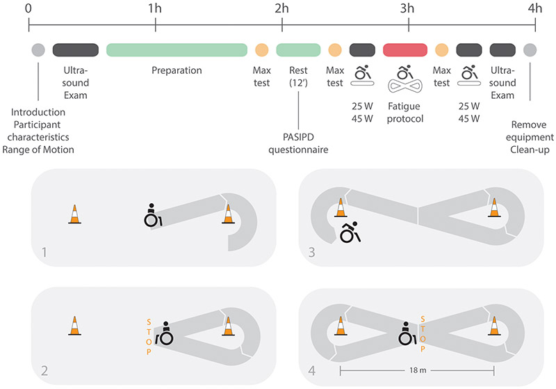
Participants were instructed to avoid strenuous exercises 48 hours prior to testing. Upon arrival to a four-hour measurement session in a biomechanical laboratory, participants received a copy of the previously read and signed informed consent and were familiarized with the methods. Several measurements were conducted before and after a fatiguing intervention (Figure 1). In-depth detail on the recruitment and study procedures has been reported previously and is briefly described below.9 This publication is part of a bigger project that aims to determine the effect of fatiguing wheelchair propulsion on risk factors for shoulder pain in persons with SCI. Compensation strategies in neuromuscular activation and propulsion biomechanics in response to fatiguing propulsion have been reported previously9, the current publication describes the ultrasound results which have not been presented elsewhere. The pre–intervention ultrasound exam took place before any kind of propulsion activity, while the post-intervention ultrasound exam took place following the completion of all propulsion tasks. Additional propulsion tasks, of which results have been published previously, in between the pre -and post intervention include two times 40 seconds (s) treadmill propulsion at 1.11 meters/s at 25 Watt and 45 Watt, three maximum push tests and a maximum 15 meter overground sprint.9
Figure 1:
Figure adjusted from Ref (9): Timeline of the assessments taken in the biomechanical laboratory including (1) introduction, self-reported participant’s characteristics and measurements of the shoulder Range of Motion (RoM), (2) pre-fatigue ultrasound exams, (3) preparation phase including a test to define individual drag force and familiarization with treadmill propulsion and figure-8 protocol, (4) three maximum push tests and a maximum 15m overground sprint test (5) passive rest phase including completion of the Physical Activity Scale for Individuals with Physical Disabilities (PASIPD), (6) treadmill propulsion at two different conditions (25Watt (W) and 45W), (7) Figure-8 protocol consisting of overground wheelchair propulsion along an 8-shaped course. Instructions given during each boat were standardized – the detailed course is presented in the figure below the timeline, and (8) post-fatigue ultrasound exams. Only results of participant characteristics, ultrasound exams, PASIPD, and heart rate and rate of perceived exertion at 25W and 45W treadmill propulsion are discussed in this manuscript.
Fatiguing intervention
The intervention, a figure-8 protocol, designed to induce fatigue included three 4-minute intervals of maximum voluntary wheelchair propulsion including right and left turns, start and stops, separated by 90 s of rest (total duration of 15 minutes) (Figure 1). Standardized instructions were given during each bout of four minutes at fixed time points. The protocol has been used previously in combination with ultrasound examinations.19
Data collection
Ultrasound examinations
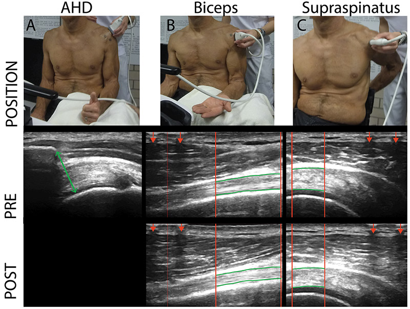
Grayscale images of the supraspinatus and long head of the biceps brachii tendons were taken by ultrasound (NextGen Logiq TM e R90.2, GE Healthcare, USA) to determine tendon appearance of the non-dominant shoulder. It was decided to investigate the non-dominant side as this project aims to investigate the shoulder most predominantly affected by wheelchair propulsion and less by activities as overhead reaching, opening doors, etc. The quantitative ultrasound protocol (QUS) has strong reliability and validity for measures of shoulder pain and tendinopathy and to determine acute changes in tendon appearance with repetitive wheelchair activity.15,18-20 As recommended to improve reliability of the measurements, a single examiner (FMB) took all ultrasound images in a randomized order.20 Image field depth was set at 4 centimeters and gain was set at 60 decibel. The protocol allows for repeated measurements before and after the propulsion tasks, with limited error in variation of probe location, because of a steel reference marker taped to the skin. For the supraspinatus tendon, two transverse images were taken in a seated position with the palm placed on the lower back, the shoulder extended, and the elbow flexed posteriorly (Figure 2). For the tendon of the long head of the biceps brachii, two longitudinal images were taken in a seated position with 90˚ elbow flexion and the hand palm facing upwards while resting on a cushion (Figure 2). Furthermore, during the pre-intervention ultrasound exam, three images of the AHD were taken during 90˚ elbow flexion with the thumb facing upwards, as has been done previously and proven good reliability (Figure 2).21
Figure 2:
Position and example images of ultrasound measurements. Each type of measurements represents a different example participant and does not relate to the person of the position images. (A) Acromio-humeral distance images were taken in a seated position with 90° elbow flexion with the thumb facing upwards. (B) Longitudinal images of long head of biceps brachii tendon were taken in a seated position with 90° elbow flexion and the hand palm facing upwards. (C) Transverse ultrasound images of supraspinatus tendon were taken in a seated position with the palm placed on the lower back, shoulder extended, and the elbow flexed posteriorly (B) The region of interest (ROI) is presented between the red vertical lines with the upper border and lower border of the tendons marked with the manually identified green horizontal lines. Lines are marked thicker as compared to the actual analysis for visualization reasons. ROI is selected based on the interference pattern that resulted from a metal marker taped to the skin (assigned with arrows).
Self-reported subject characteristics
Participants were asked to self-report socio-demographic variables (age, sex, and height), characteristics of the injury (traumatic or non-traumatic etiology, date of injury, completeness of the injury, and neurological lesion level), medication intake due to pain in the upper extremities in the last three months, and the 13-item Physical Activity Scale for Individuals with Physical Disabilities (PASIPD). The PASIPD has good construct validity and test-retest reliability.22,23 Weight was measured by subtracting the weight of the wheelchair from the total weight. Heart rate (HR, Polar V800, Electro, Kempele, Finland) and Rate of Perceived Exertion (RPE, 20-point Borg scale ranging from 6 [no perceived exertion] up to 20 [maximal exertion24]) were collected before and after every treadmill wheelchair propulsion task.
Data analysis
Susceptibility to fatigue
Participants were dichotomized into a fatigue (FG) and no fatigue group (NFG) based on a measure of perceived fatigue (RPE) and a measure of change in performance (HR). Increases in HR and RPE at the end of treadmill propulsion at a fixed power output before and after the intervention of greater than 2 times the standard error (SE) defined the FG.8 If the increase in HR and RPE was below the threshold of 2 times the SE, participants were included in the NFG. This dichotomization has been used previously and was found to be capable of identifying persons who display more changes in neuromuscular activation associated to fatigue after the Figure-8 protocol in the same study population.9
Tendon appearance
The region of interest (ROI) of each ultrasound image was defined from the interference pattern at the top of the images, created from the steel reference markers attached to the skin throughout the protocol (Figure 2). From the ROI, tendon characteristics including tendon width (mean distance between top and bottom border of the tendon), echogenicity ratio (mean pixel grayscale of tendon divided by mean pixel grayscale of muscle above tendon) and a co-occurrence matrix-derived measure contrast (intensity difference between a pixel and its neighbor in the vertical direction which is perpendicular to expected direction of the collagen fibers) were calculated. The echogenicity ratio was selected as this controls for overall brightness and differences caused by probe orientation.18 The average of the two repeated measures was used.20 If there was only one repetition available (due to bad quality of the image), only results of the available image were used. This was the case in 52 of the 600 measures (6 measures x 2 time points x 50 participants). If both repeated measurements had bad image quality, this resulted in missing measures for three participants. All ultrasound images were analysed by a single examiner (FMB) after the images were blinded and randomized using Matlab R2016b custom programs (Mathworks, Inc. Natick, MA, USA).
Acromio-humeral distance (AHD)
AHD was defined as the shortest distance between the anterior inferior edge of the acromion and the most superior aspect of the humerus (Figure 2).21 Also here, the average measure of the three repeated measures was used.20
Activity levels
The self-reported amount of days/week and hours/day of participation in recreational, household and occupational activities over the last 7 days in the PASIPD were used to calculate a total score with a metabolic equivalent value describing the activity levels of the participants.
Statistical analysis
Linear regression analyses were used to address the research questions. Dependent variables include the absolute difference between the pre- and post-measures of supraspinatus and biceps tendon thickness, echogenicity ratio and contrast and independent variables include fatigue group (FG and NFG), sex, interaction of group and sex, years since injury, AHD, activity levels, and body weight. A model was run for each of the dependent variables. The intraclass correlations (ICC) of the repeated tendon characteristics were calculated. Finally, post-hoc analyses compared subject and lesion characteristics of females in the FG, females in the NFG, males in the FG, and males in the NFG with one-way ANOVAs. If a significant difference was found, pairwise comparisons with Bonferroni corrections were used to evaluate differences between the respective individuals. Statistical analyses were defined a priori and conducted with STATA version 14 (StataCorp LP, College Station, TX, USA) (α=0.05).
Results
Fifty eligible persons who matched the selection criteria and were willing to participate were identified from a sample of 2379 persons.9 Although persons who reported pain that limited their ability to propel were excluded, in the 3 months prior to testing, 8 participants used pain medication to treat pain in the upper limbs: 4 participants used NSAIDS, one a combination of NSAIDS and Opioids (Tramadol), one Opioids only (Fentanyl Plaster) and two used medication to treat chronic neuropathic pain (Lyrica, Neurontin and Targin). Subject and lesion characteristics and activity levels are presented in Table 1. Dichotomization into FG and NFG resulted in 25 persons in FG and 25 persons in NFG. The total duration of all propulsion tasks was 50.7 (10.7) minutes. The duration between the last propulsion task and the beginning of the post ultrasound exam was 6.3 (1.6) minutes with a maximum duration of 10 minutes. The post ultrasound exam lasted 9.3 (2.7) minutes.
Table 1:
Subject and lesion characteristics for the total sample and by group (no-fatigue vs fatigue) and sex (males vs females)
| Total (n=50) | No-fatigue group (n=25) | Fatigue group (n=25) | |||
|---|---|---|---|---|---|
| Males (n=18) | Females (n=7) | Males (n=21) | Females (n=4) | ||
| Sex (% male) | 78 | 72 | 84 | ||
| Cause injury (% traumatic) | 92 | 94 | 85 | 91 | 100 |
| Completeness lesion (% incomplete) | 78 | 89 | 71 | 76 | 50 |
| Lesion level (%) | |||||
| T2-T6 | 40 | 56 | 0 | 43 | 25 |
| T7-T12 | 44 | 22 | 86 | 52 | 25 |
| L1-L2 | 16 | 22 | 14 | 5 | 50 |
| Age (years) | 50.5 (9.7) | 49.3 (12.1) | 52.3 (8.6) | 50.6 (8.8) | 52.3 (5.3) |
| Height (cm) | 173.7 (7.9) | 173.4 (5.8)* | 165.9 (6.1) | 178.5 (6.6)* | 163.5 (2.4) |
| Weight (kg) | 72.4 (13.3) | 72.7 (13.4) | 58.3 (11.9) | 77.3 (10.3)* | 70 (15.9) |
| Weight wheelchair (kg) | 14.4 (2.3) | 14.4 (2.7) | 14.8 (2.7) | 14.2 (1.8) | 14.6 (2.4) |
| Time since injury (years) | 26.6 (11.6) | 28.2 (13.3) | 35.7 (8.2) | 23.4 (9.3) | 20.4 (12.1) |
| Year at injury (years) | 23.9 (10.1) | 21.1 (9.7) | 16.6 (5.8) | 27.2 (9.8) | 31.9 (8.5) |
| Activity levels (MET) | 18.9 (12.9) | 21.0 (12.1) | 16.2 (9.3) | 18.8 (14.9) | 14.7 (13.2) |
NOTE. Significant p-values (α = 0.05) (*) corrected for multiple comparisons (Bonferroni) represent comparison of each group (male non-fatigued, male fatigued, female non-fatigued and female fatigued). The bold values are significantly different from values marked with *.
Tendon changes
The ICC of the repeated measures ranged between moderate (0.50 < ICC < 0.75) and good (ICC > 0.75) (Table 2). This is in line with previously reported intra-rater reliability of QUS measures.25 From all investigated tendon characteristics, only the supraspinatus tendon thickness changed significantly after fatiguing propulsion. More specifically, supraspinatus thickness was significantly reduced after fatiguing propulsion when controlling for all co-variables (p=0.003, Coefficient: B0 = −1.39, SE = 0.44, 95% CI: −2.28; −0.51, Table 2). The reduction in tendon thickness was larger than two times the SE (0.88 millimeters).
Table 2:
Unadjusted mean (standard deviation) and difference (diff) adjusted to all co-variables for quantitative ultrasound supraspinatus and biceps tendon measures (QUS). Intraclass correlations (ICC) for each measure were reported.
| Unadjusted | Adjusted | ||||||
|---|---|---|---|---|---|---|---|
| QUS measure | Tendon | n | Pre | Post | Diff | ||
| ICC | ICC | ||||||
| Thickness (mm) | Supraspinatus | 50 | 5.39 (1.00) | 0.94 | 5.25 (1.11) | 0.95 | −1.39* |
| Biceps | 49 | 4.21 (1.27) | 0.96 | 4.21 (1.25) | 0.97 | −0.60 | |
| Echo ratio | Supraspinatus | 49 | 1.73 (0.71) | 0.85 | 1.73 (0.71) | 0.93 | 0.26 |
| Biceps | 48 | 1.41 (0.56) | 0.88 | 1.34 (0.52) | 0.73 | −0.23 | |
| Contrast | Supraspinatus | 50 | 3.72 (0.90) | 0.73 | 3.66 (0.98) | 0.64 | 1.01 |
| Biceps | 48 | 4.83 (1.51) | 0.67 | 4.60 (1.68) | 0.78 | 0.51 | |
NOTE. Significant p-values (α = 0.05) are marked with *
Risk factors
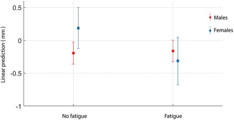
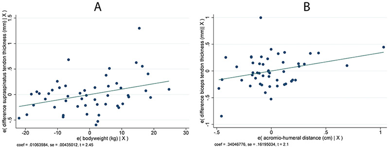
There was a significant interaction effect of group (FG vs NFG) and sex (female vs male) for the change in supraspinatus thickness (p=0.044, Coefficient: B1 = −0.53, SE = 0.26, 95% CI: −1.05; −0.02, Figure 3). Females in the FG (n=4, 16%) displayed a greater reduction in supraspinatus tendon thickness as compared to females in the NFG (n=7, 28%) who had no significant change in tendon thickness (p > 0.05), while all males (FG and NFG) displayed a similar significant reduction in supraspinatus tendon thickness. Also, there was a significant association in the change in supraspinatus tendon thickness and bodyweight with heavier persons displaying a greater increase in tendon thickness (p=0.019; Coefficients: B = 0.01, SE = 0.00, 95% CI: 0.00; 0.02, Figure 4). Finally, there was a significant association with change in biceps tendon thickness and AHD with a greater AHD displaying a greater increase in tendon thickness after the intervention (p=0.042, Coefficient: B1 = 0.34, SE =0.16, 95% CI: 0.01; 0.67, Figure 4). There were no significant associations with time since injury or activity levels. Females in FG were shorter as compared to males in FG (−14.98 meters, SE = 3.32, 95% CI: −24.12; −5.83), and as compared to males in NFG (−9.89 meters, SE = 3,36, 95% CI: −19.16; −0.62)(Table 1). Also females in NFG were shorter as compared to males in FG (−12.62 meters, SE = 2.65, 95% CI: −19.94; −5.30) and as compared to males in NFG (−7.53 meters, SE = 2.71, 95% CI: −15.00, −0.06)(Table 1). Finally, females in NFG had less body weight as compared to males in FG (−18.94 kilogram, SE = 5.30, 95% CI: −33.55; −4.34)(Table 1).
Figure 3:
Change in supraspinatus thickness (mm) after fatiguing wheelchair propulsion in females and males who fatigued and those who did not fatigue: predictive margins with 95 % confidence intervals.
Figure 4:
Association between A: change in supraspinatus tendon thickness (mm) after fatiguing wheelchair propulsion and body weight (kg) and B: change in biceps tendon thickness (mm) after fatiguing wheelchair propulsion and AHD (cm) (p < 0.05).
Discussion
Tendon changes
This study identified an acute reduction in supraspinatus tendon thickness (−1.39 mm, SE = 0.44) after fatiguing wheelchair propulsion in persons with SCI. The active role of the supraspinatus during both the push phase – by counteracting the forces of the pectoralis major – and the recovery phase of propulsion – by bringing back the hand7 – and the association of supraspinatus tendon degeneration and wheelchair use15, supports the finding of acute changes in the supraspinatus tendon after fatiguing propulsion. Indeed, greater muscular activation and muscle forces will increase tendon compressive loading and may locally reduce the tendon’s tensile strength.10 Previous studies reported that a reduction in tendon thickness may be caused by creep (i.e., alignment of collagen fibers in the direction of applied stress) or fluid transfer out of the tendon.26,27 Tendon creep in response to repetitive activity was found to alter the in-series contracting muscle and cause a shift towards less optimal force generating capacity.28 With insufficient time to recover, repetitive tendon creep may lead to micro-injuries of the collagen fibers that comprise the tendon, chronic tendon degeneration and ultimately tendon rupture.13,26 The exact mechanisms resulting in the reduced tendon thickness in this study are unclear and remain to be determined. Nevertheless, these findings warrant for improving resilience against fatigue and for providing sufficient recovery time following fatiguing propulsion.
Contrary with our hypothesis, there was no change in biceps tendon thickness, echogenicity ratio or contrast after fatiguing propulsion. This suggest that wheelchair propulsion is not the main contributor to acute changes in the biceps tendon and is in line with the biceps being less susceptible to fatigue as compared to the supraspinatus tendon during propulsion.7
Current reported findings were contradictory to two studies investigating acute changes in shoulder tendons with repetitive wheelchair activity. One study did not find a change in supraspinatus tendon thickness after the figure-8 protocol.19 The additional propulsion activities besides the figure-8 protocol included in the current study, may have placed greater demands on the shoulder as compared to the figure-8 protocol on itself. Furthermore, there were differences in study population which are expected to affect shoulder loads and tendon properties. More specifically, the aforementioned study investigated a convenience sample of 60 wheelchair users mostly (80%) athletes recruited from the National Veterans Wheelchair games (USA); participants were on average 82.8 (20.0) kilogram versus our sample of 72.4 (13.3) kilogram, and while persons who had a traumatic injury to their shoulder were excluded, persons with pain limiting their ability to propel were not excluded. Finally, our study was conducted in Switzerland with different equipment, access and other unknowns. Another study demonstrated a decrease in mean biceps echogenicity ratio after a wheelchair basketball or rugby quad game, lasting on average 28.7 (14.6) minutes.18 Furthermore, persons who played for more than 30 minutes (n=8) demonstrated an increase in biceps tendon thickness. Besides the difference in duration of the task, the diverse study population (n=34, 97 % males, 11 tetraplegia, 21 paraplegia, 2 non-SCI), included 38% persons who reported shoulder pain in the last month. Finally, other activities during the game (include ball control etc.) may have implied different loads on the biceps tendon as compared to the wheelchair propulsion activities during the current investigation and resulted in different tendon adaptations. Either way, both studies show changes with activity which lend support to the notion that acute changes in ultrasound could be used as a biomarker for injury.
Risk factors
Changes in tendon thickness were associated with susceptibility to fatigue, sex, body weight, and AHD. Susceptibility to fatigue affected the change in tendon thickness for females but not for males. Females in the FG displayed the greatest reduction in supraspinatus tendon thickness. A note of caution is due here since only four females were in the FG. Nevertheless, findings may be a result of the effect of fatigue on the reported gender differences in tendon viscoelastic properties, as female tendons have lower stiffness and hysteresis.29 Furthermore, females presented a lower rate of tendon tissue repair after exercise, expressed by reduced tendon collagen fractional synthesis, as compared to males.30 The greater reduction in tendon thickness with acute loading in females susceptible to fatigue, may give insights into the higher odds of shoulder pain in females with SCI.3
Interestingly, heavier persons had a greater increase in supraspinatus tendon thickness after the intervention. Indeed, while tendon adaptation to tensile load generally results in tendon creep and reduced tendon thickness, reactive tendinopathy – an acute influx of water increasing tendon thickness to reduce stress and increase stiffness – in response to acute overload, may have been present in heavier persons who were subject to greater loads.31 The long term consequences of this response are currently unclear. Nevertheless, this finding suggests that body weight affects acute changes in tendon appearance following repetitive activity and supports the commonly accepted recommendation to reduce body weight in persons with SCI to preserve shoulder functioning.32
We found that persons with a greater AHD during an unloaded position, have a greater increase in biceps tendon thickness after the intervention. This points towards an association between AHD and changes in tendon thickness with repetitive propulsion. It is unclear whether the AHD adapted over time to protect the tendon or if the tendon increased more because of a greater AHD. Wheelchair users were previously found to have greater AHD as compared to persons without SCI which points towards an adaptation mechanism of the AHD with propulsion.33 Identifying characteristics that predispose an individual to pathological tendon changes is a first step to tailor interventions, improve load management and limit progression of tendinopathy.
Study Limitations
This study investigated a sample of participants selected from the population-based community SwiSCI study. However, current results cannot be generalized to the entire study population based on our exclusion criteria as for example pain limiting the ability to propel. Because pain on itself may alter the tendon response to fatiguing propulsion activity, we aimed to first determine changes in tendon appearance in persons without pain. The reported use of drugs to treat upper limb pain in 8 of the participants should be acknowledged and may have affected the results. However, because the amount of persons who used drugs to treat upper limb pain were evenly distributed across the fatigue and no-fatigue group, we do not expect a significant impact. Finally, although the intervention includes overground propulsion, it remains artificial and may not represent daily-life activities of wheelchair users. The current protocol is expected to be more demanding as it requires maximum voluntary propulsion and therefore to induce greater changes in tendon appearance.
Conclusions
This is the first study demonstrating a significant reduction in supraspinatus tendon thickness after fatiguing wheelchair propulsion in a population-based, shoulder pain-free, sample of persons with SCI. Alterations in tendon thickness directly affect the muscle-tendon unit and may subsequently alter force production capacity. In contrast to the overall observed reduced supraspinatus tendon thickness following fatiguing propulsion, body weight and AHD were associated to signs of reactive tendinopathy and increased tendon thickness. Acute changes in tendon appearance related with repetitive activity and its association to subject characteristics, provide insights into the etiology and development of tendinopathy in wheelchair users with SCI. Results suggest that fatiguing wheelchair propulsion with insufficient recovery time may play a role in the development of supraspinatus tendinopathy. Future research should look into chronic adaptations resulting from the described acute changes and determine training programs that optimize tendon adaptations and limit the progression of tendinopathy. Furthermore, it needs to be determined how acute tendon changes are affected by the reduced sub-acromial space during dynamic activities as wheelchair propulsion.6 In order to answer these further questions, there is need for quantification of internal tendon stress-strain fields as has been discussed previously.10
Supplementary Material
Acknowledgements
We thank Benjamin Beirens, MSc., Angelene Fong, MSc. MA., Stephanie Marino-Wäckerlin, MA., and Ursina Minder, BSc. for their contributions to the data collection and Benjamin Beirens, MSc. and Dr. Jonviea D. Chamberlain for proofreading the manuscript.
Funding
This project is supported by the Administration on Community Living, National Institute on Disability, In- dependent Living, and Rehabilitation Research (NIDILRR) (grant no. 90SI5014). NIDILRR is a Center within the Administration for Community Living (ACL) within the Department of Health and Human Services (HHS). The contents of this paper do not necessarily represent the policy of NIDILRR, ACL, or HHS, and you should not assume endorsement by the U.S. Government. This project has been supported by the Sports Medicine Nottwil from the Swiss Paraplegic Group by providing their ultrasound device. This project has also been supported by the International Society of Biomechanics with the International Travel Grant (July 1st, 2016). This study has been financed in the framework of the Swiss Spinal Cord Injury Cohort Study (SwiSCI, www.swisci.ch), supported by the Swiss Paraplegic Foundation. The members of the SwiSCI Steering Committee are: Xavier Jordan, Fabienne Reynard (Clinique Romande de Réadaptation, Sion); Michael Baumberger, Hans Peter Gmünder (Swiss Paraplegic Center, Nottwil); Armin Curt, Martin Schubert (University Clinic Balgrist, Zürich); Margret Hund-Georgiadis, Kerstin Hug (REHAB Basel, Basel); N.N. (Swiss Paraplegic Association, Nottwil); Daniel Joggi (Swiss Paraplegic Foundation, Nottwil); Hardy Landolt (Representative of persons with SCI, Glarus); Nadja Münzel (Parahelp, Nottwil); Mirjam Brach, Gerold Stucki (Swiss Paraplegic Research, Nottwil); Christine Fekete (SwiSCI Coordination Group at Swiss Paraplegic Research, Nottwil).
Footnotes
Conflict of Interest
The authors declare that they have no conflict of interest.
Data Archiving
The datasets generated and/or analysed during the current study are available from the corresponding author on reasonable request.
Statement of Ethics
Ethical approval was granted by the Ethikkommision Nordwest-und Zentralschweiz (EKNZ, Project-ID: 2017–00355). We certify that all applicable institutional and governmental regulations concerning the ethical use of human volunteers were followed during the course of this research.
References
- 1.Veeger HEJ, van der Woude LHV, Rozendal RH. Load on the upper extremity in manual wheelchair propulsion. J Electromyogr Kinesiol 1991;1(4):270–80. [DOI] [PubMed] [Google Scholar]
- 2.Gutierrez DD, Thompson L, Kemp B, Mulroy SJ. The relationship of shoulder pain intensity to quality of life, physical activity, and community participation in persons with paraplegia. J Spinal Cord Med 2007;30:251–5. [DOI] [PMC free article] [PubMed] [Google Scholar]
- 3.Bossuyt FM, Arnet U, Brinkhof MWG, Eriks-Hoogland I, Lay V, Muller R, et al. Shoulder pain in the swiss spinal cord injury community: Prevalence and associated factors. Disabil Rehabil 2018;40(7):798–805. [DOI] [PubMed] [Google Scholar]
- 4.Michener LA, McClure PW, Karduna AR. Anatomical and biomechanical mechanisms of subacromial impingement syndrome. Clin Biomech (Bristol, Avon) 2003;18:369–79. [DOI] [PubMed] [Google Scholar]
- 5.Brose SW, Boninger ML, Fullerton B, McCann T, Collinger JL, Impink BG, et al. Shoulder ultrasound abnormalities, physical examination findings, and pain in manual wheelchair users with spinal cord injury. Arch Phys Med Rehabil 2008;89(11):2086–93. [DOI] [PubMed] [Google Scholar]
- 6.Morrow MM, Kaufman KR, An KN. Scapula kinematics and associated impingement risk in manual wheelchair users during propulsion and a weight relief lift. Clin Biomech (Bristol, Avon) 2011;26(4):352–7. [DOI] [PMC free article] [PubMed] [Google Scholar]
- 7.Mulroy SJ, Gronley JK, Newsam CJ, Perry J. Electromyographic activity of shoulder muscles during wheelchair propulsion by paraplegic persons. Arch Phys Med Rehabil 1996;77:187–93. [DOI] [PubMed] [Google Scholar]
- 8.Enoka RM, Duchateau J. Translating fatigue to human performance. Med Sci Sports Exerc 2016;48(11):2228–38. [DOI] [PMC free article] [PubMed] [Google Scholar]
- 9.Bossuyt FM, Arnet U, Cools A, Rigot S, de Vries W, Eriks-Hoogland I, Boninger ML, et al. Compensation strategies in response to fatiguing propulsion in wheelchair users: Implications for shoulder injury risk. Am J Phys Med Rehabil 2019; Published ahead of print. [DOI] [PubMed] [Google Scholar]
- 10.Maganaris CN, Chatzistergos P, Reeves ND, Narici MV. Quantification of internal stress-strain fields in human tendon: Unraveling the mechanisms that underlie regional tendon adaptations and mal-adaptations to mechanical loading and the effectiveness of therapeutic eccentric exercise. Front Physiol 2017;8:91. [DOI] [PMC free article] [PubMed] [Google Scholar]
- 11.Tardioli A, Malliaras P, Maffulli N. Immediate and short-term effects of exercise on tendon structure: Biochemical, biomechanical and imaging responses. Br Med Bull 2012;103(1):169–202. [DOI] [PubMed] [Google Scholar]
- 12.Arya S, Kulig K. Tendinopathy alters mechanical and material properties of the achilles tendon. J Appl Physiol (1985) 2010;108(3):670–5. [DOI] [PubMed] [Google Scholar]
- 13.Allen GM. Shoulder ultrasound imaging-integrating anatomy, biomechanics and disease processes. Eur J Radiol 2008;68(1):137–46. [DOI] [PubMed] [Google Scholar]
- 14.Xu Y, Murrell GAC. The basic science of tendinopathy. Clin Orthop Rel at Res 2008;466(7):1528–38. [DOI] [PMC free article] [PubMed] [Google Scholar]
- 15.Collinger JL, Fullerton B, Impink BG, Koontz AM, Boninger ML. Validation of grayscale-based quantitative ultrasound in manual wheelchair users: Relationship to established clinical measures of shoulder pathology. Am J Phys Med Rehabil 2010;89(5):390–400. [DOI] [PMC free article] [PubMed] [Google Scholar]
- 16.von Elm E, Altman DG, Egger M, Pocock SJ, Gotzsche PC, Vandenbroucke JP. The strengthening the reporting of observational studies in epidemiology (strobe) statement: Guidelines for reporting observational studies. Lancet 2007;370(9596):1453–7. [DOI] [PubMed] [Google Scholar]
- 17.Brinkhof MW, Fekete C, Chamberlain JD, Post MW, Gemperli A. Swiss national community survey on functioning after spinal cord injury: Protocol, characteristics of participants and determinants of non-response. J Rehabil Med 2016;48(2):120–30. [DOI] [PubMed] [Google Scholar]
- 18.van Drongelen S, Boninger ML, Impink BG, Khalaf T. Ultrasound imaging of acute biceps tendon changes after wheelchair sports. Arch Phys Med Rehabil 2007;88(3):381–5. [DOI] [PubMed] [Google Scholar]
- 19.Collinger JL, Impink BG, Ozawa H, Boninger ML. Effect of an intense wheelchair propulsion task on quantitative ultrasound of shoulder tendons. PM R 2010;2(10):920–5. [DOI] [PubMed] [Google Scholar]
- 20.Collinger JL, Gagnon D, Jacobson J, Impink BG, Boninger ML. Reliability of quantitative ultrasound measures of the biceps and supraspinatus tendons. Acad Radiol 2009;16(11):1424–32. [DOI] [PMC free article] [PubMed] [Google Scholar]
- 21.Mackenzie TA, Bdaiwi AH, Herrington L, Cools A. Inter-rater reliability of real-time ultrasound to measure acromiohumeral distance. PM R 2016;8(7):629–34. [DOI] [PubMed] [Google Scholar]
- 22.Washburn RA, Zhu W, McAuley E, Frogley M, Figoni SF. The physical activity scale for individuals with physical disabilities: Development and evaluation. Arch Phys Med Rehabil 2002;83:193–200. [DOI] [PubMed] [Google Scholar]
- 23.van der Ploeg HP, Streppel KR, van der Beek AJ, van der Woude LH, Vollenbroek-Hutten M, van Mechelen W. The physical activity scale for individuals with physical disabilities: Test-retest reliability and comparison with an accelerometer. J Phys Act Health 2007;4(1):96–100. [DOI] [PubMed] [Google Scholar]
- 24.Borg G. Psychophysical scaling with applications in physical work and the perception of exertion. Scand J Work Environ Health 1990;16(1):55–8. [DOI] [PubMed] [Google Scholar]
- 25.Portney LG, Watkins MP. Foundations of clinical research: Application to practice: Stamford: Appleton & Lange; 2000.
- 26.Magnusson SP, Narici MV, Maganaris CN, Kjaer M. Human tendon behaviour and adaptation, in vivo. J Physiol 2008;586(1):71–81. [DOI] [PMC free article] [PubMed] [Google Scholar]
- 27.Pearson SJ, Engel AJ, Bashford GR. Changes in tendon spatial frequency parameters with loading. J Biomech 2017;57:136–40. [DOI] [PubMed] [Google Scholar]
- 28.Maganaris CN, Baltzopoulos V, Sargeant AJ. Repeated contractions alter the geometry of human skeletal muscle. J appl Physiol 2002;93:2089–94. [DOI] [PubMed] [Google Scholar]
- 29.Kubo K, Kanehisa H, Fukunaga T. Gender differences in the viscoelastic properties of tendon structures. Eur J Appl Physiol 2003;88(6):520–6. [DOI] [PubMed] [Google Scholar]
- 30.Miller BF, Hansen M, Olesen JL, Schwarz P, Babraj JA, Smith K, et al. Tendon collagen synthesis at rest and after exercise in women. J appl Physiol 2006;102:541–6. [DOI] [PubMed] [Google Scholar]
- 31.Cook JL, Purdam CR. Is tendon pathology a continuum? A pathology model to explain the clinical presentation of load-induced tendinopathy. Br J Sports Med 2009;43(6):409–16. [DOI] [PubMed] [Google Scholar]
- 32.Paralyzed Veterans of America Consortium for Spinal Cord M. Preservation of upper limb function following spinal cord injury: A clinical practice guideline for health-care professionals. J Spinal Cord Med 2005;28(5):434–70. [DOI] [PMC free article] [PubMed] [Google Scholar]
- 33.Fournier Belley A, Gagnon DH, Routhier F, Roy JS. Ultrasonographic measures of the acromiohumeral distance and supraspinatus tendon thickness in manual wheelchair users with spinal cord injury. Arch Phys Med Rehabil 2017;98(3):517–24. [DOI] [PubMed] [Google Scholar]
Associated Data
This section collects any data citations, data availability statements, or supplementary materials included in this article.


