Abstract
Background
Tick-borne lymphadenopathy (TIBOLA) is an infectious disease, mainly caused by species from the spotted fever group rickettsiae and is characterized by enlarged lymph nodes following a tick bite. Among cases of TIBOLA, a case of scalp eschar and neck lymphadenopathy after tick bite (SENLAT) is diagnosed when an eschar is present on the scalp, accompanied by peripheral lymphadenopathy (LAP). Only a few cases of SENLAT caused by Bartonella henselae have been reported.
Case presentation
A 58-year-old male sought medical advice while suffering from high fever and diarrhea. Three weeks before the visit, he had been hunting a water deer, and upon bringing the deer home discovered a tick on his scalp area. Symptoms occurred one week after hunting, and a lump was palpated on the right neck area 6 days after the onset of symptoms. Physical examination upon presentation confirmed an eschar-like lesion on the right scalp area, and cervical palpation revealed that the lymph nodes on the right side were non-painful and enlarged at 2.5 × 1.5 cm. Fine needle aspiration of the enlarged lymph nodes was performed, and results of nested PCR for the Bartonella internal transcribed spacer (ITS) confirmed B. henselae as the causative agent.
Conclusion
With an isolated case of SENLAT and a confirmation of B. henselae in Korea, it is pertinent to raise awareness to physicians in other Asian countries that B. henselae could be a causative agent for SENLAT.
Keywords: Bartonella henselae, Spotted fever group Rickettsiosis, Tick-borne diseases, Lymph nodes
Background
Bartonella henselae is a gram-negative, facultative, intracellular bacteria that can cause various diseases, including lymphadenopathy, bacteremia, bacillary angiomatosis, and bacillary peliosis [1]. One of the typical diseases from B. henselae is cat scratch disease. People usually contract the disease from cats infected by B. henselae, but cases from flea or tick bites have been reported [2]. The infection is asymptomatic in cats, but for humans, it can result in symptoms or signs such as lymphadenopathy, red papules, fever, headache, malaise, and sometimes, in adults, fever of unknown origin (FUO) [3].
Tick-borne lymphadenopathy (TIBOLA) is an infectious disease, mainly caused by species from the spotted fever group rickettsiae (e.g. Rickettsia slovaca, Rickettsia raoultii) and is characterized by enlarged lymph nodes following a tick bite. Scalp eschar and neck lymphadenopathy after tick bite (SENLAT) occurs after a bite from a tick and key clinical features occur at the surface of the scalp and cervical lymph nodes. Therefore, we consider the TIBOLA case with eschar on the scalp as SENLAT [4]. There are some cases of SENLAT caused by B. henselae in other country [5], but there are no such case reports in South Korea, except for some other clinical syndromes [1, 6–8]. This study reports a first case of SENLAT in which B. henselae was confirmed as the etiologic agent in Korea.
Case presentation
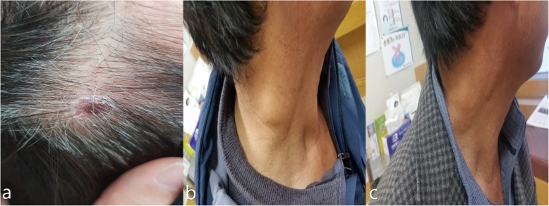
The patient was a 58-year-old male, who brought home a water deer (Hydropotes inermis argyropus) from Muan-gun, Jeollanam-do, Korea., he had hunted a week prior to his presentation. His symptoms of high fever, diarrhea, and indigestion developed after the hunting incident, and his right cervical lymph nodes suddenly became swollen 6 days following the onset of fever, which prompted him to visit the infectious diseases outpatient clinic at the Chosun University Hospital. The day after carrying the water deer home, he found a tick on his scalp, but had quickly removed and discarded it. He denied contact with cats or dogs as well as flea. On physical examination, he had a high fever of 39 °C, an eschar-like lesion was found on his right scalp area (Fig. 1a), and on palpation, non-painful peripheral lymphadenopathy (LAP) of 2.5 × 1.5 cm in size was identified in the right cervical region (Fig. 1b).
Fig. 1.
A photograph of the eschar on the scalp and right cervical area of a 58-year-old male patient with a confirmed diagnosis of Bartonella henselae, and a cytology report from fine needle aspiration of an enlarged cervical lymph node. a Eschar on the scalp at the first visit to the outpatient clinic. b Right cervical lymphadenopathy on the first visit to the outpatient clinic. c A photograph showing a marked reduction of size in the right cervical lymphadenopathy 10 days later
Blood test and fine needle aspiration were performed on the day of first visit. Laboratory investigations revealed a white blood cell count of 6.04 × 10³/uL, hemoglobin level of 17.2 g/dL, and platelet count of 313 × 103/uL on routine complete blood count. Serum biochemistry revealed the following: total protein concentration, 6.96 g/dL; albumin, 4.44 g/dL; blood urea nitrogen, 18.0 mg/dL; bilirubin, 0.55 mg/dL; alkaline phosphatase, 52 U/L; and creatinine, 1.00 mg/dL (all were within normal limits). Aspartate aminotransferase (AST) of 29.5 U/L was within normal limits, but alanine aminotransferase (ALT; 45.8 U/L) as well as lactate dehydrogenase (LDH; 421 U/L) were mildly elevated. To identify the cause of LAP, fine needle aspiration (FNA) was performed on the enlarged lymph nodes of the neck.
Cytology from the FNA demonstrated a granuloma with an unclear boundary comprised of epithelioid cells along with giant cells and some lymphocytes.
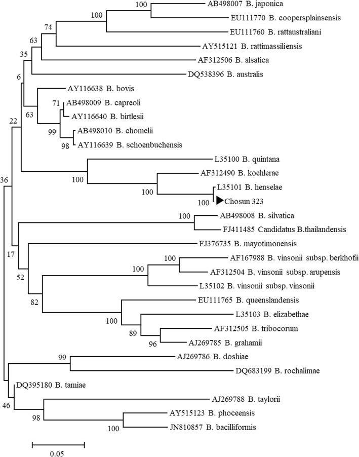
DNA was extracted from the buffy coat of the patient’s blood and from the lymph node aspirate using a QIAamp Blood Mini kit (QIAGEN, Germantown, MD). The results of the genetic detection were all negative when the 56-kDa gene from O. tsutsugamushi and the ompA gene were targeted by nested PCR for rickettsial detection [9]. Nested PCR on the Bartonella internal transcribed spacer (ITS) [10], using blood and lymph node samples, and by using B. elizabethae as a positive control, revealed a positive band by electrophoresis in only the lymph node aspirate. Sequencing of the sample was therefore requested at SolGent (Daejeon, Korea). The query output of BLASTN (NCBI) demonstrated a 100% identical sequence (703/703 bp) to the B. henselae strain BM1374165 (accession no. HG969191) previously identified in human blood (Fig. 2).
Fig. 2.
A phylogenetic tree based on Bartonella internal transcribed spacer (ITS) sequences from GenBank
Indirect immunofluorescent antibody assay (IFA) against B. henselae were conducted at at the Korea Centers for Disease Control and Prevention. The sera were examined with commercially available slides for Bartonella-IFA IgG and IgM assay (Focus Diagnostics, Cypress, CA, USA). The kit for detecting IgM and IgG antibodies utilizing Vero cells infected with either B. henselae or B. quintana was used according to the manufacturer’s instructions. Diagnostic criteria are determined to be Bartonella positive when endpoint titer of IgG ≥1:64 or IgM ≥1:20. The IFA IgM antibody titer against B. hensealse was < 1:20 at both first visit and follow up. The IFA IgG antibody titer against B. hensealse was < 1:16 at first visit (2015.12.07), and 1:16 after follow up (2016.01.04). He was treated by doxycycline for first 5 days and then with azithromycin for 5 days. Ten days later, the LAP resolved (Fig. 1c). Serological tests of Orientia and other Rickettsia species were performed together and the results were all negative.
Discussion and conclusion
The incidence of disease caused by B. henselae has been previously reported, especially in association with contact with cats or dogs. The main route of infection is thought to be from scratching the site of a cat bite or a flea bite. The prevalence of Bartonella infection in Korea, identified by PCR, is estimated to be 0–44.1% [11, 12] in animals, and 0–19.1% [11, 13, 14] in arthropod vectors, but case reports of Bartonella infections in Korea have been rare. Just a few studies have been published about case of B. henselae infection in South Korea [1, 7, 8]. Kwon et al. reported 5 cases in which B. henselae was identified from cultures of blood or bone marrow [6]. However, there have been no reports of SENLAT by B. henselae in Korea.
TIBOLA commonly occurs in women and young people and has been reported in European countries such as France, Spain, and Hungary, particularly in cold seasons. Rickettsia slovaca is known to be the most commonly confirmed etiologic agent of TIBOLA, and the most frequently identified vector is Dermacentor marginatus [15]. The scalp area is recognized as the most common site for tick bites. One possible explanation for this is that Dermacentor ticks can stick to long hair, which plays a role as a shelter. Among TIBOLA entities, disease entity with both the eschar in the scalp and the neck lymphadenopathy are recognized as a new clinical entity named by SENLAT [8], as in our case. SENLAT has characteristic epidemiological findings that occur frequently in females and young children and are seasonal bimodality (spring and autumn) [4]. Although Rickettsia slovaca is the most common pathogen in this syndrome, other several agents like Rickettsia raoultii, Rickettisa sibirica subsp. mongolitimonae, Coxiella burnetii, Borrelia burgdorferi, and Candidatus Rickettsia rioja are also known as etiological pathogens [4]. The patient in our case had an eschar lesion on his right scalp, and a superficial enlarged right cervical lymph node, consistent with SENLAT. The result of the nested PCR, using a sample from the enlarged node, was positive for Bartonella species in a genus-specific ITS gene, and the sequencing results confirmed Bartonella henselae to be the cause of infection. And then, based on previous several literature showing that water deer living in Korea serve as a reservoirs of Bartonella species [16, 17], we believed that B. henselae identified in our patient originated from a water deer contacted prior to our hospital visit. In our case, the tick residing on the water deer may have bitten the patient, and hence infected him with B. henselae. As the patient had discarded the tick, we could not investigate the role of the tick as a vector. Dermacentor ticks are, however, known to be absent in Korea [18]. Cotte et al. showed in their experimental study that potential transmission of B. henselae is possible with Ixodes ricinus ticks [19]. Therefore, one cannot exclude a possibility that SENLAT could have been caused by Ixodes nipponensis, which is frequently observed in Korea [20]. In addition, the water deer may be the source of the tick, but it is not clear, and the natural environment may have been the tick source. However, further study is needed to confirm this.
Angelakis et al. have reported SENLAT caused by B. henselae following a tick bite [5]. These three patients who are similar to our patient, but there is one big difference. That is, they are proven to be infected by B.henselae through PCR test with eschar tissue or tick specimen, but we have demonstrated SENLAT by B.henselae with PCR tests using neck lymph node tissue. All 3 of their examples occurred in the colder months in Europe, and the authors suggested that Dermacentor ticks are most active during these periods. The occurrence of SENLAT has mainly been reported in Europe. However, no cases of SENLAT have been reported in Asia.
Difficulties in culturing B. henselae from pus aspirates and lymph node biopsy specimens of patients with cat scratch disease have been reported [21], and very low levels of sensitivity in serologic and PCR tests have been found against B. henselae infection. For example, among the 18 patients with a confirmed diagnosis of cat scratch disease, only 3 were noted to have positive PCR results, and the cycle thresholds were reported to be average of 38 (range: 37.7–39.3) [22]. Our patient also showed a positive PCR result for B. henselae with lymph node aspirate, but not with a blood sample. IFA antibody test for B. henselae was also negative, presumably due to the low sensitivity of the IFA IgG antibody test [23].
In conclusion, this case report demonstrated a case of SENLAT in which the patient had ipsilateral LAP and a scalp eschar, with a confirmed diagnosis of B. henselae infection from PCR of aspirate from the affected lymph node. This study should raise awareness in clinicians that, in addition to Rickettsia species, B. henselae may be a causative agent of TIBOLA or SENLAT.
Acknowledgements
We would like to thank Jeong Hee Su, Lee Chae Hun, Kim Sung Yun, Park Sang Uk, Jeong Yu Min, Kang Hye Ju, and Choe Yu Seop for helping to review this manuscript.
Abbreviations
- ALT
Alanine aminotransferase
- AST
Asparate aminotransferase
- FNA
Fine needle aspiration
- FUO
Fever of unknown origin
- IFA
Immunofluorescence assay
- ITS
Internal transcribed spacer
- LAP
Lymphadenopathy
- LDH
Lactate dehydrogenase
- PCR
Polymerase chain reaction
- SENLAT
Scalp eschar and neck lymphadenopathy after a tick bite;
- TIBOLA
Tick-borne lymphadenopathy
Authors’ contributions
JWS, NRY, DMK contributed to the management of this patient. DMK was the leader of the clinical team. JWS conducted the literature review and wrote the manuscript. DMK revised the article. CMK contributed to molecular identification. SSK and SHC, HC contributed to perform isolation and identification of the pathogen and exclusion diagnosis, confirm final diagnosis. All authors read and approved the final manuscript.
Funding
This work was supported by a grant (2018-ER5201–00) from the Korea Centers for Disease Control and Prevention. Funding support for this project had no role in the study design, data collection and analysis, decision to publish, or preparation of this article.
Availability of data and materials
All the information supporting our conclusions and relevant references are included in the manuscript. There are no datasets related to this case report.
Ethics approval and consent to participate
Not applicable.
Consent for publication
Written informed consent was obtained from the patient for publication of this case report and any accompanying images.
Competing interests
The authors declare that they have no competing interests.
Footnotes
Publisher’s Note
Springer Nature remains neutral with regard to jurisdictional claims in published maps and institutional affiliations.
Contributor Information
Jun-Won Seo, Email: kaist-105@hanmail.net.
Choon-Mee Kim, Email: choonmee@chosun.ac.kr.
Na Ra Yun, Email: shine@chosun.ac.kr.
Dong-Min Kim, Email: drongkim@chosun.ac.kr.
Sung Soon Kim, Email: sungskim63@gmail.com.
Sangho Choi, Email: agas83@hanmail.net.
Hyuk Chu, Email: chuhyuk@korea.kr.
References
- 1.Im JH, Baek JH, Lee HJ, Lee JS, Chung MH, Kim M, Lee SM, Kang JS. First case of Bartonella henselae bacteremia in Korea. Infect Chemother. 2013;45(4):446–450. doi: 10.3947/ic.2013.45.4.446. [DOI] [PMC free article] [PubMed] [Google Scholar]
- 2.Mosbacher ME, Klotz S, Klotz J, Pinnas JL. Bartonella henselae and the potential for arthropod vector-borne transmission. Vector Borne Zoonotic Dis. 2011;11(5):471–477. doi: 10.1089/vbz.2010.0106. [DOI] [PubMed] [Google Scholar]
- 3.Jacobs RF, Schutze GE. Bartonella henselae as a cause of prolonged fever and fever of unknown origin in children. Clin Infect Dis. 1998;26(1):80–84. doi: 10.1086/516256. [DOI] [PubMed] [Google Scholar]
- 4.Dubourg G, Socolovschi C, Del Giudice P, Fournier PE, Raoult D. Scalp eschar and neck lymphadenopathy after tick bite: an emerging syndrome with multiple causes. Eur J Clin Microbiol Infect Dis. 2014;33(8):1449–1456. doi: 10.1007/s10096-014-2090-2. [DOI] [PubMed] [Google Scholar]
- 5.Angelakis E, Pulcini C, Waton J, Imbert P, Socolovschi C, Edouard S, Dellamonica P, Raoult D. Scalp eschar and neck lymphadenopathy caused by Bartonella henselae after tick bite. Clin Infect Dis. 2010;50(4):549–551. doi: 10.1086/650172. [DOI] [PubMed] [Google Scholar]
- 6.Kwon HY, Park YK, Lee SM, Baek JH, Kang JS, Chung MH, Kim EJ, Lee JS. Characterization of clinical isolates of Bartonella henselae strains, South Korea. Emerg Infect Dis. 2018;24(5):912–915. doi: 10.3201/eid2405.171497. [DOI] [PMC free article] [PubMed] [Google Scholar]
- 7.Chung JY, Han TH, Kim BN, Yoo YS, Lim SJ. Detection of bartonella henselae DNA by polymerase chain reaction in a patient with cat scratch disease: a case report. J Korean Med Sci. 2005;20(5):888–891. doi: 10.3346/jkms.2005.20.5.888. [DOI] [PMC free article] [PubMed] [Google Scholar]
- 8.Durey A, Kwon HY, Im JH, Lee SM, Baek J, Han SB, Kang JS, Lee JS. Bartonella henselae infection presenting with a picture of adult-onset Still's disease. Int J Infect Dis. 2016;46:61–63. doi: 10.1016/j.ijid.2016.03.014. [DOI] [PubMed] [Google Scholar]
- 9.Kim SW, Kim CM, Kim DM, Yun NR. Case report: Coinfection with rickettsia monacensis and Orientia tsutsugamushi. Am J Trop Med Hyg. 2019;101(2):332–335. doi: 10.4269/ajtmh.18-0631. [DOI] [PMC free article] [PubMed] [Google Scholar]
- 10.Ko S, Kang JG, Kim HC, Klein TA, Choi KS, Song JW, Youn HY, Chae JS. Prevalence, isolation and molecular characterization of Bartonella species in Republic of Korea. Transbound Emerg Dis. 2016;63(1):56–67. doi: 10.1111/tbed.12217. [DOI] [PubMed] [Google Scholar]
- 11.Kim CM, Kim JY, Yi YH, Lee MJ, Cho MR, Shah DH, Klein TA, Kim HC, Song JW, Chong ST, et al. Detection of Bartonella species from ticks, mites and small mammals in Korea. J Vet Sci. 2005;6(4):327–334. doi: 10.4142/jvs.2005.6.4.327. [DOI] [PubMed] [Google Scholar]
- 12.Kim YS, Seo KW, Lee JH, Choi EW, Lee HW, Hwang CY, Shin NS, Youn HJ, Youn HY. Prevalence of Bartonella henselae and Bartonella clarridgeiae in cats and dogs in Korea. J Vet Sci. 2009;10(1):85–87. doi: 10.4142/jvs.2009.10.1.85. [DOI] [PMC free article] [PubMed] [Google Scholar]
- 13.Chae JS, Yu DH, Shringi S, Klein TA, Kim HC, Chong ST, Lee IY, Foley J. Microbial pathogens in ticks, rodents and a shrew in northern Gyeonggi-do near the DMZ Korea. J Vet Sci. 2008;9(3):285–293. doi: 10.4142/jvs.2008.9.3.285. [DOI] [PMC free article] [PubMed] [Google Scholar]
- 14.Han TH, Chung JY, Seong HK, Kim SW. Molecular detection of Bartonella henselae DNA from fleas obtained from dogs Korea. Korean J Pediatr. 2006;49(9):983. doi: 10.3345/kjp.2006.49.9.983. [DOI] [Google Scholar]
- 15.Silva-Pinto A, Santos Mde L, Sarmento A. Tick-borne lymphadenopathy, an emerging disease. Ticks Tick Borne Dis. 2014;5(6):656–659. doi: 10.1016/j.ttbdis.2014.04.016. [DOI] [PubMed] [Google Scholar]
- 16.Ko S, Kim SJ, Kang JG, Won S, Lee H, Shin NS, Choi KS, Youn HY, Chae JS. Molecular detection of Bartonella grahamii and B. schoenbuchensis-related species in Korean water deer (Hydropotes inermis argyropus) Vector Borne Zoonotic Dis. 2013;13(6):415–418. doi: 10.1089/vbz.2012.1105. [DOI] [PubMed] [Google Scholar]
- 17.Kang JG, Ko S, Kim HC, Chong ST, Klein TA, Chae JB, Jo YS, Choi KS, Yu DH, Park BK, et al. Prevalence of Anaplasma and Bartonella spp. in ticks collected from Korean water deer (Hydropotes inermis argyropus) Korean J Parasitol. 2016;54(1):87–91. doi: 10.3347/kjp.2016.54.1.87. [DOI] [PMC free article] [PubMed] [Google Scholar]
- 18.Jo YS, Kang JG, Chae JB, Cho YK, Shin JH, Jheong WH, Chae JS. Prevalence of severe fever with thrombocytopenia syndrome virus in ticks Collected from National Parks in Korea. Vector Borne Zoonotic Dis. 2019;19(4):284–289. doi: 10.1089/vbz.2018.2338. [DOI] [PubMed] [Google Scholar]
- 19.Cotte V, Bonnet S, Le Rhun D, Le Naour E, Chauvin A, Boulouis HJ, Lecuelle B, Lilin T, Vayssier-Taussat M. Transmission of Bartonella henselae by Ixodes ricinus. Emerg Infect Dis. 2008;14(7):1074–1080. doi: 10.3201/eid1407.071110. [DOI] [PMC free article] [PubMed] [Google Scholar]
- 20.Im JH, Baek J, Durey A, Kwon HY, Chung MH, Lee JS. Current status of tick-borne diseases in South Korea. Vector Borne Zoonotic Dis. 2019;19(4):225–233. doi: 10.1089/vbz.2018.2298. [DOI] [PubMed] [Google Scholar]
- 21.Bergmans AM, Schellekens JF, van Embden JD, Schouls LM. Predominance of two Bartonella henselae variants among cat-scratch disease patients in the Netherlands. J Clin Microbiol. 1996;34(2):254–260. doi: 10.1128/JCM.34.2.254-260.1996. [DOI] [PMC free article] [PubMed] [Google Scholar]
- 22.Vermeulen MJ, Diederen BM, Verbakel H, Peeters MF. Low sensitivity of Bartonella henselae PCR in serum samples of patients with cat-scratch disease lymphadenitis. J Med Microbiol. 2008;57(Pt 8):1049–1050. doi: 10.1099/jmm.0.2008/001024-0. [DOI] [PubMed] [Google Scholar]
- 23.Bergmans AM, Peeters MF, Schellekens JF, Vos MC, Sabbe LJ, Ossewaarde JM, Verbakel H, Hooft HJ, Schouls LM. Pitfalls and fallacies of cat scratch disease serology: evaluation of Bartonella henselae-based indirect fluorescence assay and enzyme-linked immunoassay. J Clin Microbiol. 1997;35(8):1931–1937. doi: 10.1128/JCM.35.8.1931-1937.1997. [DOI] [PMC free article] [PubMed] [Google Scholar]
Associated Data
This section collects any data citations, data availability statements, or supplementary materials included in this article.
Data Availability Statement
All the information supporting our conclusions and relevant references are included in the manuscript. There are no datasets related to this case report.


