

Abstract
The injection of a local anesthetic in combination with a corticosteroid is an accepted choice in the treatment of plantar fasciitis with recalcitrant heel pain. When the injection is performed properly, post-injection infection is extremely rare. We are reporting a rare case of chronic calcaneal osteomyelitis that developed secondary to a local corticosteroid injection. A 56-year-old lady diagnosed with right plantar fasciitis presented with a 6-month history of pain and a persistent sinus with serous discharge of her right heel following a local infiltration of a corticosteroid. A Magnetic Resonance Imaging demonstrated right calcaneal osteomyelitis with intramuscular abscess. Surgical drainage and debridement were done, followed by antibiotic therapy. A recurrence of infection was not detected throughout the duration of follow-up. It is suggested that a plantar heel injection be done in a more controlled environment, such as in operating theatre, to reduce the risk of infection and to avoid injecting a steroid as compared to platelet-rich plasma (PRP) in view of their safety profiles. However, such an injection should only be offered after conservative treatment has failed, as 80% of patients recover well after initial conservative management.
Keywords: aseptic, calcaneus, corticosteroid, plantar fasciitis, osteomyelitis
Introduction
Inferior heel pain is commonly associated with plantar fasciitis. Its etiology is poorly understood; however, the condition frequently affects middle-aged women. In younger age groups, it is seen primarily in male runners. Plantar fasciitis is a self-limiting condition with up to a 90% resolution of symptoms using nonsurgical measures.1 Most patients with this condition achieve satisfactory outcomes with the aid of supportive measures. Injection of local anesthesia with a corticosteroid is a common treatment plan, and post-injection infection is rare. We are reporting a case of chronic calcaneal osteomyelitis that developed secondary to a local corticosteroid injection.
Case Report
Our patient, FAH is a 56-year-old Malay lady with a background of rheumatoid arthritis and diabetes mellitus on Salazopyrin 500 mg BD who visited a private orthopedic specialist with a complaint of right heel pain a year ago. She was diagnosed with plantar fasciitis and treated conservatively with analgesia and physiotherapy. After about 3 months, the pain worsened, and she opted for an injection of a local anesthetic combined with a corticosteroid in her right heel as the treatment plan. The pain worsened 2 weeks post-injection and was complicated by a right heel abscess. Incision and drainage was done twice at the same center; however, her condition did not improve. She later presented to us with pain and persistent sinus discharge from her right heel over a duration of 6 months from the initial injection. Clinically, the right medial heel was swollen, inflamed, and had a serous discharge coming from a sinus (Figure 1). There was localized tenderness over the medial heel on deep palpation.
Figure 1. Right medial heel swelling with sinus opening (white arrow).

Blood investigation results indicated that the HbA1C reading was 7.4%, whereas WCC and CRP were both within normal range. There was an elevated ESR of 51 mm/hr in view of the chronic condition. A right ankle radiograph revealed a lytic lesion over the calcaneum (Figure 2). Magnetic resonance imaging (MRI) revealed a right flexor digitorum brevis intramuscular abscess with focal osteomyelitic changes of the adjacent calcaneum (Figure 3).
Figure 2. Right ankle x-ray showing lytic lesion at the calcaneum (white arrow).

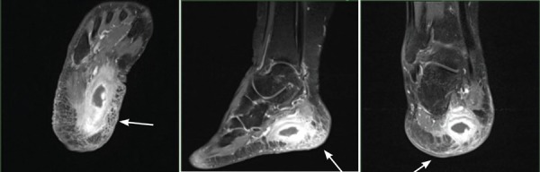
Figure 3. MRI demonstrating flexor digitorum brevis intramuscular collection (white arrow). There are also focal osteomyelitic changes to the adjacent calcaneum.
Surgical debridement and drainage was performed, with findings of 1cc intramuscular pus between the right flexor digitorum brevis plantar fascia (Figure 4). The plantar fascia also was observed to have fibrotic changes. A culture revealed Methicillin-Resistant Staphylococcus Aureus (MRSA) with a sensitivity to the antibiotic Vancomycin. The patient was treated with Vancomycin intravenously for 1 week and later completed 6 weeks of oral Fusidic acid and Rifampicin. She recovered well and could ambulate in regular shoes with a silicone heel pad without pain. The wound healed with a painless thin scar. A follow-up radiograph nearly one year of surgery showed a healed calcaneus (Figure 5).
Figure 4. Intraoperative findings of an intramuscular abscess between the flexor digitorum brevis and plantar fascia.

Figure 5. Plain radiograph taken 5 months post-surgery showing a healed calcaneum.

Discussion
The plantar fascia is a thickened fibrous aponeurosis that originates from the medial tubercle of the calcaneus. It inserts into the deep transverse ligaments of the metatarsal heads and forms the fibrous flexor sheaths on the plantar aspect of the toes.2 Pain sensation is registered and mediated by the small plantar nerves in and around the plantar fascia. Plantar fasciitis pain is caused by the degenerative irritation at the insertion of the plantar fascia on the medial process of the calcaneal tuberosity. The typical presentation is a sharp pain localized at the anterior aspect of the calcaneus. The etiology of this condition is multifactorial; however, most cases result from overuse stresses. Risk factors of developing plantar fasciitis include having pes planus or heel spurs, obesity, as well as occupations that require prolonged standing.3
The conventional therapeutic options for treating plantar fasciitis have been directed at decreasing the presumed inflammation and include non-steroidal anti-inflammatory drugs, ice therapy, rest and activity modification, corticosteroids, splinting, shoe modifications, orthosis, and physiotherapy. Other forms of treatment aim to create an acute inflammatory response to restart the healing process and include a platelet-rich plasma (PRP) injection, extracorporeal shock-wave therapy (ESWT), and surgical procedures.1 These treatment modalities may be used in combination.
The complications of corticosteroid injection into the superficial fat are fat pad necrosis and skin atrophy. Less commonly reported side effects include increased risk of spontaneous plantar fascia rupture.4 Although rare, infection is the most catastrophic possible outcome of a corticosteroid injection (risk of 1 in 1,000)5, as highlighted in this case report. To reduce the risks of infection, a sterile technique is used, and the patient is advised to maintain good foot hygiene post injection. Injection of soft tissues or peritendinous injections can lead to elevations in blood glucose that can persist for at least five days.5 Therefore, diabetic patients need to have closer glycemic monitoring and follow-up post injections to ensure a good clinical outcome.
Iatrogenic calcaneal osteomyelitis following a heel injection is rarely documented and reported. The current case was first seen in our center, and, based on the literature review, there is no local data available concerning the occurrence of this particular complication of a steroid injection in Malaysia. The calcaneum is invested by the closely adhering periosteum, over which muscles are attached. In calcaneal osteomyelitis, pus collected within the bone cannot clear the periosteum; instead, the periosteum is perforated.6 Therefore, typically, a draining sinus is formed and may be located over the plantar, medial or lateral surface of the heel.
Acute osteomyelitis is usually treated by intravenous antibiotics and drainage. Our patient recovered well following surgical drainage and antibiotic therapy, with no residual complications. However, management of intractable chronic osteomyelitis can be a challenge. Definitive surgery can proceed when optimal soft tissue conditions and normalized infective parameters are present. Other aspects to look for are co-morbidities and nutrition status, as the patient's condition needs to be optimized prior to surgery.7 Recalcitrant chronic osteomyelitis of calcaneum can be treated via different surgical procedures, for example, a partial or total calcanectomy, a split heel approach, or vascularized flaps to cover the ulcer.8 Due to the significant morbidities associated with partial or total calcanectomies, such as fat pad instability, loss of Achilles tendon insertion and action, and failure leading to proximal amputation, one should reserve these procedures as last resorts in the sensate patient without diabetes.9
In a study done by Shetty et al.10 involving 60 patients, a comparison of the effectiveness of platelet-rich plasma (PRP) and corticosteroid injections found no significant difference at 6-month follow-ups. Monto11 conducted a study of 40 patients and found that a PRP injection was more effective than a corticosteroid injection at 2-year follow-ups. Aksahin et al.12 concluded that both treatments were equally effective; however, PRP injections appeared to be safer when the potential complication of corticosteroid treatment was taken into consideration. However, such treatments should be offered only after conservative treatments have failed, as 80% of patients recovered well after initial conservative management.13
Conclusion
This case highlights the significant risk of infection following steroid injection in plantar fasciitis, especially in diabetic and immunosuppressed patients. As prevention is better than the cure, a skin preparation regimen must be adhered to, and a sterile environment must be maintained during the procedure to limit contamination. Therefore, it is important that the risks involved be explained to patient when obtaining consent and that close follow-up is carried out to detect infection early.
Acknowledgement
We would like to thank Dr. Nazatul Shima Nayan for her contribution and technical help in preparing this manuscript.
References
- 1.Gill LH, Kiebzak GM. Outcome of nonsurgical treatment for plantar fasciitis. Foot Ankle Int. 1996 Sep;17(9):527–32. doi: 10.1177/107110079601700903. [DOI] [PubMed] [Google Scholar]
- 2.William PL, Warwick R, Gray H. Gray's anatomy. Vol. 36. Philadelphia: WB Saunders; 1980. pp. 612–613. [Google Scholar]
- 3.Buchbinder R. Clinical practice. Plantar fasciitis. The New England Journal of Medicine. 2004 May;350(21):2159–66. doi: 10.1056/NEJMcp032745. [DOI] [PubMed] [Google Scholar]
- 4.Acevedo JI, Beskin JL. Complications of plantar fascia rupture associated with corticosteroid injection. Foot Ankle Int. 1998;19(2):91–97. doi: 10.1177/107110079801900207. [DOI] [PubMed] [Google Scholar]
- 5.Wang AA, Hutchinson DT. The effect of corticosteroid injection for trigger finger on blood glucose level in diabetic patients. J Hand Surg Am. 2006;31(6):979–981. doi: 10.1016/j.jhsa.2006.03.022. [DOI] [PubMed] [Google Scholar]
- 6.Gaenslen FJ. Split-heel approach in osteomyelitis of os calcis. JBJS. 1931;13(4):759–772. [Google Scholar]
- 7.Bajuri MY, Razak KA. Chronic osteomyelitis of the femur with segmental bone defect: concepts and treatment. Journal of Krishna Institute of Medical Sciences University. 2017;6(2):127–130. [Google Scholar]
- 8.Bhattacharyya A, Das R. Gaeslen's split heel approach for the treatment of chronic osteomyelitis of the calcaneus: a series of three cases. The Foot and Ankle Online Journal. 2010;3(11):3. [Google Scholar]
- 9.Canales M, Bowen M, Gerhard J. Treating iatrogenic calcaneal osteomyelitis following a plantar heel injection. Podiatry Today. 2014;27(7) [Google Scholar]
- 10.Shetty VD, Dhillon M, Hegde C, et al. A study to compare the efficacy of corticosteroid therapy with platelet-rich plasma therapy in recalcitrant plantar fasciitis: a preliminary report. Foot Ankle Surg. 2014;20(1):10–13. doi: 10.1016/j.fas.2013.08.002. [DOI] [PubMed] [Google Scholar]
- 11.Monto RR. Platelet-rich plasma efficacy versus corticosteroid injection treatment for chronic severe plantar fasciitis. Foot Ankle Int. 2014;35(4):313–318. doi: 10.1177/1071100713519778. [DOI] [PubMed] [Google Scholar]
- 12.Aksahin E, Dogruyol D, Yüksel HY, et al. The comparison of the effect of corticosteroids and platelet-rich plasma (PRP) for the treatment of plantar fasciitis. Arch Orthop Trauma Surg. 2012;132(6):781–785. doi: 10.1007/s00402-012-1488-5. [DOI] [PubMed] [Google Scholar]
- 13.Ragab EM, Othman AM. Platelets rich plasma for treatment of chronic plantar fasciitis. Arch Orthop Trauma Surg. 2012;132(8):1065–1070. doi: 10.1007/s00402-012-1505-8. [DOI] [PubMed] [Google Scholar]